
T.P. Kalantaripour
Faculty of Midwifery and Nursing, Islamic Azad University, Branch of Kerman, Kerman, Iran
M. Asadi-Shekaari
Neuroscience Research Center, Kerman Medical University, Kerman, Iran
M. Basiri
Neuroscience Research Center, Kerman Medical University, Kerman, Iran
A. Gholaamhosseinian Najar
Department of Biochemistry, Afzali Pour Medical Faculty, Kerman Medical University, Kerman, Iran
Journal of Biological Sciences
Year: 2012 | Volume: 12 | Issue: 3 | Page No.: 180-185


ABSTRACT
In the present study, we investigate the role of Date Seed Extract (DSE) in protection against cerebral ischemic damages. Focal cerebral ischemia was induced by using Middle Cerebral Artery Occlusion (MCAO) method in male rats. DSE was administrated at the dose of 80 mg kg-1 i.p. 30 min after MCAO. Superoxide Dismutase (SOD), Malondialdehyde (MDA) and Total antioxidant Activity (TAS) were measured. Morphological studies and behavioral activity were also done. MCAO significantly decreased SOD and TAS activities. MDA content significantly was elevated by MCAO. Motor coordination was also decreased due to transient ischemia. Treatment with DSE attenuated all of alterations and neuronal damage induced by MCAO in male rats. The data showed that treatment with DSE could protect cortical neurons against cerebral-induced injuries most probably due to its antioxidant properties.
PDF Abstract XML References Citation
Received: December 08, 2011;
Accepted: March 12, 2012;
Published: June 12, 2012
How to cite this article
T.P. Kalantaripour, M. Asadi-Shekaari, M. Basiri and A. Gholaamhosseinian Najar, 2012. Cerebroprotective Effect of Date Seed Extract (Phoenix dactylifera) on Focal Cerebral Ischemia in Male Rats. Journal of Biological Sciences, 12: 180-185.
DOI: 10.3923/jbs.2012.180.185
URL: https://scialert.net/abstract/?doi=jbs.2012.180.185
DOI: 10.3923/jbs.2012.180.185
URL: https://scialert.net/abstract/?doi=jbs.2012.180.185
INTRODUCTION
The date palm (Phoenix dactylifera) is a palm in the genus Phoenix, widely cultivated for edible fruit. Date seed constitutes approximately 15% of date fruit (Hussein et al., 1998). At the present time, date seeds are used mostly for animal feed, while most regarded as waste. Use of such waste is very important to date agriculture and to increase the income to this part. Date seed (pit) contains relatively high amounts of fat (9.0 g/100 g) and protein (5.1 g/100 g) compared to date fruit. They are a very rich source of antioxidants (80400 μmol 100 g), phenolics (3942 mg/ 100 g) and dietary fiber (73.1 g/100 g). The seed could potentially be considered as an economical source of natural antioxidants and dietary fiber (Al-Farsi and Lee, 2008).
Iranian date seeds are potent antioxidants and strong free radical scavengers (Ardekani et al., 2010). They contain several antioxidant components including phenolic compounds (Phenolic acids, anthocyanins and flavonoids), selenium (coenzyme of GPx), vitamin C, Oleic acid and carotenoids (Al-Farsi and Lee, 2008; Al-Qarawi et al., 2004). A recent study showed that the antioxidants of DSE are approximately 27 fold of date fruit (Al-Farsi and Lee, 2008). It supports its noticeable antioxidant activity.
Stroke is a leading cause of death in developed countries. Current therapeutic strategies for stroke have been largely unsuccessful (Caplan, 2004). Many studies have shown the neuroprotective effects of herbal extracts on different animal models (Waggas, 2009; Tehranipour and Javaheri, 2009; Tehranipour and Ghadamyari, 2010). Therefore, there is a great interest in prevention and treatment of the disease. In the present study, we investigate the cerebroprotective effect of DSE in male rats.
MATERIALS AND METHODS
Animals: Healthy NMRI male rats (weighing 225-280 g) were used in the study. This study was performed in accordance with neuroscience research center guidelines for care and use of laboratory animals (EC/KNRC/87-29). All of efforts were taken to minimize distress, discomfort, and pain to the animals.
Date seed extract preparation: Date fruits (Bamy Mozafaty rutab) were purchased from authenticated market. Date seeds were isolated from date fruit and were soaked in distilled water, washed to get rid of any adhering flesh and dried at room temperature. The Seeds were milled in a heavy-duty grinder. The fine powder (50 g) was extracted with distilled water (1: 3 ratio, w/v) and centrifuged at 4°C for 20 min at 4000 g and the supernatant was collected, lyophilized and stored at -20°C until use.
Experimental design: Rats were randomly divided into 3 groups: The ischemic group suffered 30 min MCAO followed by 48 h reperfusion (MCAO) (n = 9), the sham group suffered the same as ischemic group without MCAO (control) (n = 10) and the experimental group suffered 30 min MCAO treated with the dose of DSE 80 mg kg-1 i.p. 30 min after MCAO induction and 48 h reperfusion (DSE) (n = 10). The dose of DSE was selected based on a pilot study.
Middle cerebral artery occlusion: The right middle cerebral artery occlusion was induced using intraluminal filament model (Longa et al., 1989). In brief, the animals were anesthetized with chloral hydrate (360 mg kg-1). In supine position, a midline ventral incision was made the right Common Carotid Artery (CCA) was exposed and separated carefully from Vagus nerve. All of External Carotid Artery (ECA) branches and extra cranial of Internal Carotid Artery (ICA) were blocked. Then 4-0 nylon suture was introduced into ICA and advancing in intracranially to block blood flow into MCA. After thirty min ischemia, suture was withdrawn to restore the blood flow (reperfusion). The rectal temperature was maintained at 37±0.5°C with a thermistor coupled to a heating blanket during surgery. After recovery from the anesthesia, the animals were returned to their cages. After full recovery, neurological examination (Garcia et al., 1995) was performed to ensure MCAO occurred and the animals without clinical signs were excluded from the experiment.
Sample preparation method
Light microscopy: Under deep anesthesia, the animals were perfused intracardially with 10% formalin in phosphate buffered saline (pH 7.4, M 0.1%). Brains were removed and immersed in the same fixative overnight at 4°C. After, the brains were processed for light microscopy studies according to routine procedures. Coronal sections 1.6-2.8 mm posterior to bregma were cut at a thickness of 5 μm using a microtome. Neuronal damage in the cortex was assessed by staining sections with hematoxylin and eosin using the standard method (Sakurai-Yamashita et al., 2006).
Electron microscopy: Rats were killed by perfusion transcardially with 4% paraformaldehyde in 0.1 M phosphate buffer solution (pH 7.4). The brains were removed and immersed in buffered 2.5% glutaraldehyde overnight. A small piece of cortical area was dissected and fixed in the same fixative for an additional 24 h. Specimens were postfixed in 1% (w/v) OsO 4/1% (w/v) of phosphate buffer. After dehydration in ethanol, slices were embedded in Epon 812 resin. A section (300 nm) was stained with toluidine blue to locate areas of interest. Subsequently, 70-80 nm sections were cut and stained with uranyl acetate and lead citrate stain. The sections were examined with a Philips (EM 300, Eindhoven, Netherlands) transmission electron microscope (Zangiabadi et al., 2011a).
Biochemical analysis: Forty-eight h after reperfusion onset, the rats were sacrificed and brains removed immediately. Homogenization was performed according to Aydin et al. (2002) with some modification.
Briefly, piece of the cortical tissue was removed and washed with saline and homogenized by sonication (4 °C) in 0.15 M NaCl for lipid peroxidation and antioxidant parameters. Homogenate was centrifuged at 5000 rpm for 15 min. aliquots for each sample were poured in small Eppendorf tubes and kept frozen at -70°C until use. Malondialdehyde (MDA) levels were determined spectrophotometrically by modified method of Uchiyama and Mihara (1997). Superoxide Dismutase (SOD) activity and Total antioxidants level (TAS) were measured by spectrophotometric methods using Randox assay kits (Randox Laboratories, Antrim, UK) based on the manufacturer protocols. In order to compensate the effect of size and amount of used sample in the homogenization step, the protein content of each preparation was measured by Folin phenol reagent of Lowry et al. (1951).
Rotarod activity: The animals were evaluated for balance and grip strength using the rotarod. Each animal was given a former training session before beginning of therapy to adapt them on a rotarod apparatus (Technical and Scientific Equipment, GmbH, Germany). Rats were placed on the rotating rod with a diameter of 7 cm (speed 20 rpm). Three trials were given to each animal at 10 min interval and cut off time (300 sec) was maintained throughout the trial. The average results were recorded as fall of time (Gaur and Kumar, 2010a).
Statistical analysis: The statistical analysis was done using SPSS software v13.5. All values are expressed as Mean±SEM.
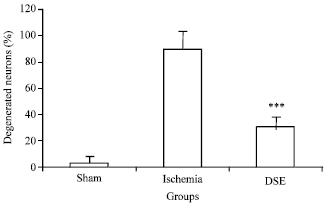 | |
| Fig. 1: | Cerebroprotective effect of treatment with DSE on ischemic-reperfusion injury in male rat, Neuronal damage in the cerebral cortex 48 h after reperfusion was assessed using hematoxylin and eosin (H and E), Results are expressed as Mean±SE and data were analyzed by one-way ANOVA |
Differences in measured parameters among different groups were analyzed by one-way ANOVA followed by post hoc Tukey’s test. A statistical difference was determined by a value of p<0.05.
RESULTS
Effects of DSE on neuronal damage: According to the obtained data_ 30 min MCAO followed by 48 h reperfusion induced 89.37% neuronal death in MCAO group. Treatment with DSE (80 mg kg-1) significantly decreased the neuronal damage (30.33%) (Fig. 1).
Electron microscopy: Cortical neurons were examined by transmission electron microscope 48 h after 30 min MCAO. Morphology of neurons in control group was normal. Degenerative changes including darkening of nucleus, chromatin aggregation, organelles swelling were observed in MCAO group. The ultrastructure of most cortical neurons was preserved in DSE group (Fig. 2).
Effect of DSE on fall-off time in rotarod after MCAO in rats: The fall-off time is measured for rotarod evaluation to measure motor in coordination. A significant decrease observed in fall-off time in MCAO group as compared to control group which show motor in coordination and muscle weakness. DSE significantly (p<0.05) improved the fall-off latency time as compared to MCAO group (Fig. 3).
Effect of DSE on oxidative stress in brain after MCAO in rats: MDA as an oxidative marker was significantly increased after ischemia reperfusion damage in MCAO group as compared to control group.
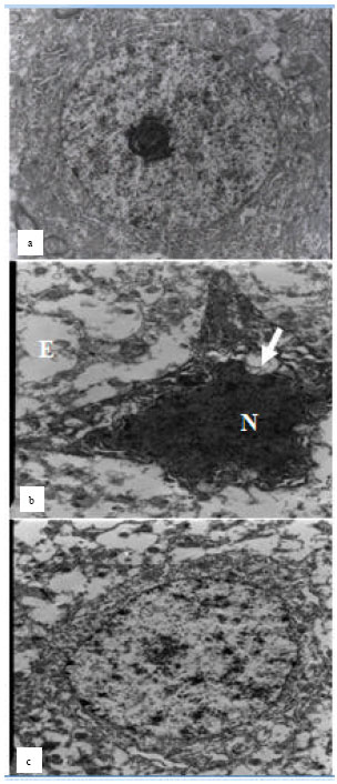 | |
| Fig. 2 (a-c): | Electron micrograph of cortical neurons from control, MCAO and DSE groups, (a) The normal ultrastructure is visible in intact neuron, (b) Ischemia reperfusion results in severe degenerative changes in cortical neuron. (c) Cortical neurons in DSE group have some degenerative changes like extracellular edema, chromatin aggregation but the whole ultrastructure was maintained (part c), Magnification in (a) and (b) is 8900x and in (c) is 3900 |
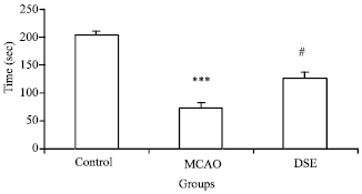 | |
| Fig. 3: | Effect of DSE on fall-off time from rotarod in ischemic-reperfusion injury in male rat, ***p<0.001 as compared to control group, #p<0.05 as compared to MCAO |
| Table 1: | *Levels of MDA, SOD and TAS in control, MCAO and MCAO + DSE groups in rats |
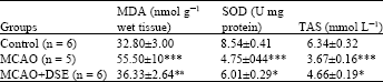 | |
| *p<0.05 as compared to MCAO, *ap<0.001 as compared to MCAO, ***p<0.001 as compared to control, MDA: Malondialdehyde, SOD: Superoxide dismutase, TAS: Total Antioxidant Status, MCAO: Middle cerebral artery occlusion | |
DSE administration (80 mg kg-1) significantly decreased MDA level after ischemia reperfusion as compared to MCAO group (p<0.05).
Effect of DSE on SOD activity and total antioxidants level in brain after MCAO in rats: Antioxidant enzyme activity (SOD) and total antioxidants level were attenuated after ischemia reperfusion damage in MCAO group. A significant increase in SOD activity and TAS level was observed in DSE treated group (p<0.05) (Table 1).
DISCUSSION
Oxidative stress is involved in the pathophysiology of stroke. A large number of studies have shown that oxidative stress contributes to brain injury due to ischemia reperfusion (Ikeda and Langn, 1990; Yavuz et al., 1997; Cuzzocrea et al., 2001; Guizzo et al., 2005; Oboh, 2009). Ischemia reperfusion causes a significant increase in oxidative stress markers such as: nitrite, MDA, and reactive oxygen species concentration. In addition, there is a considerable decrease observed in antioxidant enzymes such as catalase and SOD activity, in the brain (Levine, 2004; Siesjo, 2008; Gaur and Kumar, 2011; Nandagopal et al., 2011). In the present study we have evaluated oxidative stress parameters (MDA, SOD and TAS) in the brain. However, a marked increase in MDA concentration was observed in MCAO group. A decrease in SOD as well as TAS activity was also seen 48 h after reperfusion indicating oxidative stress caused by ischemic-reperfusion damage. DSE significantly attenuated the oxidative stress and restored the antioxidant enzyme activity.
Motor disorders are one of the most destructive outcomes of cerebral ischemia due to MCAO because most of the pyramidal tract and motor cortex lie inside territory supplied by MCA. Motor disorder may arise from failure of cortical excitability and/or inhibition of electrical impulses at the subcortical area. After ischemia and reperfusion, axonal conduction readily recovers. However, a constant failure at cortical synapses leads to motor dysfunction (Bolay and Dalkara, 1998). Several studies have shown that the neurological and locomotor deficits after ischemia in rats (Gaur and Kumar, 2010b; Aggarwal et al., 2010; Gupta et al., 2005). The fall-off time from rotarod was significantly decreased when compared to control group verifying the deficit in muscle co-ordination and grip strength. Decrease in muscle co-ordination and grip strength has also been shown by various studies in MCAO (Jafari et al., 2011; Maheshwari et al., 2011) as well as the BCCAO (bilateral common carotid artery occlusion) model of cerebral ischemia (Gaur et al., 2009; Gaur and Kumar, 2010c). Treatment with DSE attenuated muscle weakness. This supports its protective effect against ischemia reperfusion damage.
On the other hand, histological and ultrastructural findings showed that DSE could protect cortical neurons against ischemia reperfusion induced insults. According to our data, 30 min MCAO followed by 48 h reperfusion results in severe damage to neurons (89.37%) in MCAO group. This finding is in agreement with other studies (Asadi-Shekaari et al., 2010; Asadi-Shekaari et al., 2008). DSE treatment can protect them against ischemic injury. In line with above findings, electron microscopy examination showed degenerative changes in cortical neurons as shown in Fig. 3 after ischemia reperfusion and DSE had protective effects against these changes.
Previously we demonstrated that aqueous extract of date fruit has neuroprotective action against cerebral ischemic injuries and diabetic neuropathy most probably due to its antioxidant effects (Asadi-Shekaari et al., 2008; Panahi et al., 2008; Zangiabadi et al., 2011b). Here we showed the DSE significantly improved the outcome in male rats after ischemia reperfusion. At the present time, the exact mechanism by which the DSE induces its cerebroprotective activity against ischemia reperfusion injury is not known. But it is possible that its antioxidant components present in the DSE are responsible for this protection.
In conclusion, the present study demonstrated that DSE has a cerebroprotective role for the period of brain ischemia followed by reperfusion in male rats. This implies that use of DSE may have beneficial effects in cerebral ischemia. Further studies are needed to be able to propose the potential therapeutic use of DSE in preventing the brain from ischemic-induced oxidative damage.
ACKNOWLEDGMENT
This study was supported by the research grant from Kerman Neuroscience Research Center (KNRC).
REFERENCES
- Aggarwal, A., V. Gaur and A. Kumar, 2010. Nitric oxide mechanism in the protective effect of naringin against Post-Stroke Depression (PSD) in mice. Life Sci., 86: 928-935.
CrossRefPubMedDirect Link - Al-Farsi, M.A. and C.Y. Lee, 2008. Nutritional and functional properties of dates: A review. Crit. Rev. Food Sci. Nutr., 48: 877-887.
CrossRefDirect Link - Al-Qarawi, A.A., H.M. Mousa, B.E.H. Ali, H. Abedl-Rahman and S.A. El-Mougy, 2004. Protective effect of extracts from dates (Phoenix dactylifera L.) on carbon tetrachloride-induced hepatotoxicity in rats. Int. J. Rev. Vet. Med., 2: 176-180.
Direct Link - Asadi-Shekaari, M., H.E. Vaghefi, A. Talakoub and H.R.K. Khorshid, 2010. Effects of semelil (Angipars™) on focal cerebral ischemia in male rats. DARU., 18: 265-269.
Direct Link - Asadi-Shekaari, M., P. Marzieh, D. Shahriar, S.K. Zahed and K.T. Pari, 2008. Neuroprotective effects of aqueous date fruit extract on focal cerebral ischemia in rats. Pak. J. Med. Sci., 24: 661-665.
Direct Link - Aydin, S., R. Ozaras, H. Uzun, A. Belce and E. Uslu et al., 2002. N-acetylcysteine reduced the effect of ethanol on antioxidant system in rat plasma and brain tissue. Tohoku J. Exp. Med., 198: 71-77.
CrossRef - Caplan, L.R., 2004. Treatment of acute stroke: Still struggling. J. Am. Med. Assoc., 292: 1883-1885.
CrossRefDirect Link - Cuzzocrea, S., D.P. Riley, A.P. Caputi and D. Salvemini, 2001. Antioxidant therapy: A new pharmacological approach in shock, inflammation and ischemia/reperfusion injury. Pharmacol. Rev., 53: 135-159.
PubMedDirect Link - Garcia, J.H., S. Wagner, K.F. Liu and X.J. Hu, 1995. Neurological deficit and extent of neuronal necrosis attributable to middle cerebral artery occlusion in rats. Statistical validation. Stroke, 26: 627-634.
PubMedDirect Link - Gaur, V., A. Aggarwal and A. Kumar, 2009. Protective effect of naringin against ischemic reperfusion cerebral injury: Possible neurobehavioral, biochemical and cellular alterations in rat brain. Eur. J. Pharmacol., 616: 147-154.
CrossRefPubMedDirect Link - Gaur, V. and A. Kumar, 2010. Behavioral, biochemical and cellular correlates in the protective effect of sertraline against transient global ischemia induced behavioral despair: Possible involvement of nitric oxide-cyclic guanosine monophosphate study pathway. Brain Res. Bull., 82: 57-64.
CrossRefPubMedDirect Link - Gaur, V. and A. Kumar, 2010. Protective effect of desipramine, venlafaxine and trazodone against experimental animal model of transient global ischemia: Possible involvement of NO-cGMP pathway. Brain Res., 1353: 204-212.
CrossRefPubMedDirect Link - Guizzo, R., M.A.R. Cairrao, J. Coutinho-Netto, A.R. Meirelles e Silva, N.C. Coimbra and W.F. dos Santos, 2005. Neuroprotection in acute ischemia and ischemia/reperfusion in rat retina. Int. J. Pharmacol., 1: 369-375.
CrossRefDirect Link - Gupta, Y.K., S. Briyal, U. Sharma, N.R. Jagannathan and A. Gulati, 2005. Effect of endothelin antagonist (TAK-044) on cerebral ischemic volume, oxidative stress markers and neurobehavioral parameters in the middle cerebral artery occlusion model of stroke in rats. Life Sci., 77: 15-27.
CrossRefDirect Link - Hussein, A.S., G.A. Alhadrami and Y.H. Khalil, 1998. The use of dates and date pits in broiler starter and finishers diets. Bioresour. Tehnol., 66: 219-223.
Direct Link - Ikeda, Y. and D.M. Long, 1990. The molecular basis of brain injury and brain edema the role of oxygen radicals. Neurosurgery, 27: 1-11.
PubMed - Longa, E.Z., P.R. Weinstein, S. Carlson and R. Cummins, 1989. Reversible middle cerebral artery occlusion without craniectomy in rats. Stroke, 20: 84-91.
CrossRefDirect Link - Lowry, O.H., N.J. Rosebrough, A.L. Farr and R.J. Randall, 1951. Protein measurement with the folin phenol reagent. J. Biol. Chem., 193: 265-275.
CrossRefPubMedDirect Link - Maheshwari, A., L. Badgujar, B. Phukan, S.L. Bodhankar and P. Thakurdesai, 2011. Protective effect of Etoricoxib against middle cerebral artery occlusion induced transient focal cerebral ischemia in rats. Eur. J. Pharmacol., 667: 230-237.
CrossRefPubMedDirect Link - Nandagopal, M., P. Muralidharan and G. Thirumurugan, 2011. Cerebroprotective effect of root extract of Asparagus racemosus Willd. in global cerebral ischemia in rats. J. Pharmacol. Toxicol., 6: 49-61.
CrossRefDirect Link - Oboh, G., 2009. The neuroprotective potentials of sour (Hibiscus sabdariffa, Calyx) and green (Camellia sinensis) teas on some pro-oxidants induced oxidative stress in brain. Asian J. Clin. Nutr., 1: 40-49.
CrossRefDirect Link - Sakurai-Yamashita, Y., H. Kinugawa and N. Niwa, 2006. Neuroprotective effect of pentosan polysulphate on ischemia-related neuronal death of hippocampus. Neurosci. Lett., 409: 30-34.
PubMedDirect Link - Jafari, S.S.S., A.A. Aghaei, M. Asadi-Shekaari, S.N. Nematollahi-Mahani and V. Sheibani, 2011. Investigating the effects of adult neural stem cell transplantation by lumbar puncture in transient cerebral ischemia. Neurosci. Lett., 495: 1-5.
CrossRefPubMedDirect Link - Ardekani, M.R.S., M. Khanavi, M. Hajimahmoodi, M. Jahangiri and A. Hadjiakhoondi, 2010. Comparison of antioxidant activity and total phenol contents of some date seed varieties from Iran. Iran. J. Pharm. Res., 9: 141-146.
PubMedDirect Link - Tehranipour, M. and T. Ghadamyari, 2010. The effects of root aquatic extract of Salvia staminea on neuronal density of alpha motoneurons in spinal cord anterior horn after sciatic nerve compression in rats. J. Biol. Sci., 10: 48-52.
CrossRefDirect Link - Tehranipour, M. and R. Javaheri, 2009. Neuroprotetive effect of Curcuma longa alcoholic extract on peripheral nerves degeneration after sciatic nerve compression in rats. J. Biol. Sci., 9: 889-893.
CrossRefDirect Link - Siesjo, B.K., 2008. Pathophysiology and treatment of focal cerebral ischemia. Part I: Pathophysiology. (1992). J. Neurosurg., 108: 616-631.
PubMedDirect Link - Uchiyama, M. and M. Mihara, 1979. Determination of malonaldehyde precursor in tissues by thiobarbituric acid test. Anal. Biochem., 86: 271-278.
CrossRefDirect Link - Waggas, A.M., 2009. Neuroprotective evaluation of extract of ginger (Zingiber officinale) root in monosodium glutamate-induced toxicity in different brain areas of male albino rats. Pak. J. Biol. Sci., 12: 201-212.
CrossRefPubMedDirect Link - Zangiabadi, N., V. Sheibani, M. Asadi-Shekaari, M. Shabani and M. Jafari et al., 2011. Effects of melatonin in prevention of neuropathy in STZ-induced diabetic rats. Am. J. Pharmacol. Toxicol., 6: 59-67.
Direct Link - Zangiabadi, N., M. Asadi-Shekaari, V. Sheibani, M. Jafari and M. Shabani et al., 2011. Date fruit extract is neuroprotective agent on diabetic peripheral neuropathy in streptozotocin-induced diabetic rats: A multimodal analysis. Oxid. Med. Cell. Long., (In Press).
Direct Link - Gaur, V. and A. Kumar, 2011. Neuroprotective potentials of candesartan, atorvastatin and their combination against stroke induced motor dysfunction. Inflammopharmacology, 19: 205-214.
CrossRefPubMedDirect Link