
A.Y. Coulibaly
Universite Norbert Zongo, UFR/ST, BP 376, Koudougou, Burkina Faso
LiveDNA: 226.30688
J.H. Bationo
Laboratoire de Biochimie and Chimie Appliquees, UFR/SVT, University Joseph Ki-zerbo, 09 PB 848 Ouagadougou 09, Burkina Faso
Fatouma Mohamed Abdoul-Latif
Institut de Recherche Medicale, Centre d�Etude et de Recherche de Djibouti, P.O. Box 486, Djibouti
Bakasso Sahabi
Laboratory of Natural Products and Organic Synthesis, Department of Chemistry, Faculty of Science and Technology, Abdou Moumouni University of Niamey, BP 10662, Niamey, Niger
M. Kiendrebeogo
Laboratoire de Biochimie and Chimie Appliquees, UFR/SVT, University Joseph Ki-zerbo, 09 PB 848 Ouagadougou 09, Burkina Faso
Journal of Biological Sciences
Year: 2020 | Volume: 20 | Issue: 1 | Page No.: 1-6
DOI: 10.3923/jbs.2020.1.6


ABSTRACT
Background and objectives: Scoparia dulcis is a medicinal herb traditionally used to relieve dementia disorders in Burkina Faso. A female Wistar rats model was used against hydroacetonic extract of Scoparia dulcis to assess the effect of neuroprotective, hepatoprotective and renoprotective. Materials and Methods: The rats were divided into 5 groups with 6 animals/group. Carboxymethyl cellulose (CMC, 0.5%) was used as vehicle to treat the animals by oral route with silymarin or the plant extract. Seven days later, they were intoxicated by intra-peritoneal injection of carbon tetrachloride (2 mL kg–1) for 24 h. Then blood, brain, kidney and liver were taken for further analysis. Results: It resulted on a neuroprotective effect noted by an increase in acetylcholinesterase level, a hepatoprotective effect was also registered with a significant decrease in triglycerides, cholesterol, alanine amino-transferase (ALT), aspartate amino-transferase (AST), superoxide dismutase (SOD) and malondialdehyde (MDA). The renoprotective potential was marked by a decrease of urea, uric acid and creatinine. Conclusions: Scoparia dulcis can be a suitable candidate for investigation of neuroprotective agents.
PDF Abstract XML References Citation
Copyright: © 2020. This is an open access article distributed under the terms of the creative commons attribution License, which permits unrestricted use, distribution and reproduction in any medium, provided the original author and source are credited.
How to cite this article
A.Y. Coulibaly, J.H. Bationo, Fatouma Mohamed Abdoul-Latif, Bakasso Sahabi and M. Kiendrebeogo, 2020. A Female Wistar Rats Model for Assessing the in vivo Protective Role of Scoparia dulcis on Liver, Kidney and Brain. Journal of Biological Sciences, 20: 1-6.
DOI: 10.3923/jbs.2020.1.6
URL: https://scialert.net/abstract/?doi=jbs.2020.1.6
DOI: 10.3923/jbs.2020.1.6
URL: https://scialert.net/abstract/?doi=jbs.2020.1.6
INTRODUCTION
Scoparia dulcis L. (Scrophulariaceae) is a widespread herb in tropical and subtropical regions where it is well known as a folk-medicinal plant1.
Previous investigations evidenced a number of its medicinal properties against diabetes, inflammation and oxidation in vivo2,3 and its impact on lipid peroxidation4,5. Moreover, its protective role on insulinoma cell line RINm5F6 and on kidney, heart and liver of rats exposed to cadmium2 were demonstrated. A few studies were carried on the hepatoprotective role of the plant7-10. Along with its protective effect on liver and kidney, S. dulcis is used to help with the management of memory impairments in local populations in Burkina Faso11 and our previous research related its positive impact on brain protection in vitro12.
Indeed, the brain and nervous system have limited antioxidant capacity and are most vulnerable to oxidative stress13. Acetylcholine (Ach) is a neurotransmitter in neuronal cells and its deficiency in cerebral cortex was found to be a major cause of neurodegenerative disorder such as Alzheimer’s disease14. Its action is stopped by acetylcholinesterase (AChE), a key enzyme catalyzing the hydrolysis of acetylcholine (ACh) in the nervous system of animals and insects. The use of AChE inhibitors can help for neuroprotection in neurodegenerative disorders by enhancing the Ach level in the brain15.
Previous study stated a markedly inhibition of acetylcholinesterase by scoparia dulcis in vitro 12.
This study was then carried out to contribute to the understanding of the in vivo protective role of Scoparia dulcis particularly on brain based biomarker AChE activity along with the assessing of its hepatoprotection and renoprotection on rats exposed to carbon tetrachloride toxicity.
MATERIAL AND METHODS
Plant material: The whole plant of Scoparia dulcis L. was harvested around the dam of Loumbila in August, 2011. The plant was authenticated by the Plant Ecology and Botany Laboratory/University of Ouagadougou where a specimen was deposited in the herbarium under the identification code SD_ac01. The sample was dried at room temperature (25°C) and then reduced to powder and kept for extraction.
Extraction: A mass of 25 g of plant powder was macerated for 24 h in 250 mL of a mixture constitute by acetone and water (80/20, v/v). After filtration, the acetone was evaporated in a rotavapor apparatus and the aqueous phase obtained was lyophilized to constitute the extract12.
Experimental design
Acute toxicity: The toxicity of the plant extract was evaluated according to the protocol described by OECD16. A group of 3 Wistar rats was used and fasted for 18 h. Each rat then received a single dose of 3000 mg kg–1 of body weight orally. They were then observed constantly during the 1st 30 min and then regularly for the next 24 h to note any sign of toxicity (contortions, panic, moribund state, death .). The observation continued beyond 14 days after which the animals were sacrificed and buried according to the rules of ethics.
Animals treatment conditions: The animals were treated based on previous study by Murthy et al.17 with slight modifications.
Wistar female rats (223-302 g), 3 months old, were acclimated at 25°C with free access to water and food. They were then divided into 5 groups of 6 rats each. Each group receives for 7 days, a single daily dose of carboxyl methylcellulose (CMC, 0.5%) as vehicle alone, either silymarin (100 mg kg–1 b.wt.) or the extract of Scoparia dulcis at the respective doses of 200 and 400 mg kg–1 as follow:
| • | Group 1 (negative control): CMC alone |
| • | Group 2 (normal): CMC alone |
| • | Group 3 (positive control): Silymarin dissolved in the CMC |
| • | Group 4 (test): Extract of Scoparia dulcis at 200 mg kg–1 dissolved in the CMC |
| • | Group 5 (test): Extract of Scoparia dulcis at 400 mg kg–1 dissolved in the CMC |
After 7 days of treatment, the rats of all groups (except those in group 2) are intoxicated by intra-peritoneal injection with carbon tetrachloride (2 mL kg–1 b.wt.). About 24 h later, the rats are asleep under the anesthetic effect of diethyl ether administered by inhalation. The blood is then collected by cardiac puncture using a sterile syringe and stored in 2 types of sterile tubes containing EDTA (acide Ethylène Diamine Tetra Acetique) or not. The liver, kidneys and brain are also collected for the preparation of homogenates (5%) by grinding the organs in a solution of potassium chloride (KCl, 0.15 M) followed by centrifugation at 4000 rpm for 10 min. The collected supernatant constitutes the homogenate.
Quantification of biochemical parameters: The serum obtained by centrifugation of the tubes without EDTA at 1500 rpm is used to determine the biochemical parameters.
Alanine aminotransferase (ALT or GPT), aspartate aminotransferase (AST or GOT), triglycerides, HDL-cholesterol, uric acid and urea are quantified using a kit (LABKIT) according to the manufacturer’s instructions.
Creatinine is quantified using the spinreact kit according to the manufacturer’s instructions.
Total proteins are assayed from liver, kidney and brain homogenates using the spinreact kit according to the manufacturer’s instructions.
Superoxide dismutase estimation: Superoxide dismutase (SOD) was assayed as described by Murthy et al.17. It is based on the reduction of nitroblue tetrazolium (NBT) in blue formazan crystals. Briefly, the liver or kidney homogenate (0.5 mL) was mixed with 1 mL of sodium carbonate (125 mM), 0.4 mL of NBT (24 μM) and 0.2 mL of EDTA (0.1 mM). The reaction is then initiated by the addition of 0.4 mL of hydroxylamine hydrochloride (1 mM). After 5 min of incubation at 25°C, the absorbance is measured at 560 nm against a blank without hydroxylamine hydrochloride. A control without homogenate is prepared under the same conditions. One unit of SOD activity is the amount of enzyme that inhibits 50% of NBT. The specific activity of SOD was expressed as units per milligram of protein.
Lipid peroxidation: Peroxidation of lipids was evaluated as described by Murthy et al.17. It is based on the reaction between thiobarbituric acid (TBA) and malondialdehyde (MDA) to form a detectable chromogen at 532 nm. A volume of 0.5 mL of homogenate of liver or kidney is mixed with 1 mL of a solution of potassium chloride (KCl, 0.15 M). Peroxidation is then initiated by adding 250 μL of iron tri-chloride (FeCl3, 0.2 mM) and then incubated at 37°C for 30 min. The reaction is then stopped by adding 2 mL of an ice-cold mixture of a hydrochloric acid solution (HCl, 0.25 N) containing 15% trichloroacetic acid, 0.3% thiobarbituric acid and 0.05% ascorbic acid. Then the mixture was incubated in a hot bath at 80°C for 60 min. After centrifugation at 4000 rpm for 10 min, the absorbance was read at 532 nm. Lipid peroxidation measures the reactive substances of thiobarbituric acid (TBARS) in MDA equivalent using a molar extinction coefficient of 1.56×10⁵ M–1 cm–1. Results are expressed in nmole mg–1 of protein.
Acetylcholinesterase activity: This method was based on that described by Ellman et al.18 with some modifications. The reaction mixture was constituted by 300 μL of buffer (Tris-HCl 50 mM pH 8.008, 0.1% BSA) and 100 μL of brain homogenate. About 500 μL of DTNB (3 mM) and 100 μL of ATCI substrate are then added and the kinetics was monitored at 405 nm on a spectrophotometer for 5 min against a blank without homogenate. The activity of acetylcholinesterase (AChE) is expressed by Beer Lambert’s law using a molar extinction coefficient of 13.6 mM–1 cm–1 in μmole min–1 mg–1 of protein.
Statistical analysis: All the reactions were performed in triplicate and data are presented as mean±standard deviation. Data were analyzed by one-way analysis of variance followed by the Tukey multiple-comparison test. Analysis were done by using XLSTAT7.1 software. A p-value <0.05 was used as the criterion for statistical significance.
RESULTS
Acute toxicity: Acute toxicity of the hydroacetonic macerate of Scoparia dulcis was determined according to the protocol described by OECD16.
After 14 days of observation, no sign of poisoning was seen on rats after ingestion of a single dose of 3000 mg kg–1 b.wt. of S. dulcis extract. The lethal dose of this extract is then greater than 3000 mg kg–1 and according to the scale of toxicity of the OECD, S. dulcis extract is non-toxic.
The variation in organ weights is not significant between groups 4 and 5 (Table 1) and the relative organ weights indicate liver hypertrophy and kidney atrophy as compared to the normal group (group 2).
Biochemical parameters: Preliminary administration of the extract of Scoparia dulcis in rats showed a protective effect against acute carbon tetrachloride toxicity, characterized by a significant decrease in triglyceride, cholesterol, urea and uric acid levels as compared to the negative control group (Table 2). This hepato-protective effect of the extract is also characterized by a significant decrease in malondialdehyde (MDA) and in the enzymes alanine-amino-transferase (ALT), aspartate amino transferase (AST) and superoxide dismutase (SOD) in comparison to the negative control group as showed in Table 3.
However, creatinine and protein levels were significantly higher in the groups treated with the extract comparatively to the negative control group (Table 3).
The neuro-protective effect of the extract against the toxicity of CCl4 is also expressed by the increase of the acetylcholinesterase level of the groups treated with the plant extract as compared to the negative control group.
| Table 1: | Animals and organ weights |
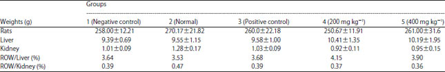 | |
Values are Means±Standard deviation, ROW: Relative organ weight, n = 6 | |
| Table 2: | Chemical components in blood and organs |
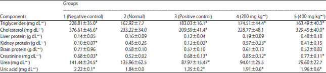 | |
Values are Means±Standard deviation, n = 6, *Significantly different as compared to normal group (p<0.05) | |
| Table 3: | Biochemical parameters in blood and organs |
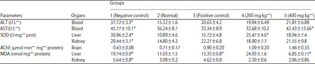 | |
Values are Mean±Standard deviation, n = 6, *Significantly different as compared to normal group (p<0.05), ALT: Alanine amino transferase, AST: Aspartate amino transferase, SOD: Superoxide dismutase, AChE: Acetylcholinesterase, MDA: Malondialdehyde | |
In addition, the positive control group that received silymarin also showed a hepatoprotective and neuroprotective effect, resulting in a decrease in the various measured chemical and biochemical parameters as compared to the negative control group. In some cases (ALT, AST) the level of the decrease is comparable to that of S. dulcis extract at a dose of 200 mg kg–1.
DISCUSSION
The protective effect of S. dulcis extract was evaluated in an animal model using female wistar rats that are in general slightly more sensitive than males19.
The poisoning of the rats was made using carbon tetrachloride (CCl4), a known hepatotoxic agent. After administration, CCl4 is metabolized in hepatocytes by cytochrome p450 with generation of highly reactive derivatives such as trichloromethyl (CCl3) and trichloromethyl peroxyl (CCl3O2) which initiate lipid peroxidation causing fibrosis of the liver20. Generation of free radicals is important, as it occur in acute intoxication, the oxidative damages spread to other organs including kidney and brain17.
Pretreatment of the rats for seven days with the extract or silymarin before CCl4 intoxication tend to bring the rates of the different chemical and biochemical parameters to the normal level (normal group). Accordingly, in comparison with the control group (group 1), animals in groups 3, 4 and 5 that received extract or silymarin were significantly protected from CCl4 intoxication leading to a decrease in transaminases (ALT, AST), MDA, triglycerides, cholesterol, uric acid, urea and creatinine and also an increase of the levels of proteins, SOD and AChE.
These findings indicate the protective role of S. dulcis extract on liver, kidney and brain. These protective components in the plant extract may prevent from the CCl4-induced oxidative changes. For indeed, CCl4 intoxication of rats in the negative control group (group 1) leads to a significant decrease in the level of antioxidant enzymes such as superoxide dismutase (SOD) as compared to the normal group (group 2), that is therefore increased in the treated groups (groups 3-5).
The decrease of SOD due to CCl4 intoxication could be a result of either their oxidative modification21 or their bio-unavailability due to their strong mobilization to neutralize CCl4-induced radicals. SOD removes superoxide radicals by converting them into hydrogen peroxides which are then rapidly converted by catalase and glutathione peroxidase.
The hepato-protective role of S. dulcis was previously demonstrated against other toxic agents such as streptozotocin4,6 and cadmium2.
The probable mechanism could be explained by the overproduction of radicals resulting from CCl4 intoxication that leads to an attack of the polyunsaturated fatty acids of the endoplasmic reticulum raising the blood levels in transaminases as observed. This radical attack of the reticulum results in a functional loss and therefore a decrease in protein synthesis followed by an accumulation of triglycerides22, which justifies the decrease of total proteins and the elevation of triglycerides in the negative control group with a disruption in the treated groups (groups 3-5). Also, the elevation of cholesterol level could be explained by the inhibitory effect of CCl4 on the synthesis of bile acids from cholesterol, hence its accumulation23.
The renal function impairment by CCl4 toxicity in intoxicated rats (negative control) results in a decrease in the elimination of some waste products resulting from the degradation of proteins such as urea, uric acid and creatinine which accumulate in the blood, this could justify the high levels of these substances observed in these intoxicated rats (group 1) in comparison to normal rats and their reduction in presence of S. dulcis extract. The elevation of the blood levels in these substances could indicate kidney failure caused by CCl4 intoxication.
The neuroprotective effect of the hydro-acetone extract of S. dulcis against the neurotoxicity of carbon tetrachloride was evaluated through the acetylcholinesterase (AChE) activity in the rats brains. In fact, the initiation of lipid peroxidation by the acute toxicity of CCl4 is followed by a massive production of radicals which propagates and reaches the nerve cells, altering their neurotransmitter function. It results in a non-significant decrease in AChE levels in intoxicated rats (negative control group) as compared to normal rats. This could be explained by an inhibitory effect of CCl4 on AChE24 or by an alteration of the synthesis of this enzyme as a consequence of the hepato-toxic effect on the synthesis of total proteins as mentioned above. Some minor anticholinergic symptoms such as mud on the pupils have been observed in these rats in the negative control group. Inhibition of AChE causes accumulation of its natural substrate acetylcholine in synapses leading to overexcitation of postsynaptic receptors, resulting in neurotoxicity24.
CONCLUSION
Scoparia dulcis hydro-acetone extract significantly impacted on liver, kidney and brain protection against carbon tetrachloride toxicity by modulating the different chemical and biochemical parameters. This finding strengthens the medicinal use of this herb for the management of these organ-related diseases. Then Scoparia dulcis is a suitable candidate to investigate the neuroprotective agents.
SIGNIFICANCE STATEMENT
This study discovered the multi-protective role of S. dulcis hydro-aceton extract on liver, kidney and brain against acute toxicity of carbon tetrachloride that can be beneficial for everyone interested in traditional medicine and researchers in natural products.
This study will help the researchers to uncover the critical areas of formulation and/or isolation of neuroprotectives, hepatoprotectives and/or renoprotectives from natural products as S. dulcis that many researchers were not able to explore.
ACKNOWLEDGMENT
We are grateful to International Foundation for Science (IFS) for providing basic chemicals through the project IFS_E/4811-1.
REFERENCES
- Adaikpoh, M.A., N.E.J. Orhue and I. Igbe, 2007. The protective role of Scoparia dulcis on tissue antioxidant defense system of rats exposed to cadmium. Afr. J. Biotechnol., 6: 1192-1196.
Direct Link - De Farias Freire, S.M., J.A. da Silva Emim, A.J. Lapa, C. Souccar and L.M.B. Torres, 1993. Analgesic and antiinflammatory properties of Scoparia dulcis L. extracts and glutinol in rodents. Phytother. Res., 7: 408-414.
CrossRefDirect Link - Pari, L. and M. Latha, 2005. Antidiabetic effect of Scoparia dulcis: Effect on lipid peroxidation in streptozotocin diabetes. Gen. Physiol. Biophys., 24: 13-26.
PubMedDirect Link - Ratnasooriya, W.D., J.R.A.C. Jayakody, G.A.S. Premakumara and E.R.H.S.S. Ediriweera, 2005. Antioxidant activity of water extract of Scoparia dulcis. Fitoterapia, 76: 220-222.
CrossRefDirect Link - Latha, M., L. Pari, S. Sitasawad and R. Bhonde, 2004. Insulin-secretagogue activity and cytoprotective role of the traditional antidiabetic plant Scoparia dulcis (Sweet Broomweed). Life Sci., 75: 2003-2014.
CrossRefDirect Link - Praveen, T K., S. Dharmaraj, J. Bajaj, S.P. Dhanabal, S. Manimaran, M.J. Nanjan and R. Razdan, 2009. Hepatoprotective activity of petroleum ether, diethyl ether and methanol extract of Scoparia dulcis L. against CCl4-induced acute liver injury in mice. Indian J. Pharmacol., 41: 110-114.
CrossRefPubMedDirect Link - Sahoo, A.K. and V. Madhavan, 2009. Hepatoprotective evaluation of Scoparia dulcis L. J. Complement. Integr. Med., Vol. 6.
CrossRefDirect Link - Tsai, J.C., W.H. Peng, T.H. Chiu, S.C. Huang and T.H. Huang et al., 2010. Hepatoprotective effect of Scoparia dulcis on carbon tetrachloride induced acute liver injury in mice. Am. J. Chin. Med., 38: 761-775.
CrossRefDirect Link - Patra, P.K., S.K. Shete and S. Dange, 2018. Phytochemical investigation and hepatoprotective effect of Scoparia dulcis against carbon tetrachloride induced liver damage in rats. Int. J. Pharmaceut. Sci. Res., 9: 1086-1092.
CrossRefDirect Link - Coulibaly, A.Y., P.A.E.D. Sombie, A. Tibiri, M. Kiendrebeogo, M.M.Y. Compaore and O.G. Nacoulma, 2011. Protective effect of Scoparia dulcis on brain and erythrocytes. Curr. Res. J. Biol. Sci., 3: 254-261.
Direct Link - Vega-Naredo, I., B. Poeggeler, V. Sierra-Sanchez, B. Caballero and C. Tomas-Zapico et al., 2005. Melatonin neutralizes neurotoxicity induced by quinolinic acid in brain tissue culture. J. Pineal Res., 39: 266-275.
CrossRefPubMedDirect Link - Bierer, L.M., V. Haroutunian, S. Gabriel, P.J. Knott and L.S. Carlin et al., 1995. Neurochemical correlates of dementia severity in Alzheimer's disease: Relative importance of the cholinergic deficits. J. Neurochem., 64: 749-760.
CrossRefPubMedDirect Link - Enz, A., R. Amstutz, H. Boddeke, G. Gmelin and J. Malanowski, 1993. Brain selective inhibition of acetylcholinesterase: A novel approach to therapy for Alzheimer's disease. Progr. Brain Res., 98: 431-438.
CrossRefPubMedDirect Link - Murthy, K.N.C., A. Vanitha, J. Rajesha, M.M. Swamy, P.R. Sowmya and G.A. Ravishankar, 2005. In vivo antioxidant activity of carotenoids from Dunaliella Salina-a green microalga. Life Sci., 76: 1381-1390.
CrossRefPubMedDirect Link - Ellman, G.L., K.D. Courtney, V. Andres Jr. and R.M. Featherstone, 1961. A new and rapid colorimetric determination of acetylcholinesterase activity. Biochem. Pharmacol., 7: 88-90.
CrossRefDirect Link - Lipnick, R.L., J.A. Cotruvo, R.N. Hill, R.D. Bruce and K.A. Stitzel et al., 1995. Comparison of the up-and-down, conventional LD50 and fixed-dose acute toxicity procedures. Food Chem. Toxicol., 33: 223-231.
CrossRefPubMedDirect Link - Fang, H.L., J.T. Lai and W.C. Lin, 2008. Inhibitory effect of olive oil on fibrosis induced by carbon tetrachloride in rat liver. Clin. Nutr., 27: 900-907.
CrossRefDirect Link - Augustyniak, A., E. Waszkiewicz and E. Skrzydlewska, 2005. Preventive action of green tea from changes in the liver antioxidant abilities of different aged rats intoxicated with ethanol. Nutrition, 21: 925-932.
CrossRefPubMedDirect Link - Ravikumar, S. and M. Gnanadesigan, 2011. Hepatoprotective and antioxidant activity of a mangrove plant Lumnitzera racemosa. Asian Pac. J. Trop. Biomed., 1: 348-352.
CrossRefDirect Link - Kim, J.R., H.J. Kim and O.S. Kwon, 2005. Acetylcholinesterase and neuropathy target esterase activity in female and male rats exposed to pesticide terbufos. Environ. Toxicol. Pharmacol., 20: 149-156.
CrossRefDirect Link