
S. Nor Afifah
School of Biological Sciences, Universiti Sains Malaysia, Penang, Malaysia
I. Darah
School of Biological Sciences, Universiti Sains Malaysia, Penang, Malaysia
S. Shaida Fariza
School of Biological Sciences, Universiti Sains Malaysia, Penang, Malaysia
M.K. Mohd Jain Nordin
School of Chemical Sciences, Universiti Sains Malaysia, Penang, Malaysia
Z. Nurul Aili
School of Biological Sciences, Universiti Sains Malaysia, Penang, Malaysia
Journal of Applied Sciences
Year: 2010 | Volume: 10 | Issue: 23 | Page No.: 3007-3013


ABSTRACT
The antimicrobial activities of methanol, diethyl ether, ethyl acetate, butanol, hexane and chloroform extracts of Halimeda discoidea were studied. These extracts were screened for the presence of antimicrobial activities against a broad spectrum of Gram-negative, Gram-positive bacteria, yeast and fungi. Hexane extract possessed the highest activities, followed by ethyl acetate and chloroform extracts, respectively. The hexane extract was active against eight bacterial and two yeast strains while ethyl acetate extract was active against four bacterial strains and chloroform extract was active against one yeast strain. Only the tested microorganisms that were susceptible to the extracts were further tested for the Minimum Inhibitory Concentration (MIC) and Minimum Bactericidal Concentration (MBC) and Minimum Fungicidal Concentrations (MFC) test by the tube dilution method. Minimum Inhibitory Concentrations for the tested microorganisms were between 0.25-1.00 mg mL-1 while the Minimum Bactericidal and Minimum Fungicidal Concentrations were 1.00 mg mL-1 to 2.00 mg mL-1. No fungal isolates tested showed any susceptibility against any of the extracts. The activities were compared to known commercialized antibiotics, chloramphenicol for the bacteria and ketoconazole for the yeasts and fungi.
PDF Abstract XML References Citation
Received: May 22, 2010;
Accepted: July 27, 2010;
Published: October 09, 2010
How to cite this article
S. Nor Afifah, I. Darah, S. Shaida Fariza, M.K. Mohd Jain Nordin and Z. Nurul Aili, 2010. Antimicrobial Activity of Various Extracts of a Tropical Chlorophyta Macroalgae, Halimeda discoidea. Journal of Applied Sciences, 10: 3007-3013.
DOI: 10.3923/jas.2010.3007.3013
URL: https://scialert.net/abstract/?doi=jas.2010.3007.3013
DOI: 10.3923/jas.2010.3007.3013
URL: https://scialert.net/abstract/?doi=jas.2010.3007.3013
INTRODUCTION
The interest in finding new drugs from natural sources, including the marine macroalgae has increased in these recent years (Smith, 2004), even though the production of inhibitory substances by marine algae was noted long time ago. Phenomenon like increment of antibiotic resistance of many pathogenic microorganisms has lead to this natural-drug research. Those primary and secondary metabolites from various marine macroalgae could have potential bioactive compounds of interest for the pharmacological as well as pharmaceutical industries. The antimicrobial compounds derived from the marine macroalgae consist of diverse group of chemical compounds. The extracts and active constituents of various macroalgae have been shown to have antibacterial activity against Gram positive and Gram negative bacteria (Nicholas and Philips, 2006; Paul et al., 2006).
According to Phang (1998), the variedness of marine macro algae (also known as seaweed) species found in Malaysia are due to the suitable habitat, such as extensive coastal area with stony and sandy shores and coral reefs that present in Malaysia waters. The second highest taxa found in Malaysia waters were the Chlorophyta (green algae) and several of Halimeda sp. have been identified to dominate the coral reefs region (Phang, 2006). Halimeda Lamouroux, is a type of green algae that can grow widely on coral reefs in tropical and subtropical waters (Hillis-Colinvaux, 1980), but it is not used as a food source (Yoshie et al., 2002). A bioactive compound called clionasterol, a triterpenoid has been reported from the Kenyan marine green macroalga, Halimeda macroloba (Dzeha et al., 2003). A research done by Yoshie et al. (2002) was successfully discovering the polyphenolic contents of two different Halimeda sp. However, to date there is no report on the Halimeda discoidea that can be found abundantly in Malaysian waters. Therefore, the present study is undertaken in order to provide a detailed study of the antimicrobial capability of a Malaysia’s algae, Halimeda discoidea.
MATERIALS AND METHODS
Microorganisms: Eighteen bacterial strains which were divided into two groups, ATCC and clinical isolate groups were used in the study. The ATCC group consisted of Bacillus cereus ATCC 10876, Bacillus licheniformis ATCC 12759, Bacillus spizizenii ATCC 6633, Staphylococcus aureus ATCC 12600, Staphylococcus epidermidis ATCC 12228, Shigella boydyii ATCC 9207, Klebsiella pneumoniae ATCC 13883, Pseudomonas aeruginosa ATCC 27853 and P. stutzeri ATCC 17588. The clinical isolates used were Acinetobacter anitratus, Bacillus subtilis, Citrobacter freundii, Escherichia coli, Erwinia sp., Klebsiella pneumoniae, Methicillin resistance-Staphylococcus aureus (MRSA) and Yersinia sp.
Three yeast strains which were Candida utilis, Candida albicans, Saccharomyces cererisae and six fungal strains which were Aspergillus niger, Microsporum gypseum, Penicillium sp., Rhizopus sp., Trichoderma viridae and Trichophyton rubrum were also studied. All of the bacterial, yeast and fungal strains were obtained from the Industrial Biotechnology Research Laboratory and Phytochemistry Laboratory, School of Biological Sciences, Universiti Sains Malaysia. The bacterial cultures were maintained on Nutrient Agar (NA) slants incubated at 37°C for 24 h whilst Potato Dextrose Agar (PDA) slants were used to maintain the yeast (at 30°C for 24 h) and fungal cultures at 30°C for five days (until sporulation). All the cultures were kept at 4°C until further used.
Algal sample: A fresh sample of Halimeda discoidea was collected from Kera Island coastal region, Penang, Malaysia in August and October 2008. The sample was identified through its morphological characteristics according to the book of Rumpai Laut Malaysia (Ismail, 1995) and authenticated by Associate Professor Dr. Shaida Fariza Sulaiman from the School of Biological Sciences, Universiti Sains Malaysia.
Extraction procedures: At the sampling site, the fresh samples of the algae were rinsed with seawater several times to remove associated debris and epiphytes. Once arrived at the laboratory, the samples were later rinsed thoroughly under running tap water to remove all the unwanted epiphytes, necrotic parts and the salt from the seawater. The clean samples were then dried in an oven at 45°C for 4-7 days until they were completely dried. The dried algae sample was then ground into a powder form. Two extraction methods were applied to the samples. The first procedure of the extraction was the soaking method. Approximately 40 g of dried powder form algae was soaked in 400 mL of 100% methanol at room temperature (30±2°C) for 3 consecutive days. The mixture was filtered using a muslin cloth and followed by Whatman No.1 filter paper. A part of the methanol extract was concentrated using a rotary evaporator and the remainder extract was further partitioned with diethyl ether in the separating funnel. Subsequently the aqueous residue formed from the partitioning was further partitioned in ethyl acetate. Finally, the aqueous residue from the ethyl acetate partitioning was mixed with butanol solvent. From the first method, soaking and partitioning method, four extracts namely methanol, diethyl ether, ethyl acetate and butanol extracts were obtained. The second extraction method that was using Soxhlet apparatus, approximately 20 g of dried powder formed algae was wrapped properly in a Whatman. No 1 filter paper and was placed in the extraction chamber. Then, 400 mL of hexane as a solvent was filled in a 500 mL round flask. The temperature controller of the apparatus was set properly with appropriate temperature (50-60°C) for 2-3 days. The hexane extract was collected. The residue of the algal sample was allowed to dry off before being extracted subsequently with other solvents which were chloroform, ethyl acetate and methanol. From the second method (soxhlet apparatus) of extraction, four extracts also obtained, which were hexane, chloroform, ethyl acetate and methanol extracts.
Antimicrobial activity: The screening of the antimicrobial activity was done according to the disk diffusion method (El-Masry et al., 2000) with slight modifications.
Disk diffusion method: Bacterial and yeast suspensions were prepared by inoculating one loopful of a pure colony into 10.0 mL of sterile distilled water. The inoculum size was standardized by matching its turbidity with the Mc Farland 0.5 standard that equivalent to 1.5x10⁸ cells mL-1 for the bacteria and 1.0x10⁶ cells mL-1 for the yeast, while fungal spore suspension was determined using Haemositometer slide (Neubauer, Germany) and the density was 1x10⁶ cells mL-1. The test was conducted in triplicate. Then, 1.0 mL of the suspension was pipetted into 15.0 mL of sterilized molten NA or PDA aseptically. The plate was then swirled well. The agar medium was left to solidify. For the fungus, the spore suspension was swabbed homogenously onto the PDA medium. The disks of Whatman’s No. 1 filter paper, 6 mm in diameter were used to screen the antimicrobial activity. Each sterile disk was impregnated with 20 μL of the extracts (corresponding to 100 mg mL-1 extract stock). Chloramphenicol or ketoconazole (30 μg mL-1, as positive controls for bacteria and yeast/fungi, respectively) and 100% methanol (as negative control). All the disks were air dried before being placed on the surface of the agar. The plates were incubated at 37°C for 24 h for bacteria and 48 h for yeast and incubated at 30°C for 72 h for fungi. The antimicrobial activity was determined by measuring the diameter of the inhibition zones formed around the disks.
Determination of Minimum Inhibitory Concentrations (MIC) and Minimum Bactericidal Concentration (MBC)/ Minimum Fungicidal Concentration (MFC): The MIC values were studied for the microorganisms that were susceptible in the previous screening. A serial of 2-fold dilutions of the extracts were set up using sterile nutrient broth or Sabouraud dextrose broth medium as diluents in 10 mL sterile test tubes containing 500 μL of the inoculum, to give final crude extract concentrations within the range of 0.04-2.00 mg mL-1. All the tubes were incubated at 37°C for 24 h for bacteria and yeast. MIC value was determined by comparing the turbidity of the whole tubes with negative control (nutrient broth inoculated with bacteria and without extract) and two positive control test tubes (nutrient broth only and nutrient broth with the extract only). The lowest dilution of the tube that showed no visual turbidity was taken as the MIC value. To determine the MBC/MFC value, one loopful of inoculum from all the nonturbid tubes was subcultured onto the agar and incubated overnight at 37°C. Following the overnight incubation, the plates for each dilution subcultured were examined for colony growth. The lowest concentration at which there was no visible growth was regarded as the MBC/MFC.
RESULTS AND DISCUSSION
The yield of extracts obtained from the partitioning method were 1.53, 0.23, 0.13 and 0.53% for methanol, diethyl ether, ethyl acetate and butanol, respectively. From the soxhlet method, the fractions yielded were 0.3, 5.4, 10.0 and 2.4% for the hexane, chloroform, ethyl acetate and methanol, respectively.
Three out of eight different crude extracts tested exhibited antimicrobial activities against some of the tested microorganisms. Table 1 shows the antimicrobial activity results for the extracts extracted using the soxhlet apparatus, the results revealed that soxhlet method extraction was a good method to extract the antimicrobial compounds in this macroalgae.
| Table 1: | Antimicrobial activity test of extracts (2.00 mg disk-1) of Halimeda discoidea (using Soxhlet apparatus extraction method) |
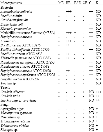 | |
Activity was classified according to the diameter of the inhibition zone around the point of application of the sample (+++: 15.00 mm; ++: 10.00-14.00 mm; +: 9.00 mm; -: No activity; ND: Not done). ME: Methanol extract, HE: Hexane extract, EAE: Ethyl acetate extract, CE: Chloroform extract, DEE: Diethyl ether extract, BE: Butanol extract, C: Chloramphenicol (30.0 μg mL-1), K: Ketoconazole (30.0 μg mL-1) | |
The most potent extract was hexane extract, followed by ethyl acetate and Gram positive bacteria (Bacillus subtilis, MRSA, Bacillus cereus ATCC 10876, Bacillus spizizenii ATCC 6633 and Staphylococcus aureus ATCC 12600), three Gram negative bacteria (Acinetobacter anitratus, Klebsiella pneumoniae ATCC 13883 and Erwinia sp.) and two yeasts (Candida utilis and Saccharomyces cerevisiae) within the range of 8.0-18.0 mm in diameter of the inhibition zones. Ethyl acetate extract was active against four Gram positive bacteria (Staphylococcus epidermis ATCC 12228, Bacillus spizizenii ATCC 6633, MRSA and Bacillus cereus ATCC 10876) and the inhibition zones were in the range of 7.0-9.0 mm in diameter. In comparison to the other extracts, chloroform extract only active against Candida albicans with the inhibition zone of 7.0 mm in diameter.
Table 2 shows the antimicrobial activities results from the partitioning method. All the extracts exhibited no antimicrobial activity except one extract, which was the ethyl acetate extract. Bacillus spizizenii ATCC 6633 and chloroform extract. Hexane extract was active against five Staphylococcus epidermis ATCC 12228 were susceptible to this extract, with the inhibition zones of 7.0-9.0 mm.
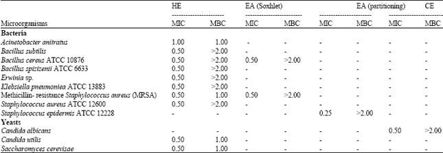
Table 3 shows the MIC and MBC values for each of the microorganisms that were susceptible to the extracts.
| Table 2: | Antimicrobial activity test of extracts (2.00 mg disk-1) of Halimeda discoidea (using partitioning extraction method) |
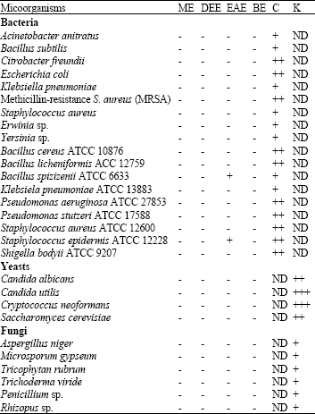 | |
Activity is classified according to the diameter of the inhibition zone around the point of application of the sample (+++: 15.00 mm; ++: 10.00-14.00 mm; +: 9.00 mm; : No activity; ND: Not done). ME: Methanol extract, HE: Hexane extract, EAE: Ethyl acetate extract, CE: Chloroform extract, DEE: Diethyl ether extract, BE: Butanol extract, C: Chloramphenicol (30.0 μg mL-1), K: Ketoconazole (30.0 μg mL-1) | |
One microorganism showed MIC values of 0.25 mg mL-1 (Staphylococcus epidermidis ATCC 12228 from the ethyl acetate using partitioning method), ten microorganisms showed MIC values of 0.50 mg mL-1 (Bacillus spizizenii ATCC 6633, MRSA, B. cereus ATCC 10876, Klebsiella pneumoniae ATCC 13883, Bacillus subtilis, Erwinia sp., Staphylococcus aureus ATCC 12600, Candida albicans, Candida utilis, Saccharomyces cerevisiae and Cryptococcus neoformans from the hexane extract using Soxhlet method) and one microorganism showed MIC values of 1.00 mg mL-1 (Acinetobacter anitratus from the hexane extract using Soxhlet method). Only five out of 13 microorganisms exhibited the MBC values which were 1.0 mg mL-1 (MRSA, Acinetobacter anitratus, Candida utilis, Saccharomyces cerevisiae). The remaining eight micoorganisms showed MBC values bigger than 2.00 mg mL-1.
The main objective of the present study was to evaluate and to compare the antimicrobial ability of different crude extract preparations from a green macroalgae, Halimeda discoidea collected from Penang coastal region, with the interest of finding new bioactive compounds. Several types of organic solvents with a wide polarity range were used for extraction in order to obtain a diverse set of possible bioactive compounds. The large variety of extracted bioactive compounds may possess the antimicrobial activity (Quinn et al., 2007). Two methods of extraction were applied to see which method was the more convenient and gave more quality extracts. It seemed that Soxhlet apparatus method was more convenient and produced better antimicrobial activity results. There are several advantages in using the soxhlet apparatus, including repeatedly bringing fresh solvent into direct contact with the dried sample, maintaining the required temperature according to the solvent boiling point because of the applied heat, better extraction process and lastly there was no need of filtration requirement after extraction process completed (Luque de Castro and Garcia-Ayuso, 1998).
| Table 3: | MIC and MBC values (mg mL-1) of various extracts of Halimeda discoidea |
 | |
This is in contrast with the partitioning method which needs filtration in order to get rid of the unnecessary debris and also the solvents could not come into direct contact with the sample itself.
In the present study, initial antimicrobial screening of different crude extracts from H. discoidea proved that this macroalgae exhibited wide range of antimicrobial property, by showing varying degrees of antimicrobial activity against some tested Gram-positive and Gram-negative bacteria and also some fungi. This result is in support with the findings on antimicrobial activities of some macroalgae against a number of pathogenic microorganisms that have been previously reported (Tuney et al., 2006; Devi et al., 2008) and also the varying degrees of sensitivity to the extracts tested (Neogi et al., 2008). The results indicated that there are antimicrobial compounds present in this macroalgae and the compounds were successfully extracted. Hexane, ethyl acetate and chloroform extracts consecutively inhibit the growth of 11, 4 and 1 species of microbes respectively. It can be said that hexane was the best solvent for extracting the bioactive compounds from the algal species used in this study. This result was in contrast with some other investigations that showed diethyl ether (Tuney et al., 2006) methanol: chloroform and methanol extracts (Choudhury et al., 2005) of some marine macroalgae were the best extracts to have the antimicrobial capability. In addition, this present results showed that the antimicrobial activity only exhibited by the non-polar extracts (n-hexane, ethyl acetate and chloroform), indicating that this macroalgae is rich with the non-polar compounds that are responsible in the antimicrobial activity. Non-polar extracts such as hexane usually comprised of non-polar compounds such as fatty acids, alkaloids and terpenoids. These compounds are known for antimicrobial property. Quinn et al. (2007) also reported the presence of compounds such as terpenes, terpenic alcohols, terpenic aldehydes and ketones in the hexane extract of Conradina etonia. There are several mechanisms of action possess by the terpenoids in inhibiting the microbial growth. Most terpenoids will distract the structure of the cell walls and cell membranes, therefore increased the permeability of the cell and caused the cell to release its constituents. Three types of monoterpenes and their mechanisms of antibacterial action such as the penetration and the damages caused by the compounds have been revealed by Trombetta et al. (2005). Thus, it proves the capability of the terpenes compounds to have higher antibacterial activity. There was also a phytochemical study done by Ramkumar et al. (2007) on the ethanol and hexane extract that showed the presence of alkaloids that caused the plant to have the antimicrobial properties.
A study carried-out on the same species by Gonzalez del Val et al. (2001) showed that the methanol extract of this species possessed antimicrobial activity against Staphylococcus aureus and Bacillus subtilis. The result was totally different from the present study that showed methanol extract of the same species exhibited no antibacterial activity against any bacteria tested, including both Staphylococcus aureus and Bacillus subtilis. In addition, the present study also showed that Candida albicans was susceptible to chloroform extract while Saccharomyces cerevisiae was susceptible to the hexane extract. The results again were in contrary to the earlier study done by Gonzalez del Val et al. (2001) that showed both Candida albicans and Saccharomyces cerevisiae were not susceptible to the methanol extract of the same algal species. The differences of the results obtained from the present and past studies were due to many factors, such as the differences in seasons, assay method, extraction method and also the organic solvents used for extraction of bioactive compounds (Kandhasamy and Arunachalam, 2008) as well as the algal species (Tuney et al., 2006). The presence of different bioactive compounds in different extracts (Ali-Shtayeh et al., 1998) and the capability of the extracts to penetrate the microbes and perform certain metabolic actions also contribute to the variation of the antimicrobial effectiveness (Ramkumar et al., 2007).
In this study, the extracts were more effective against the Gram-positive bacteria and less on the Gram-negative bacteria. The differences in the antibacterial activities may be related to the differences in the bacterial cell walls. The cell wall of Gram positive bacteria only consists of an outer peptidoglycan layer that makes it more permeable to other substances. In contrast, the cell wall of Gram negative bacteria comprised of lipopolysaccharides in their outer membrane. This structure makes the cell less permeable to other substances. This pattern of Gram positive bacteria being more susceptible to most antibiotic compounds is very common in many researches (Taskin et al., 2007; Lothfipour et al., 2008; Kandhasamy and Arunachalam, 2008). The susceptibility of the microorganisms towards the antibiotic depends on the mechanism of action of the compound and the differences in the cell wall structure of both types of bacteria. The less susceptibility of the Gram negative bacteria is due to the presence of an outer membrane surrounding the cell wall, which can hinder the access of active compound through its lipopolysaccharide covering (Tian et al., 2009). According to Vannuffel and Cocito (1996) the mechanism of actions of the antibiotic against bacteria including the interfering with nucleic acid synthesis and modifying the vital structure formation such as the membrane, cell wall, lipopolysaccharide and peptidoglycan. In addition, the differences in the antimicrobial activity among the different type of microorganisms are also due to the antimicrobial chemical defenses that are widespread among the marine macroalgae (Puglisi et al., 2007).
According to Andrews (2001) MIC test is done to further confirm the antimicrobial activity of new antimicrobial compound and as an alternative method to test for the susceptibility of organisms towards the extracts, whenever disk diffusion method is inappropriate. MIC is vital in determining the exact dose needed to inhibit the growth of particular microorganisms, especially when it comes to treating infected person. Overall, the MIC values for the 13 tested microorganisms ranged from 0.25 to 1.00 mg mL-1 and this shows that different microorganisms have different range of susceptibility in responding to particular concentrations of the extracts. In addition, different extracts with different antimicrobial compounds tend to possess different mechanisms of actions towards the microorganisms thus inhibit the growth. There is a wide range of MIC values obtained in the antimicrobial research done, ranging from as low as 0.24 mg mL-1 (Hellio et al., 2001) and as high as 6.25 mg mL-1 (Sasidharan et al., 2009) depending on the type of plant extracts used and the types of tested microorganisms. A low MIC value suggested that the compound is a strong antimicrobial compound as it can inhibit the microbial growth at lower concentration. The MBC is the extension part of the MIC. Only four of them showed MBC values that were less than 2.00 mg mL-1. The remaining tested microbes that showed no MBC values suggested that the extracts either do not possess the bactericidal property or higher concentration of the extract is needed to fully kill the bacterial cells, rather than only inhibiting the growth of those cells.
The remarkable part of this study was that chloroform and hexane extracts of this macroalgae successfully showed antimicrobial activity against first class pathogens, Candida albicans and Methicillin-resistance S. aureus (MRSA), respectively. The chloroform extract of this species that successfully inhibited C. albicans was in accordance with the study done by Nasimul Islam et al. (2003). Nasimul Islam et al. (2003) reported that the growth of C. albicans was inhibited because the presence of an antimicrobial terpenoid in the chloroform extract of Caesalpinia pulcherrima. In addition, hexane extract turned out to have greater inhibition zone than the positive control itself, against the MRSA strain. C. albicans and MRSA are well-known as the multi-drug resistant strains and this phenomenon started to worsen during the decade of 1980s (Schaberg et al., 1991) and therefore a more convenient method is developed in finding new drugs that are active against these microorganisms (Zgoda and Porter, 2001). C. albicans can easily infected patients with immunocompromised resulting from the AIDS pandemic and cause candidosis (Shai et al., 2008). The most common candidosis that infected approximately 75% of women at least once in their lifetime is the vaginal candidosis.
Finally, it can be concluded that the algal species under investigation could be effective and suitable for general use against some Gram positive and Gram negative bacteria and also yeast species. Further purification of the antimicrobial compounds should be done in order to isolate and to identify the pure bioactive compound involved. This will be very useful in searching for new natural antibiotics from natural sources.
ACKNOWLEDGMENT
The authors would like to thank National Oceanography Directorate, MOSTI for their financial support.
REFERENCES
- Ali-Shtayeh, M.S., R.M.R. Yaghmour, Y.R. Faidi, K. Salem and M.A. Al-Nuri, 1998. Antimicrobial activity of 20 plants used in folkloric medicine in the Palestinian area. J. Ethnopharmacol., 60: 265-271.
CrossRefPubMedDirect Link - Andrews, J.M., 2001. Determination of minimum inhibitory concentrations. J. Antimicrob. Chemother., 48: 5-16.
CrossRefDirect Link - Devi, P.K., N. Suganthy, P. Kesika and S.K. Pandian, 2008. Bioprotective properties of seaweeds: In vitro evaluation of antioxidant activity and antimicrobial activity against food borne bacteria in relation to polyphenolic content. BMC Complement. Alternat. Med., Vol. 8.
CrossRefDirect Link - El-Masry, H.A., H.H. Fahmy and S.H.A. Abdelwahed, 2000. Synthesis and antimicrobial activity of some new benzimidazole derivatives. Molecules, 5: 1429-1438.
CrossRefDirect Link - Del Val, A.G., G. Platas, A. Basilio, A. Cabello and J. Gorrochategui et al., 2001. Screening of antimicrobial activities in red, green and brown macroalgae from Gran Canaria (Canary Islands, Spain). Int. Microbiol., 4: 35-40.
CrossRefDirect Link - Hellio, C., D. De La Broise, L. Dufosse, Y. Le Gal and N. Bourgougnon, 2001. Inhibition of marine bacteria by extracts of macroalgae: Potential use for environmentally friendly antifouling paints. Mar. Environ. Res., 52: 231-247.
CrossRefDirect Link - Hillis-Colinvaux, L., 1980. Ecology taxonomy of Halimeda: Primary producer of coral reefs. Adv. Mar. Biol., 17: 1-327.
CrossRef - Kandhasamy, M. and K.D. Arunachalam, 2008. Evaluation of in vitro antibacterial property of seaweeds of southest coast of India. Afr. J. Biotechnol., 7: 1958-1961.
Direct Link - Lothfipour, F., H. Nazemiyeh, F.F. Azad, N. Garaei and S. Arami et al., 2008. Evaluation of antibacterial activities of some medicinal plants from North-West Iran. Iran. J. Basic Med. Sci., 11: 80-85.
Direct Link - Islam, A.K.M.N., M.A. Ali, A. Sayeed, S.M.A. Salam and A. Islam et al., 2003. An antimicrobial terpenoid from Caesalpinia pulcherrima swartz.: Its characterization, antimicrobial and cytotoxic activities. Asian J. Plant Sci., 2: 1162-1165.
CrossRefDirect Link - Neogi, U., R. Saumya, R.K. Mishra and K.C. Raju, 2008. Lipid content and in vitro antimicrobial activity of oil seeds of some Indian medicinal plants. Curr. Res. Bacteriol., 1: 1-6.
CrossRefDirect Link - Nicholas, G.M. and A.J. Phillips, 2006. Marine natural products: Synthetic aspects. Nat. Prod. Rep., 23: 79-99.
CrossRef - Phang, S.M., 2006. Seaweed resources in Malaysia: Current status and future prospects. Aquat. Ecosyst. Health Manag., 9: 185-202.
CrossRefDirect Link - Puglisi, M.P., S. Engel, P.R. Jensen and W. Fenical, 2007. Antimicrobial activities of extracts from Indo-Pacific marine plants against marine pathogens and saprophytes. Mar. Biol., 150: 531-540.
CrossRef - Quinn, B.P., U.R. Bernier and M.M. Booth, 2007. Identification of compounds from Etonia rosemary (Conradina etonia). J. Chromatography A., 1160: 306-310.
CrossRefDirect Link - Sasidharan, S., I. Darah and M.K.M.J. Noordin, 2009. Screening antimicrobial activity of various extracts of Gracilaria changii. Pharm. Biol., 47: 72-76.
CrossRefDirect Link - Schaberg, D.R., D.H. Culver and R.P. Gaynes, 1991. Major trends in the microbial etiology of nosocomial infection. Ann. J. Med., 91: 72S-75S.
PubMedDirect Link - Shai, L.J., L.J. McGraw, P. Masoko and J.N. Eloff, 2008. Antifungal and antibacterial activity of seven traditionally used South African plant species active against Candida albicans. South Afr. J. Bot., 74: 677-684.
CrossRef - Smit, A.J., 2004. Medicinal and pharmaceutical uses of seaweed natural products: A review. J. Applied Phycol., 16: 245-262.
CrossRefDirect Link - Taskin, E., M. Ozturk, E. Taskin and O. Kurt, 2007. Antibacterial activities of some marine algae from the Aegean Sea (Turkey). Afr. J. Biotechnol., 6: 2746-2751.
Direct Link - Tian, F., B. Li, B. Ji, J. Yang, G. Zhang, Y. Chen and Y. Luo, 2009. Antioxidant and antimicrobial activities of consecutive extracts from Galla chinensis: The polarity affects the bioactivities. Food Chem., 113: 173-179.
CrossRefDirect Link - Trombetta, D., F. Castelli, M.G. Sarpietro, V. Venuti and M. Cristani et al., 2005. Mechanisms of antibacterial action of three monoterpenes. Antimicrob. Agents. Chemother., 49: 2474-2478.
CrossRefDirect Link - Tuney, I., B.H. Cadirci, D. Unal and A. Sukatar, 2006. Antimicrobial activities of the extracts of marine algae from the coast of Urla (Izmir, Turkey). Turk. J. Biol., 30: 171-175.
Direct Link - Vannuffel, P. and C. Cocito, 1996. Mechanism of action of streptogramins and macrolides. Drugs, 51: 20-30.
PubMedDirect Link - Yoshie, Y., W. Wang, Y.P. Hsieh and T. Suzuki, 2002. Compositional difference of phenolic compounds between two Seaweeds, Halimeda spp. J. Tokyo. Univ. Fish., 88: 21-24.
Direct Link - Zgoda, J.R. and J.R. Porter, 2001. A convenient microdilution method for screening natural products against bacteria and fungi. J. Pharm. Biol., 39: 221-225.
Direct Link - De Castro, M.D.L. and L.E. Garcia-Ayuso, 1998. Soxhlet extraction of solid matrices: An outdated technique with a promising innovative future. Anal. Chim. Acta, 369: 1-10.
CrossRef