
Nidhi Mathur
Reproductive Physiology Section, Center for Advanced Studies, Department of Zoology, University of Rajasthan, Jaipur-302004, India
G. C. Jain
Reproductive Physiology Section, Center for Advanced Studies, Department of Zoology, University of Rajasthan, Jaipur-302004, India
Geeta Pandey
Reproductive Physiology Section, Center for Advanced Studies, Department of Zoology, University of Rajasthan, Jaipur-302004, India
International Journal of Pharmacology
Year: 2010 | Volume: 6 | Issue: 2 | Page No.: 152-156


ABSTRACT
Effects of 50% ethanolic extract of Tecoma stans leaves were investigated in adult Wistar male rats. Changes in weight of the liver, kidneys and adrenal of the rats were statistically insignificant (p<0.05). The relative weight of testes, epididymis, vas deferens, ventral prostate and seminal vesicle were decreased significantly (p<0.001). Tecoma stans treatment showed significant high cholesterol, glycogen and low tissue sialic acid and total protein in testes and epididymis and low fructose level in seminal vesicle of treated rats. Hormonal assay showed decrease in testosterone level. The epididymal sperm count, motility and fertility test (%) reduced significantly in treated rats. Histopathological study of the testes depicted marked degenerative changes in testes. The seminiferous tubules appear reduced in size. Vacuolization was observed in the sertoli cells, spermatogonia and spermatocytes. Leydig cells were atrophied. Germ cell proliferation beyond the level of the spermatocyte was affected. The lumen contained sloughed debris and few germ cells.
PDF Abstract XML References Citation
How to cite this article
Nidhi Mathur, G. C. Jain and Geeta Pandey, 2010. Effect of Tecoma stans Leaves on the Reproductive System of Male Albino Rats. International Journal of Pharmacology, 6: 152-156.
DOI: 10.3923/ijp.2010.152.156
URL: https://scialert.net/abstract/?doi=ijp.2010.152.156
DOI: 10.3923/ijp.2010.152.156
URL: https://scialert.net/abstract/?doi=ijp.2010.152.156
INTRODUCTION
Tecoma stans (family: Bignoniaceae, common name-Yellow elder) is a semi ever green ornamental shrub or tree. The shrub is planted and managed to enhance the beauty of green belts and natural forests used for recreation (Roman-Ramos et al., 1991). Tecoma stans is used for a great range of action in traditional medicines (Costantino et al., 2003). Tecoma stans L. is known for the antimicrobial (Gharib Naseri et al., 2007), anti-inflammatory (Marzouk et al., 2006), antirheumatic (Andrade-Cetto and Heinrich, 2005), antinociceptive (Alguacil et al., 2000), narcotic or antisyphilitic (Marzouk et al., 2006) activity of their extracts. According to a study the leaves of the plant contained 17% crude protein, 6% ash, 18% fat, 25% fiber and 14% total phenols. Tecoma stans contains many biologically active chemicals, the most promising compounds are monoterpine alkaloids, which have been shown to effectively reduced the symptoms of diabetes mellitus in rat, dog and mice (Aguilar-Santamaria et al., 2009; Lozoya-Meckes and Mellado-Campos, 1985; Perez et al., 1984; Hernandez and Garcia, 1958). Da La Paz-Naranjo et al. (2003) observed hypoglycemic effect of T. stans leaves at 500 mg kg-1 in mice and rats.
Male reproductive toxicology has recently become a rapidly extending area of research and testing. In the last decades there has been growing concern over the effects of either synthetic or natural products on the male reproductive health (US EPA, 1996). Phytoestrogens, which are hormone-mimicking substances naturally present in plants, are suspected of interfering with the endocrine system of animals. The purpose of the present study was to elucidate whether 50% ethanolic extract of T. stans leaves implies reproductive hazards to male rats exposed during 60 days. A variety of well-validated endpoints were considered in order to provide a comprehensive risk assessment of the effects of T. stans on the reproductive system.
MATERIALS AND METHODS
Plant extract and animals used: Tecoma stans plant leaves were collected from the Rajasthan University Campus, Jaipur and were authenticated in the Herbarium, Department of Botany, University of Rajasthan, under the specimen voucher No. RUBL 20256. The leaves were shade-dried, crushed and were extracted with 50% ethanol in a soxhalet apparatus to obtain a solid viscous dark green mass, that is, extract.
Colony bred adult healthy male and female Wistar rats weighing 170-200 g were used in the present investigation. The rats were housed in standard rat cages and maintained under standard conditions (12 h light/dark cycle; 25"3°C temperature) and provided a standard laboratory chow (Aashirwad Food Industries, Chandigarh, India) and water ad libitum. Extract and/or vehicle were administered to all male rats by oral intubation.
This research project was conducted from 8th March 08 to 6th May 08. The duration of experiment was 60 days. The study was approved by ethical committee of the Centre for Advanced Studies, University Department of Zoology, Jaipur, Rajasthan, India. The Indian National Science Academy (2000), New Delhi Guidelines, were followed for maintenance and use of the experimental animals.
Dose and duration of treatment: Male rats of proven fertility were divided into two groups of 6 rats each. The daily dose of the plant extract was freshly dissolved in 0.5 mL of distilled water and orally administered to each experimental animal every morning for 60 days.
| • | Group A: Control rats received 0.5 mL day-1 of the vehicle, that is, distilled water |
| • | Group B: Rats treated with Tecoma stans extract 500 mg/kg/day |
Autopsy schedule: The animals were weighed and autopsied under light ether anesthesia 24 h after last dose of the treatment.
Body and organ weight: Initial and final body weights of the animals were recorded. The testes, epididymides, vas deferens, seminal vesicle ventral prostate, kidney, adrenal and liver were dissected out, freed from adherent tissues and blood and weighed to the nearest milligram.
Tissue biochemistry: Testes, epididymis and accessory sex organs were frozen at -20°C for the biochemical estimations. Testes and epididymis were assayed for protein (Lowry et al., 1951), sialic acid (Warren, 1959), glycogen (Montgomery, 1957) and cholesterol (Zlatkis et al., 1953). Fructose in seminal vesicle was also estimated (Mann, 1964).
Radioimmunoassay of testosterone: Blood samples were also collected for estimations of serum testosterone by radioimmunoassay (RIA) (WHO Method Manual, 1987). Serum samples were separated by standard procedures and stored at -20°C for subsequent analysis.
Sperm analysis and fertility test: Cauda epididymal sperm motility and count were made according to the procedure of Prasad et al. (1972). One hundred milligram of each tissue was minced in 1 mL of physiological saline. For sperm motility, one drop of evenly mixed sample was applied to a glass slide under a cover glass. The percent motility was determined by counting both motile and immotile spermatozoa per unit area (WHO, 1983). Cauda epididymal sperm counts expressed as million mm-3 of suspension. Male rats were introduced to parous females, 170-200 g (male:female ratio, 1:2) at 17:00 h after 55 days of treatment. The successful mating was confirmed in the forthcoming mornings from 56 to 61 days by vaginal plug and spermatozoa in the vaginal smear. The inseminated females were separated and allowed to deliver at term. The number of pups delivered and their characteristics were noted (WHO, 1983).
Histological examination: Testes were fixed in Bouin’s fluid. Paraffin section were cut (5 μm) and stained with hematoxylin and eosin. Mean seminiferous tubular diameter was determined by measuring 100 round sections of seminiferous tubule with the help of ocular micrometer. Diameter of Leydig cells nuclei were measured at H800.
Statistical analysis: Data are expressed as Mean±SEM Student's t test was used for statistical comparison.
RESULTS
Body and organ weight: Fifty percent ethanolic extract of T. stans leaves did not cause any significant adverse effect on the body weight of treated rats. However, the weight of testes, epididymides, vas deferens, seminal vesicle and ventral prostate was reduced significantly (p≤0.001) as compared to control. The weight of other vital organs remained significantly unchanged as compared to the control rats (p≤0.05) (Table 1).
Tissue biochemistry: Protein content of testes (p≤0.01) and epididymides (p≤0.001) was reduced after T. stans treatment as compared with controls. Sialic acid content of the testes (p≤0.01) and epididymides (p≤0.001) was also significantly depleted. However, glycogen content of the testes (p≤0.001) and epididymides (p≤0.001) was increased significantly. The cholesterol content of testes was highly significantly increased (p≤0.001), while in epididymides remained unchanged after the administration of T. stans extract (Table 2).
Radioimmunoassay of testosterone: Significant decline in serum testosterone levels was observed in treated group when compared with group A (p≤0.001) (Table 3).
Sperm analysis and fertility test: Tecoma stans leaves extract significantly reduced sperm count (p≤0.01) and sperm motility (p≤0.001) in cauda epididymis. Tecoma stans reduced the fertility of male rats (Table 3).
| Table 1: | Effect of Tecoma stans treatment on the body and organ weight of male albino rats |
 | |
| Data are expressed as Mean±SEM of 6 animals, Groups B was compared with Group A, aNon significant (p≤0.05), bSignificant (p≤0.01), cHighly significant (p≤0.001) | |
| Table 2: | Effect of Tecoma stans treatment on the tissue biochemistry of male albino rats |
 | |
Data are expressed as Mean±SEM of 6 animals, Groups B was compared with Group A, aNon significant (p≤0.05), bSignificant (p≤0.01), cHighly significant (p≤0.001) | |
| Table 3: | Effect of Tecoma stans on serum testosterone, cauda epididymis sperm analysis, fertility test and morphometry of male albino rats |
 | |
Data are expressed as Mean±SEM of 6 animals, Groups B was compared with Group A, aNon significant (p≤0.05), bSignificant (p≤0.01), cHighly significant (p≤0.001) | |
 | |
| Fig. 1: | Photomicrograph of testes of a rat of group A (vehicle treated control) showing normal features with successive stages of transformation of seminiferous epithelium to spermatozoa. H and E x200 |
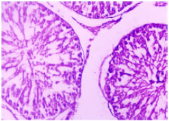 | |
| Fig. 2: | Photomicrograph of testes of a rat of group B (500 mg kg-1 b.wt., T. stans) after 60 days of treatment showing reduced seminiferous tubular diameter and cellular damage of tubular elements. H and E x200 |
Histopathology of testes: Histological studies of control rat testes showed all successive stages of spermatogenesis, where the lumen was filled with sperm. Leydig cells were situated in between the tubules with prominent nuclei (Fig. 1). The testes of the treated animals revealed arrest of spermatogenesis. The seminiferous tubules appeared reduce in size. Vacuolization was observed in the sertoli cells, spermatogonia and spermatocytes. Germ cell proliferation beyond the level of the spermatocyte was affected. The lumen contained sloughed debris and few germ cells. Leydig cell nuclei diameter area (p≤0.01) and seminiferous tubular diameter were significantly reduced in treated rats (p≤0.001) (Table 3, Fig. 2).
DISCUSSION
The herbal medicines are being used by up to 80% of the population in developing countries. Despite widespread use, few scientific studies have been undertaken to ascertain the safety and efficacy of traditional remedies. The present study was undertaken to evaluate the male reproductive toxicity of the 50% ethanolic extract of T. stans leaves, which is an herbal medicine (Costantino et al., 2003). The impact of the extract on the reproductive system, particularly in males has not previously been investigated in detail. The toxic effects of plant extracts are analyzed by monitoring alterations in body and organ weight in animals. In the male reproductive system weight loss of the gonads, epididymides and accessory sex organs as well as histology of gonads, biochemistry of testes and epididymides are considered standard criteria for the characterization of toxic agents that may cause fertility problems in the treated subject (Da Silveira et al., 2003; Mishra and Singh, 2009).
In the subchronic toxicity study of rats given the extract orally at doses of 500 mg kg-1 b.wt., we did not notice any abnormal behavior and body weight gains were not significantly different in the treated rats when compared to controls. Since, changes in body weight have been used as an indicator of good health (Chauhan and Agarwal, 2008; Singh and Singh, 2009), the present results suggest that at the doses administered orally, the T. stans extracts are non-toxic in rats. Furthermore, the mean weight of the vital organs was not affected by the extract, suggesting that the extract is relatively non-toxic to these organs. The present data show that the administration of T. stans brought about a highly significant loss in testes and accessory sex organ weights, which are known to be mostly related to the number of spermatids and spermatozoa in the tissue. The decreasing weight of the reproductive organs in the extract-treated male rats clearly indicated that the extract caused structural and functional alteration in the testes, epididymis, seminal vesicle, ventral prostate and vas deferens (Sarkar et al., 2000). Reduction in the weight of testes and other accessory sex organs might be due to low level of androgen, which was not enough to maintain the weight of gonads and accessories (Singh and Singh, 2009). Testosterone is produced by Leydig cells in the testes and decreased number of Leydig cells and their nuclear diameter in the treated rats diminished the production of testosterone (Bhatt et al., 2007). Testosterone level is depleted in serum of treated animals. The number and nuclei diameter of Leydig cells were reduced in T. stans treated rats Fig. 2. The negative impact of T. stans on the male structural and functional integrity of testicular tissues was evidenced by the histopathological data highlighting the seminiferous tubules appear reduced in size. The vacuolization was observed in the sertoli cells, spermatogonia and spermatocytes. Germ cell proliferation beyond the level of the spermatocyte was affected. The lumen contained sloughed debris and few germ cells (Fig. 2), which may be due to wide spread cellular damage and androgen deprivation (Gupta et al., 2006). Reduced testicular and epididymal protein content could be correlated with absence of spermatozoa in the lumen (Zhen et al., 1995). The reduction in sperm count and motility in cauda epididymides is of importance with regard to fertilization (Bedford, 1983; Raji et al., 2006). Low level of sialic acid in testes, epididymides may be correlated with loss of androgen (Gupta et al., 2001). An increase in the concentration of cholesterol in testes of extract-treated male rats may reflect reduced conversion of cholesterol to testosterone (Vijaykumar et al., 2004). Glucose is the major substrate for metabolism in speramatids and spermatocytes in rat testes. In the testes and epididymis glycogen levels were increased suggesting an inhibitory action of glycolysis (Das and Dasgupta, 1997). In the present study, reduction in the number of fertile males was observed and fertility depleted after 60 days of treatment. The reduced number of fertile males may be due to decreased sperm counts and motility.
In conclusion, the oral administration of 50% ethanol extract of T. stans leaves to male rats produced effects on reproduction. The effects may have an inhibitory influence on gonadotrophin release which may be responsible for the decline in testosterone production, leading to changes in spermatogenesis.
ACKNOWLEDGMENTS
The authors are thankful to Prof. Ashok Kumar, coordinator, CAS and Prof. RS Bedwal, Head of the department for providing necessary facilities.
REFERENCES
- Roman-Ramos, R., J.L. Flores-Saenz, G. Partida-Hernandez, A. Lara-Lemus and F. Alarcon-Aguilar, 1991. Experimental study of the hypoglycemic effect of some antidiabetic plants. Arch. Invest. Med., 22: 87-93.
PubMedDirect Link - Marzouk, M., A. Gamal-Eldeen, M. Mohamed and M. El-Sayed, 2006. Anti-proliferative and antioxidant constituents from Tecoma stans. Z. Naturforsch. C., 61: 783-791.
PubMedDirect Link - Andrade-Cetto, A. and M. Heinrich, 2005. Mexican plants with hypoglycemic effect used in the treatment of diabetes. J. Ethnopharmacol., 99: 325-348.
CrossRefPubMedDirect Link - Lozoya-Meckes, M. and V. Mellado-Campos, 1985. Is the Tecoma stans infusion an antidiabetic remedy? J. Ethnopharmacol., 14: 1-9.
CrossRefPubMedDirect Link - Hernandez, J.R. and C.G. Garcia, 1958. Research on the hypoglycemic action of the active fraction of Tecoma stans. Med. (Mex)., 38: 25-34.
PubMedDirect Link - Lowry, O.H., N.J. Rosebrough, A.L. Farr and R.J. Randall, 1951. Protein measurement with the folin phenol reagent. J. Biol. Chem., 193: 265-275.
CrossRefPubMedDirect Link - Warren, L., 1959. The thiobarbituric acid assay of sialic acids. J. Biol. Chem., 234: 1971-1975.
PubMedDirect Link - Zlatkis, A., B. Zak and A.J. Boyle, 1953. A new method for the direct determination of serum cholesterol. J. Lab. Clin. Med., 41: 486-492.
PubMedDirect Link - Prasad, M.R.N., N.J. Chinoy, and K.M. Kadam, 1972. Changes in succinate dehydrogenase levels in rat epididymis under normal and altered physiological conditions. Fert. Ster., 23: 186-190.
PubMedDirect Link - Sa, R.D.C.D.S., M.N. Leite, M.D.M. Reporedo and R.N. de Almeida, 2003. Evaluation of long-term exposure to Mikania glomerata (Sprengel) extract on male Wistar rats reproductive organs, sperm production and testosterone level. Contraception, 67: 327-331.
PubMed - Mishra, R.K. and S.K. Singh, 2009. Antispermatogenic and antifertility effects of fruits of Piper nigrum L. in mice. Indian J. Exp. Biol., 47: 706-714.
Direct Link - Bhatt, N., S.L. Chawla and M.V. Rao, 2007. Contraceptive evaluation of seed extract of Abrus precatorius (L.) in male mice (Mus musculus). J. Herb. Med. Toxicol., 1: 45-48.
Direct Link - Gupta, R.S., M. Kanwar, H. Rehwani, S.K. Verma and M.P. Dobhal, 2006. Contraceptive efficacy of Strychnos potatorum seed extract in male albino rats. Asian J. Exp. Sci., 20: 181-187.
Direct Link - Bedford, J.M., 1983. Significance of the need for sperm capacitation before fertilization in eutherian mammals. Biol. Reprod., 28: 108-120.
PubMedDirect Link - Raji, Y., M.A. Gbadegesin, O.A. Osonuga, R.A. Adisa and O.S. Akinsomisoye et al., 2006. Reproductive, haematologic and biochemical profile of male rats treated with aqueous extract of Spondias mombin bark. Int. J. Pharmacol., 2: 126-130.
CrossRef - Vijaykumar, B., I. Sangamma, A. Sharanabasappa and S.B. Patil, 2004. Antispermatogenic and hormonal effects of Crotalaria juncea Linn. seed extracts in male mice. Asian J. Androl., 6: 67-70.
PubMedDirect Link - Gharib-Naseri, M.K., M. Asadi-Moghaddam and S. Bahadoram, 2007. Antispasmodic effect of Tecoma stans (L.) Juss leaf extract on rat ileum. DARU, 15: 123-128.
Direct Link - De La Paz-Naranjo, M.C.J., A.C. Salvado, G.R. Jimenez, T.M.F. Menendez and P.E. Perez-Santoya, 2003. Efecto hipoglicemiante del extracto fluido de Tecoma stans Linn en roedores. Rev. Cubana Med. Milit. 32(1).
Direct Link - Chauhan, A. and M. Agarwal, 2008. Reversible changes in the antifertility induced by Aegle marmelos in male albino rats. Syst. Biol. Reprod. Med., 54: 240-246.
Direct Link