
Ismiarti
Faculty of Animal Science, Universitas Gadjah Mada, Jl Fauna No. 3, Bulaksumur, Yogyakarta 55281, Indonesia
Yustina Yuni Suranindyah
Faculty of Animal Science, Universitas Gadjah Mada, Jl Fauna No. 3, Bulaksumur, Yogyakarta 55281, Indonesia
Widodo
Faculty of Animal Science, Universitas Gadjah Mada, Jl Fauna No. 3, Bulaksumur, Yogyakarta 55281, Indonesia
LiveDNA: 62.23055
International Journal of Dairy Science
Year: 2019 | Volume: 14 | Issue: 1 | Page No.: 29-35


ABSTRACT
Background and Objective: In Indonesia, smallholder dairy farms contribute 20.37% of domestic market. A small portion of these farms is from goat dairy farms that are traditionally managed with poor sanitation during milking and improper storage management. This system causes contamination that can affect consumer health and cause financial loss. This study aimed to evaluate the microbiological quality of goat milk obtained under different milking systems at a smallholder dairy farm in Yogyakarta, Indonesia. Materials and Methods: Samples were collected from 20 crossbred dairy goats divided into two groups: Group A and B. In Group A, milking was conducted manually. In Group B, milking was conducted using a bucket milking machine. Total Plate Count (TPC) and the presence of Enterobacteriaceae (EB), Shiga toxin-producing Escherichia coli (STEC) and Salmonella were assessed. Data obtained were analyzed using a t-test. Results: The results showed that TPC and EB were higher in Group A than in Group B. In Groups A and B, 80% (8/10) of samples contained STEC and 30% (3/10) contained Salmonella. Phylogenetic analysis showed that partial sequencing of amplified genomic DNA using stx1 primers had more than 90% similarity with several sequences of Escherichia coli O157:H7 strain Shiga toxin subunit 1A (stx1A) and Shiga toxin subunit 1B (stx1B). Moreover, partial sequencing of amplified genomic DNA using 16S rRNA primers had more than 90% similarity with several sequences of S. enterica. Conclusion: The results conclude that hygienic and sanitary practices in smallholder dairy goat farming are still poor as shown by the presence of pathogenic bacteria.
PDF Abstract XML References Citation
Copyright: © 2019. This is an open access article distributed under the terms of the creative commons attribution License, which permits unrestricted use, distribution and reproduction in any medium, provided the original author and source are credited.
How to cite this article
Ismiarti, Yustina Yuni Suranindyah and Widodo, 2019. Microbiological Qualities of Goat Milk Obtained under Different Milking Systems at a Smallholder Dairy Farm in Yogyakarta, Indonesia. International Journal of Dairy Science, 14: 29-35.
DOI: 10.3923/ijds.2019.29.35
URL: https://scialert.net/abstract/?doi=ijds.2019.29.35
DOI: 10.3923/ijds.2019.29.35
URL: https://scialert.net/abstract/?doi=ijds.2019.29.35
INTRODUCTION
Among milk from other species, goat milk has special characteristics; it can be easily digested because its fat globules are smaller with more short chain fatty acids and it is less allergenic than other milk and safe to be consumed by people with lactose intolerance1,2. Microbiological qualities of goat milk are determined by its composition and hygienic practices during milking and condition during storage and distribution3. Microbial contamination of milk can occur from direct contact between fresh milk and contaminated equipment surfaces during milking4. Improper handlings or storage of fresh milk can represent a transmission hazard for bacterial pathogens that responsible for foodborne illness4. A number of bacterial pathogens was previously reported to be responsible for milk-borne illness and it includes Shiga toxin-producing Escherichia coli (STEC)5 and Salmonella sp.6. Global milk-borne illness caused by STEC and Salmonella sp. has previously been reported6-8. In 2010, the estimated global diseases of STEC was 2,841,511 cases with 269 people were deaths9. Healthy dairy ruminants commonly carrying STEC in their feces10. Transmission of this bacterial pathogen to human mainly occurs through less-cooked meat, unpasteurized milk, feces-contaminated water and improper hygienic and sanitary practices during milking.
Smallholder dairy farms in Indonesia contribute 20.37% of milk needed domestically11 of which, a small portion is from goat dairy farms. Generally, smallholder dairy farms are traditionally managed such as milking by hand with improper storage management12. In many developing countries, poor sanitation during milking causes contamination that can affect consumer health and cause financial loss13. It is therefore, important to investigate microbiological quality of goat milk harvested from two different milking systems from smallholder dairy farm in Indonesia. The data obtained during this study will be used as a recommendation for better practices in managing smallholder dairy farm and improving goat milk qualities.
This study was conducted to evaluate microbiological qualities of goat milk based on total plate count (TPC), Enterobacteriaceae (EB) count and the presence of pathogens such as STEC and Salmonella in milk from a smallholder dairy goat farm managed under different milking systems. The TPC shows all living mesophilic microbes in goat milk that can grow as forming colonies on plate count agar (PCA) medium. The EB are indicators of fecal contamination and an effective measurement for the environmental sanitary program, such as in powdered milk and ready-to-serve food14.
MATERIALS AND METHODS
Sampling of goat milk farms: Goat milk was sampled in January to April, 2018 from two goat farms in Yogyakarta Indonesia that applied different milking systems. In Group A, milking was carried out after cleaning around the stall. Milking in Group A was conducted by hand without cleaning the udders and dipping and farmers washed hands before milking. The first milk flow in Group A was discarded, the milk was collected in receptacle drinking-water bottles. Milk samples in Group A were collected from the bottles and moved to a sterile plastic container. In Group B, milking was carried out after cleaning around the stall and goats from colony stalls were moved to milking stalls. Farmers washed their hands before milking and the goat’s udder was cleaned using warm water. The first milk flows were discarded. Milking was conducted using a bucket milking machine connected to tubes on a vacuum pump that fastens on teats, so milk flowed from tubes and was collected in a milk bucket. Samples were carried out from the milk bucket to a sterile plastic container. Samples from Groups A and B were put in a box with ice, which was quickly transported to the laboratory to be analyzed.
Microbiological analysis of goat milk: Microbiological qualities of goat milk were determined using three parameters, TPC to measure total amount of mesophilic aerobic microbes was conducted using plate count agar (PCA) media15 isolation of EB to measure enteric bacteria using violet red bile glucose (VRBG) media16, isolation of STEC17-19 and Salmonella19 to measure the presence of pathogen. Isolates found were identified using specific primers stx1 and stx2 for STEC and 16S rRNA sequencing for Salmonella determination.
TPC: Goat milk samples were serially diluted from 10–1-10–3 and then plated onto PCA medium and incubated at 37°C for 24 h. Total colonies on PCA medium were counted15 as CFU mL–1.
EB: Goat milk samples were serially diluted from 10–1-10–3 and then plated onto VRBG medium and incubated at 37°C for 24 h. Predictive EB colonies that were reddish or purplish in color were counted.
Pathogen isolation
STEC: Isolation of Escherichia coli was carried out using an enrichment broth medium of Brilliant Green Lactose Bile (BGLB) and incubated at 37°C for 24 h. A positive sample was detected with turbidity and then inoculated onto a selective medium, Eosin Methylene Blue (EMB) agar and incubated at 37°C for 24 h. A positive sample was detected when a colony appeared with a metallic sheen or dark chocolate color. Positive samples on EMB were then streaked onto a selective medium, Sorbitol MacConkey (SMAC) agar and incubated at 37°C for 24 h. A positive sample of STEC was detected as a colorless colony on the surface of SMAC medium. Predictive STEC colonies were inoculated onto Lactose Broth medium.
Salmonella: Isolation of Salmonella was carried out with pre-enrichment using buffered peptone water (BPW) and incubated at 37°C for 24 h. Turbid samples were enriched into rappaport-vassiliadis soy (RVS) broth and incubated at 37°C for 24 h. Turbid samples from the enrichment were then streaked onto xylose lysine deoxycholate (XLD) agar, hektoen enteric (HE) agar and bismuth sulfite agar (BSA) and incubated at 37°C for 24 h. Positive samples were pink with or without black spots on XLD, green or dark green with or without dark spots on HE and gray or dark gray on BSA. Positive isolates were tested for their biochemical characteristics using tryptic soy iron (TSI) agar and lysine iron agar (LIA). Positive isolates were then transferred into nutrient broth.
Molecular detection: One milliliter of culture of putative STEC and Salmonella isolates were transferred into DNA isolation kit (Favorgen™). Genomic DNA of STEC isolates was amplified using specific primers for stx1 (F: 5 -ATAAATCGCCATTCGTT GACTAC-3 and R: 5 -AGAACGCCCACTGAGATCATC-3) and stx2 (F: 5 -GGCACTGTCTGAAACTGCTCC-3 and R: 5 -TCGCCAGTTAT CTGACATTCTG-3). Genomic DNA of putative Salmonella isolates were amplified using universal primers of 16S rRNA [F: 5-AGA GTTTGAT(C/T)(A/C)TGGCTCAG-3 and R: 5-CA(G/T)AAAGGAGGTGATCC-3 ]. Amplification of STEC genomic DNA using stx1 primers has a 180 bp target product and stx2 has a 255 bp target product16. Amplification of Salmonella genomic DNA using universal 16S rRNA primers has a 1500 bp target product. Amplification was conducted using a thermal cycler 2720 (Applied BiosystemsTM). Amplicons produced were visualized using agarose gel electrophoresis. The PCR products were sequenced using ABI PRISMTM 3730-XL 1406-022.
| Table 1: | The TPC of goat milk from Groups A and B |
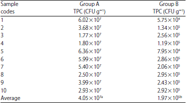 | |
| TPC: Total plate count. The number followed by different superscript means different number (p<0.05) | |
| Table 2: | The EB count in goat milk from Groups A and B |
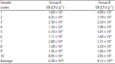 | |
| EB: Enterobacteriaceae. The number followed by different superscript means different number (p<0.05) | |
RESULTS
Total plate count (TPC): The TPC of goat milk from Groups A and B is presented in Table 1. Table 1 shows that TPC was higher in Group A than in Group B (p<0.05). The average TPC in Groups A and B was 4.05×10⁷ and 1.97×10⁵ CFU g–1, respectively. The highest amount of TPC in Group A and B were 6.36×10⁷ and 2.95×10⁵ CFU g–1, respectively. The lowest were 1.77×10⁷ and 5.75×10⁴ CFU g–1, respectively.
Enterobacteriaceae (EB): There was different amount of EB between Group A and B. Table 2 shows that the EB count was higher in Group A than in Group B (p<0.05). The average EB count in Groups A and B was 6.38×10⁴ and 9.15×10³ CFU g–1, respectively. According to Standar Nasional Indonesia, the maximum total EB count in raw milk is 1×10³ CFU mL–1. A high EB count in the two groups indicated poor sanitation.
Shiga toxin-producing Eschericia coli (STEC) and Salmonella: Regarding the detection of STEC, all samples from both groups showed positive growth in BGLB and EMB, while samples grown in SMAC showed 8 positive growth.
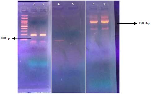 | |
| Fig. 1: | Electrophenogram of amplified genomic DNA; 1: DNA ladder 100 bp, 2: Amplified DNA from Group A with primer stx1,3: Amplified DNA from Group B with primer stx1, 4: Unamplified DNA from Group A with primer stx2, 5: Unamplified DNA from Group B with stx2 primer, 6: Amplified DNA from Group A with 16S rRNA primer, 7: Amplified DNA from Group B with primer 16S rRNA |
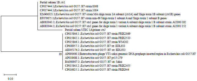 | |
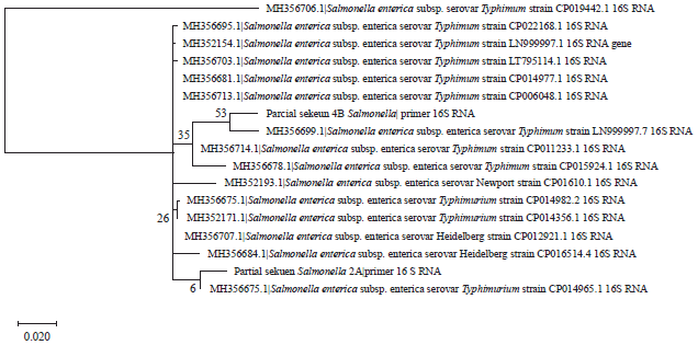
| Fig. 2: | Phylogenetic tree of partial sequence of STEC Groups A and B using primer stx1 |
| Note: Sequence accession number provided before culture identity | |
Meanwhile, the detection of Salmonella in BPW and RVS showed all samples with positive growth. In Group A, 1 sample was positive in XLD, another 1 sample was positive in HE and 2 samples were positive in BSA. In Group B, no sample was positive in XLD, 3 samples were positive in HE and no sample was positive in BSA. Based on biochemical tests, all putative Salmonella isolates were positive in TSI and all isolates were negative in LIA.
Molecular detection of STEC and Salmonella was carried out by PCR and the products were visualized using 2% agarose gel electrophoresis (Fig. 1). Figure 1 shows that amplification with stx1 primers resulted in a 180 bp product; however, stx2 primers had no amplification products, indicating that these primers did not recognize the corresponding sequence of the STEC genomic DNA. BLASTN analysis showed that amplified DNA using stx1 primers in Groups A and B had more than 90% similarity with several identified sequences as E. coli O157:H7. Further analysis using a phylogenetic tree showed that amplified sequences in Groups A and B were aligned with the sequence of E. coli O157:H7 strain Shiga toxin subunit 1A (stx1A) and 1B (stx1B) (Fig. 2).
 | |
| Fig. 3: | Phylogenetic three of partial sequence of Salmonella Groups A and B using primer 16s rRNA |
| Note: Sequence accession number is provided before culture identity | |
Genomic DNA of putative Salmonella species was amplified using 16S RNA primers and resulted in a 1500 bp product (Fig. 1). The BLASTN analysis showed that amplified DNA using 16S RNA primer of putative Salmonella in Groups A and B had more than 90% similarity with several sequences of S. enterica (Fig. 3).
DISCUSSION
Different milking systems between Group A and B had different amount of TPC and EB. Based on Standar Nasional Indonesia, good-quality milk has TPC <1×10⁶ CFU mL–1, hence, goat milk from Group A did not meet this standard20. The higher TPC in Group A was due to low hygienic and sanitation practices, such as no cleaning of the udder and teats before milking. Improper hygienic practices trigger microbial contamination during milking. Mohammadi et al.8 reported that milk quality is determined by its composition and hygienic practices that are applied during milking processes, such as cleanliness of milking equipment, conditions of storage and transportation and cleanliness of the udder of the individual goat. Suranindyah et al.12 also reported that improving environmental sanitation during milking and dipping of teats can reduce total microbes in raw milk. This is in line with the present study results. Smallholder dairy farms with better sanitation produce raw milk with lower total microbes. The TPC in raw milk indicated growth of total microbes14.
The lower EB in Group B was due to better hygienic practices such as conducting milking in a milking stall and cleaning the udder before milking. In Group A, milking was conducted in an individual stall without cleaning the udder before milking. This finding was in line with a study conducted by Kyozaire et al.21, which reported that milking with a bucket-system milking machine produces milk with the lowest TPC and Coliform, compared with manual milking and pipeline system milking. Good handling practices, especially for fresh milk are essential in controlling microbial contamination before (internal) and after (external) milking22. Hence, improving sanitation is important to reduce EB contamination in milk. According to Farrokh et al.10, poor hygiene and sanitation of the milking system caused STEC from feces to adhere in teats, leading to contamination during milking.
The presence of pathogenic Salmonella sp., indicated that the stall environment had low hygiene and sanitation. This brings the possibility of Salmonella sp., to transfer from feces to the fresh milk harvested through air, equipment and during milking. The presence of Salmonella in fresh milk leads to food-borne disease known as Salmonellosis23. Several S. enterica isolates from warm-blooded animals were known to cause fever, gastroenteritis, bacteremia and other symptoms23,24. Based on these findings, better hygienic and sanitary practices have to be applied in dairy farms to prevent pathogenic transmission from ruminants to humans. The presence of STEC and Salmonella sp. in goat milk obtained from smallholder farms in Indonesia confirms the necessity of milk processing, such as pasteurization, before consumption.
CONCLUSION
Goat milk obtained in a milking system with better hygiene and sanitation results in a better microbiological qualities. However, the presence of STEC, Salmonella sp. and high EB count shows the hygiene and sanitation during milking is not good enough to prevent pathogenic contamination and needs to be improved.
SIGNIFICANCE STATEMENT
This study discovers that pathogenic bacteria were present in goat milk obtained under poor hygienic and sanitation system in smallholder dairy farms. This study recommend a better milking systems with better hygienic and sanitation condition have to be applied in smallholder dairy farms in Indonesia to avoid milk-borne illness.
ACKNOWLEDGMENT
Ismiarti was a recipient of the Indonesian Endowment Fund for Education from the Ministry of Finance, Republic of Indonesia.
REFERENCES
- Park, Y.W., M. Juarez, M. Ramos and G.F.W. Haenlein, 2007. Physico-chemical characteristics of goat and sheep milk. Small Rumin. Res., 68: 88-113.
CrossRefDirect Link - Damunupola, D.A.P.R., W.A.D.V. Weerathilake and G.S. Sumanasekara, 2014. Evaluation of quality characteristics of goat milk yogurt incorporated with beetroot juice. Int. J. Scient. Res. Pub., 4: 515-519.
Direct Link - Mohamed, A.F., M.K. Somda, A.E. Fourreh, A.A. Okieh, C.N. Said, A. Merito and S. Yagi, 2017. Evaluation of microbiological quality of raw milk from farmers and dairy producers in six districts of Djibouti. J. Food Microbiol. Saf. Hyg., Vol. 2.
CrossRefDirect Link - Oliver, S.P., B.M. Jayarao and R.A. Almeida, 2005. Foodborne pathogens in milk and the dairy farm environment: Food safety and public health implications. Foodborne Pathog. Dis., 2: 115-129.
CrossRefPubMedDirect Link - Bender, J.B., C.W. Hedberg, J.M. Besser, D.J. Boxrud, K.L. MacDonald and M.T. Osterholm, 1997. Surveillance for Escherichia coli O157: H7 infections in Minnesota by molecular subtyping. New Engl. J. Med., 337: 388-394.
CrossRefDirect Link - Zeinhom, M.M. and G.K. Abdel-Latef, 2014. Public health risk of some milk borne pathogens. Beni-Suef Univ. J. Basic Applied Sci., 3: 209-215.
CrossRefDirect Link - Dhanashekar, R., S. Akkinepalli and A. Nellutla, 2012. Milk-borne infections. An analysis of their potential effect on the milk industry. Germs, 2: 101-109.
Direct Link - Mohammadi, P., R. Abiri, M. Rezaei and S. Salmanzadeh-Ahrabi, 2013. Isolation of Shiga toxin-producing Escherichia coli from raw milk in Kermanshah, Iran. Iran. J. Microbiol., 5: 233-238.
Direct Link - Farrokh, C., K. Jordan, F. Auvray, K. Glass and H. Oppegaard et al., 2013. Review of Shiga-toxin-producing Escherichia coli (STEC) and their significance in dairy production. Int. J. Food Microbiol., 162: 190-212.
CrossRefDirect Link - Suranindyah, Y., E. Wahyuni, S. Bintara and G. Purbaya, 2015. The effect of improving sanitation prior to milking on milk quality of dairy cow in farmer group. Procedia Food Sci., 3: 150-155.
CrossRefDirect Link - Swai, E.S. and L. Schoonman, 2011. Microbial quality and associated health risks of raw milk marketed in the Tanga region of Tanzania. Asian Pac. J. Trop. Biomed., 1: 217-222.
CrossRefDirect Link - Buchanan, R.L. and R. Oni, 2012. Use of microbiological indicators for assessing hygiene controls for the manufacture of powdered infant formula. J. Food Prot., 75: 989-997.
CrossRefDirect Link - Borneman, D.L. and S. Ingham, 2014. Correlation between standard plate count and somatic cell count milk quality results for Wisconsin dairy producers. J. Dairy Sci., 97: 2646-2652.
CrossRefDirect Link - Owen, M., C. Willis and D. Lamph, 2010. Evaluation of the TEMPO® most probable number technique for the enumeration of Enterobacteriaceae in food and dairy products. J. Applied Microbiol., 109: 1810-1816.
CrossRefDirect Link - Paton, A.W. and J.C. Paton, 1998. Detection and characterization of Shiga toxigenic Escherichia coli by using multiplex PCR assays for stx1, stx2, eaeA, enterohemorrhagic E. coli hlyA, rfbO111 and rfbO157. J. Clin. Microbiol., 36: 598-602.
PubMedDirect Link - Bandyopadhyay, S., C. Lodh, H. Rahaman, D. Bhattacharya and A.K. Bera et al., 2012. Characterization of shiga toxin producing (STEC) and enteropathogenic Escherichia coli (EPEC) in raw yak (Poephagus grunniens) milk and milk products. Res. Vet. Sci., 93: 604-610.
CrossRefDirect Link - Nguyen, T.T., V. van Giau and T.K. Vo, 2016. Multiplex PCR for simultaneous identification of E. coli O157: H7, Salmonella spp. and L. monocytogenes in food. 3 Biotech, Vol. 6.
CrossRefDirect Link - Kyozaire, J.K., C.M. Veary, I.M. Petzer and E.F. Donkin, 2005. Microbiological quality of goat's milk obtained under different production systems. J. S. Afr. Vet. Assoc., 76: 69-73.
PubMedDirect Link - De Silva, S.A.S.D., K.A.N.P. Kanugala and N.S. Weerakkody, 2016. Microbiological quality of raw milk and effect on quality by implementing good management practices. Proc. Food Sci., 6: 92-96.
CrossRefDirect Link - Wong, D.L.F., T. Hald, P.J. van Der Wolf and M. Swanenburg, 2002. Epidemiology and control measures for Salmonella in pigs and pork. Livestock Prod. Sci., 76: 215-222.
CrossRefDirect Link - Pui, C.F., W.C. Wong, L.C. Chai, R. Tunung and P. Jeyaletchumi et al., 2011. Salmonella: A foodborne pathogen. Int. Food Res. J., 18: 465-473.
Direct Link
Daniel Kahsu Reply
thank you for help me