
V. Sodde
Department of Pharmacognosy, Manipal College of Pharmaceutical Sciences, Manipal University, Manipal-576 104, Karnataka, India
N. Dashora
Department of Pharmacognosy, Manipal College of Pharmaceutical Sciences, Manipal University, Manipal-576 104, Karnataka, India
K.S. Prabhu
Department of Pharmacognosy, Manipal College of Pharmaceutical Sciences, Manipal University, Manipal-576 104, Karnataka, India
R. Lobo
Department of Pharmacognosy, Manipal College of Pharmaceutical Sciences, Manipal University, Manipal-576 104, Karnataka, India
International Journal of Cancer Research
Year: 2011 | Volume: 7 | Issue: 2 | Page No.: 135-143


ABSTRACT
The present study was undertaken is to explore the anticancer activity of the methanolic and aqueous stem extract of the Macrosolen parasiticus in Swiss albino mice against Ehrlich Ascites Carcinoma (EAC) cell line. The activity of ethanolic and aqueous extracts of Macrosolen parasiticus was evaluated in Swiss albino mice against Ehrlich Ascites Carcinoma (EAC) cell line at the doses of 150 and 300 mg kg-1 body weight orally. Both extracts at both doses were administered for 13 consecutive days. Twenty four hours of last dose and eighteen hours of fasting, the mice were sacrificed and anti cancer effect of methanolic and aqueous extracts assessed by evaluating tumor volume, viable and nonviable tumor cell count, tumor weight and hematological parameters of EAC bearing host. The both the extracts showed significant decrease in (p<0.0001) tumor volume, viable cell count, tumor weight and elevated the life span of EAC tumor bearing mice. Haematological profile such as RBC, haemoglobin and WBC count reverted to normal level in treated mice. From the result it was demonstrated that the extract has potent dose dependent anticancer activity and that is comparable to that of cisplatin. Aqueous extract at both doses (150 and 300 mg kg-1) and methanolic extract at 300 mg kg-1 dose showed potent anticancer activity. Further investigation to explore the potential of the aqueous extracts of M. parasiticus in tumor treatment may prove to be worthwhile as a natural source of the drug.
PDF Abstract XML References Citation
Received: October 24, 2010;
Accepted: February 16, 2011;
Published: April 22, 2011
How to cite this article
V. Sodde, N. Dashora, K.S. Prabhu and R. Lobo, 2011. Evaluation of Anticancer Activity of Macrosolen parasiticus (L.) Danser on Ehrlich's Ascites Carcinoma Treated Mice. International Journal of Cancer Research, 7: 135-143.
DOI: 10.3923/ijcr.2011.135.143
URL: https://scialert.net/abstract/?doi=ijcr.2011.135.143
DOI: 10.3923/ijcr.2011.135.143
URL: https://scialert.net/abstract/?doi=ijcr.2011.135.143
INTRODUCTION
Cancer continues to represent the largest cause of mortality in the world and claims over 6 million lives every year. An extremely promising strategy for cancer prevention today is chemoprevention which is defined as the use of synthetic or natural agents (alone or in combination) to block the development of cancer in humans (Karthikeyan et al., 2007).
Cancer is one of the most dreaded diseases of the 20th century and spreading further continuously and increasing incidence in 21st century. Cancer is a group of more than 100 different diseases, characterized by uncontrolled cellular growth, local tissue invasion and distant metastases (Kaufman and Chabner, 1996). Cancer cells divide more rapidly than do normal cells and usually do not function normally. Eventually, cancer cell may begin to pile on top of nearby cells, forming mass of tissue called tumour. The process by which a normal cell becomes a cancerous tumour usually takes years (Mackay et al., 2006).
Cancer treatment is usually accompanied by diverse side effects to different body organs (Saalu et al., 2009). Multidisciplinary scientific investigations are making best efforts to combat this disease but the sure-shot, perfect cure is yet to be brought into world medicine. Cancer is caused by internal factors (tobacco, chemicals, radiations and infectious organisms) and external factors (mutation, hormones and immune conditions) (English et al., 1997; Kuper et al., 2002) and can be treated with surgery, radiation, chemotherapy, hormone therapy and biological therapy. Plants have a long history of use in the treatment of cancer. Over 60% of currently used anti-cancer agents are derived in one-way or another from natural sources, including plants, marine organisms and microorganisms (Newman et al., 2003). It is estimated that more than 50% of all the patients diagnosed with cancer explore complementary and alternative medicine-especially herbal medicine (Nelson et al., 2004). Since the increase in the use of synthetic chemicals in cancer therapy has led to many side effects and undesirable hazards, there is a worldwide trend to go back to natural resources (medicinal plants) which are therapeutically effective, culturally acceptable and economically within the research the poor people. Medicinal plants have become a major component of human health care as they have no or less side effects. Surveys conducted in Australia and US indicate that almost 48.5 and 34% of respondents had used at least one form of unconventional therapy including herbal medicine (Jeyachandran et al., 2007).
One of such plants is, Macrosolen parasiticus belongs to the family Loranthaceae, has a widespread occurrence in India and believed to have good cytotoxic activity.
The Loranthaceae is a family in the group commonly known as mistletoes. The family comprises about 60 genera and 700 species and is widely distributed in all tropical regions and also in the southern land masses. Its species are mostly woody perennials which occur as aerial parasites of other dicotyledons (Sodde et al., 2011). Macrosolen parasiticus (Loranthaceae) is a perennial climbing woody parasitic plant. Growing on different host plants. Preliminary phytochemical screening revealed the presence of carbohydrates, phytosterols, fixed oils and phenolic compounds, saponins, proteins and falvonoides (Sodde et al., 2011). In the present study anticancer effect of M. parasiticus against Ehrlichs Ascites Carcinoma (EAC) was evaluated.
MATERIALS AND METHODS
Plant collection and extraction: The stem of Macrosolen parasiticus was collected from in and around Manipal, Karnataka, India during the month of September 2009. The plant was authenticated by Dr. Gopalakrishna Bhat, Department of Botany, Poorna Prajna College Udupi, Karnataka, India. A voucher specimen (PP 565) has been deposited in the Department of Pharmacognosy, Manipal College of Pharmaceutical Sciences (Manipal, India).
The stems were shade dried, coarsely powdered and about 500 g of powder was extracted with methanol by hot extraction process (soxhlet). After completion of the extraction the solvent was recovered by distillation and concentrated in vacuo. The aqueous extract was prepared by maceration process with 500 g of the stem powder using Chloroform: Water (1:99) for seven days, after completion of the extraction the solvent was recovered by distillation and concentrated in vacuo till use. The percentage yield of methanolic and aqueous extracts were calculated and found to be 11.9 and 14.4% w/w, respectively.
Acute toxicity study: Acute toxicity study was carried out for methanolic and aqueous extracts of M. parasiticus according to the method described by Litchfield and Wilcoxon, 1949 using male Swiss albino mice orally. The LD50 values were found to be 3 g kg-1 body weight, respectively.
Chemicals: Sodium chloride, propylene glycol, tryphan blue, methyl violet, sodium sulphate, methylene blue (MERCK Limited, Mumbai, India), cisplatin (Sigma Aldrich, USA). All other chemicals and reagents used were of pure analytical grade.
Animals: Male Swiss albino mice weighing 22-28 g were used in the experiment. They were obtained from Manipal Central animal house and were acclimatized to the experimental room having temperature 23±2°C, controlled humidity conditions and 12-h light-dark cycle. Animals were caged in poly acrylic cages (38x23x10 cm) with maximum of four animals per cage. The mice were fed with standard food pellets and water ad libitum. Before commencement of the experiment the mice were acclimatized to laboratory conditions for 7 days. All procedures described were reviewed and approved by the University Animal Ethical Committee and study was conducted after obtaining ethical committee clearance from the Institutional Animal Ethics Committee of KMC, Manipal (IAEC/KMC/74/2009-2010).
Transplantation of tumor: The EAC induced mice were originally obtained from animal house in Manipal. The EAC cells propagated for 12-14 days were used in experiment. The ascitic fluid from mice was drawn using an 18 gauge needle into sterile syringe and was tested for microbial contamination. Tumor viability was determined by Tryphan blue exclusion test and cells were counted using Haemocytometer. The ascitic fluid was suitably diluted in normal saline to get a concentration of 10x106 cells mL-1 of tumor cell suspension. From this stock suspension 0.25 mL (2.5x10⁶ cells mic-1) was injected intraperitonially (i.p.) to obtain ascitic tumor.
Treatment schedule: One hundred and forty Swiss albino mice were used in the experiment which were divided into seven groups (n = 20) they were fed with food and water ad libitum. All the animals in each groups received EAC Cells (2.5x10⁶ cells/mouse i.p.) except Group-I. This was taken as day 0. Group-I animals served as normal saline control (5 mL kg-1 i.p.) and group-II animals served as EAC control without any drug. Twenty four hours after EAC transplantation, groups-III and IV animals received methanolic extract of M. parasiticus at a dose of 150 and 300 mg kg-1 orally, groups-V and VI animals received aqueous extract at a dose of 150 and 300 mg kg-1 orally for 13 alternative days respectively. Group-VII animals received reference drug cisplatin (3.5 mg kg-1 i.p) on first day (Gopal et al., 2003). After 24 h of last dose and 18 h of fasting, ten animals of each group were sacrificed by cervical dislocation to measure tumor volume, tumor weight cell viability and haematological parameters and the rest of the animals were kept with food and water ad libitum to check percentage increase in life span (%ILS) of the tumor host. The antitumor activity of the methanolic and aqueous extracts of M. parasiticus was measured in EAC animals using the following parameters:
| • | Tumor volume: The ascitic fluid was collected from the peritoneal cavity of Swiss albino mice and the volume was measured by using a graduated centrifuge tube |
| • | Tumor weight: The tumor weight was measured by taking the weight of the mice before and after collection of the ascitic fluid from peritoneal cavity |
| • | Percentage increase in life span: The effect of methanolic and aqueous extracts of M. parasiticus on percentage increases in life span (%ILS) of the animals was calculated on the basis of mortality of the experimental mice (Sur and Ganguly, 1994) |
| • | Tumor cell count: The ascitic fluid withdrawn from the peritoneal cavity of the mice was taken in a WBC pipette and diluted 100 times with normal saline. A drop of the diluted cell suspension was placed on the Neubauers counting chamber and the numbers of cells in the 64 squares were counted |
| • | Viable/nonviable tumor cell count by tryphan blue assay: The viability and nonviability of the cells was checked by tryphan blue assay. The cells were stained with tryphan blue (0.4% in normal saline) dye. Upon staining, the viable cells did not take the stain while the non viable cells were stained blue |
Blood parameters: At the end of the experimental period, the next day after an overnight fasting blood was withdrawn from retro-orbital plexus and used for the estimation of Haemoglobin (Hb) content, Red Blood Cell (RBC) count and White Blood Cell (WBC) count by using an automatic analyzer (ERMA INC Tokyo, Veterinary Blood cell counter Model- PEC-21 OVET).
Statistical analysis: Statistical significance (p) was calculated by one-way ANOVA between the treated groups and the EAC control group followed by Dunnetts post hoc test of significance where p<0.05, p<0.01 and p<0.0001 considered being significant, very significant and highly significant, respectively. All datas are expessed as Mean±SEM (n =10 mice per group).
RESULTS AND DISCUSSION
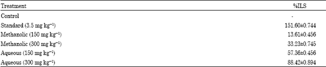
The methanolic and aqueous extracts of M. parasiticus at the doses of 150 and 300 mg kg-1 body weight when administered orally elevated the MST 18.36±0.356 and 21.53±0.357 days for methanolic extracts and 25.43±0.323 and 30.45±0.664 days for aqueous extract, respectively the extracts also increased the %ILS 13.61±0.456 and 33.23±0.745 for methanolic extract and 57.36±0.456, 88.42±0.894 for aqueous extract, respectively of EAC tumor bearing mice (ILS) (Table 1, 2). The haematological profile such as RBC count increased to5.95±0.68, 6.53±0.54 for methanolic extract and 6.89±0.42, 8.32±0.58 for aqueous extract when compared with control 3.69±0.58. Haemoglobin content was increased to 9.92±0.37, 10.85±0.25 for methanolic extract and 11.14±0.28, 12.24±0.49 for aqueous extract when compared with control 8.75±0.24. (Fig. 1a, c) but the WBC count was decreased to 27.83±0.46, 24.24±0.75 for methanolic extract and 22.54±0.67, 16.37±0.87 aqueous extract as compared to that of EAC control 39.93±0.54 (Fig. 1b). The extracts showed increased nonviable cell count as 2.4±0.07, 2.46±0.15 for methanolic extract and 2.34±0.05, 2.28±0.08 for aqueous extract as compared to that of control 0.3±0.21. The viable cell count was decreased as 2.1±0.10, 1.84±0.06 for methanolic extract and 1.56±0.24, 1.22±0.19 for aqueous extract as compared to that of EAC control 7.50±0.05 (Fig. 2a, b).
| Table 1: | Effect of various extracts on Mean Survival Time (MST) in EAC Inoculated Mice, Mean±SEM |
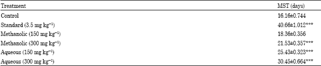 | |
| Each value represents the Mean±SEM (n = 10 mice per groups). *p<0.05, **p<0.01 and ***p<0.0001 when treated is compared with control | |
The administration of extracts also decreased tumor volume 3.37±0.189 and 2.74±0.241 for methanolic extract and 2.55±0.393 to 1.85±0.227 for aqueous extract when compared to control 5.66±1.203 (Fig. 3). It was also observed that Tumor weight was decreased 3.82±0.205 to 3.28±0.985 for methanolic extract and 2.85±1.455 to 2.20±0.773 (Table 3) when compared to that of EAC control mice 6.79±0.132.
The reliable criteria for judging the value of any anticancer drugs are prolongation of life span, inhibition of gain in average body weight and decrease of WBC from blood (Kumar et al., 2007).
| Table 2: | Effect of various extracts on percent increase in life span |
 | |
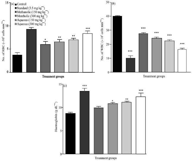 | |
| Fig. 1: | (a) Effect of methanolic and aqueous extracts on (a) RBC count, (b) WBC count and (c) hemoglobin count. Each point represents the Mean±SEM (n = 10 mice per groups). *p<0.05, **p<0.01 and ***p<0.0001 when treated is compared with control |
| Table 3: | Effect of various extracts on tumor weight |
 | |
| Each values represents the mean±SEM (n =10 mice per groups). *p<0.05, **p<0.01 and ***p<0.0001 when treated is compared with control | |
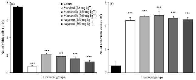 | |
| Fig. 2: | Effect of methanolic and aqueous extracts on (a) Viability of cells and (b) non viable. Each point represents the Mean±SEM (n = 10 mice per groups). *p<0.05, **p<0.01 and ***p<0.0001 when treated is compared with control |
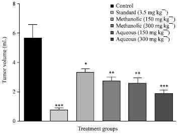 | |
| Fig. 3: | Effect of methanolic and aqueous extracts on tumor volume. Each point represents the Mean±SEM (n = 10 mice per groups). *p<0.05, **p<0.01 and ***p<0.0001 when treated is compared with control |
The methanolic and aqueous extracts delayed the cell division, thereby suggesting the reduction in EAC volume and increased survival time in mice.
Aqueous extract at both the doses (150 and 300 mg kg-1) and methanolic extract (300 mg kg-1) significantly improved the MST in tumor bearing mice. No. visible sign of toxicity and changes in vital functions were observed in any of treated animals. The prolongation of life span is a reliable criterion for judging efficacy of anticancer drugs (Hogland, 1982) and the extracts were able to meet this criterion. Myelosuppression and anemia have been frequently observed in ascites carcinoma (Price and Greenfield, 1950; Mitsuaki et al., 1981). In EAC control mice elevated WBC count; reduced haemoglobin and RBC count were observed. Anemia (reduced haemoglobin) encountered in ascites carcinoma mainly due to iron deficiency, either by haemolytic or myelopathic conditions which finally lead to reduced RBC number (Fenninger and Mider, 1954). In this research work it was observed that the oral administration of M. parasiticus extracts restored haemoglobin content and maintained normal values of RBC and WBC thus supporting its haematopoietic protecting activity without inducing myelotoxicity the most common side effects of cancer chemotherapy.
Recent physiological, pharmacological and biochemical studies appear to support the wisdom of the traditional dietary practices. They proved that phytochemicals from fruits and vegetables have shown to exert varied beneficial biological functions (Hafidh et al., 2009). Preliminary phytochemical screening revealed the presence of carbohydrates, phytosterols, fixed oils and phenolic compounds, saponins, proteins and flavonoids (Sodde et al., 2011). Flavonoids have been shown to posses antimutagenic and antimalignant effect (Fotsis et al., 1997). Furthermore, flavonoids have a chemopreventive role in cancer through their effect on signal transduction in cell proliferation and angiogenesis. The cytotoxicity and anticancer activity of methanolic extract is probably due to presence of flavonoids. Aqueous extracts reported the presence of phytosterols. Phytosterols are able to get incorporated in the cell membrane, alters membrane fluidity and the activity of membrane-bound enzymes. They also alter signal transduction in pathways leading to tumor growth and stimulate apoptosis in tumor cell lines. They also have been shown to enhance in-vitro human peripheral blood lymphocyte and T-cell proliferation in vitro which suggests a possible stimulation of the immune system function (Jones and AbuMweis, 2009). However, further investigation to explore the potential of the aqueous extracts of M. parasiticus in tumor treatment may prove to be worthwhile.
CONCLUSION
In the present study anticancer effect of methanolic and aqueous extracts assessed by evaluating tumor volume, viable and nonviable tumor cell count, tumor weight and hematological parameters of EAC bearing host. The both the extracts showed significant decrease in tumor volume, viable cell count, tumor weight and elevated the life span of EAC tumor bearing mice. Haematological profile such as RBC, haemoglobin and WBC count reverted to normal level in treated mice. From the result it was demonstrated that the extract has potent dose dependent anticancer activity and that is comparable to that of cisplatin. Aqueous extract at both doses (150 and 300 mg kg-1) and methanolic extract at 300 mg kg-1 dose showed potent anticancer activity However, further investigation to explore the potential of the aqueous extracts of M. parasiticus in tumor treatment may prove to be worthwhile.
ACKNOWLEDGMENT
The authors sincerely thank Manipal University, Manipal, India for providing all facilities to carry out this study.
REFERENCES
- English, D.R., B.K. Armstrong, A. Kricker and C. Fleming, 1997. Sunlight and cancer. Cancer Causes Control, 8: 271-283.
PubMedDirect Link - Fotsis, T., M.S. Pepper, E. Aktas, S. Breit and S. Rasku et al., 1997. Flavonoid, dietary-derived inhibitors of cell proliferation and in vitro angiogenesis. Cancer Res., 57: 2916-2921.
PubMedDirect Link - Hafidh, R.R., F. Abas, A.S. Abdulamir, F. Jahanshiri, F. Abu Bakar and Z. Sekawi, 2009. A review: Cancer research of natural products in Asia. Int. J. Cancer Res., 5: 69-82.
CrossRefDirect Link - Jeyachandran, R., A. Mahesh and L. Cindrella, 2007. DEN-Induced cancer and its alleviation by Anisomeles malabarica (L.) R.Br. ethanolic leaf extract in male albino mice. Int. J. Cancer Res., 3: 174-179.
CrossRefDirect Link - Jones, P.J. and S.S. AbuMweis, 2009. Phytosterols as functional food ingredients: Linkages to cardiovascular disease and cancer. Curr. Opin. Clin. Nutr. Metab. Care, 12: 147-151.
PubMedDirect Link - Karthikeyan, R., S. Karthigayan, M. Sri Balasubashi, S. Vijayalakshni and T. Balasubramanian, 2007. Antitumor effect of snake venom (Hydrophis spiralis) on ehrlich ascites carcinoma bearing mice. Int. J. Cancer Res., 3: 167-173.
CrossRefDirect Link - Kuper, H., P. Boffetta and H.O. Adami, 2002. Tobacco use and cancer causation: Association by tumour type. J. Internal Med., 252: 206-224.
CrossRefDirect Link - Litchfield, Jr. J.T. and F. Wilcoxon, 1949. A simplified method of evaluating dose-effect experiments. J. Pharmacol. Exp. Ther., 96: 99-113.
PubMedDirect Link - Mitsuaki, M., N. Ikuo, H. Masako, T. Yutaka and Y. Kunio, 1981. Lipid peroxide levels and lipid content of serum lipoprotein fractions of pregnant subjects with or without pre-eclampsia. Clin. Chim. Acta, 115: 155-161.
CrossRef - Nelson, D.A., T.T. Tan, A.B. Rabson, D. Anderson, K. Degenhardt and E. White, 2004. Hypoxia and defective apoptosis drive genomic instability and tumorigenesis. Genes Dev., 18: 2095-2107.
PubMedDirect Link - Newman, D.J., G.M. Cragg and K.M. Snader, 2003. Natural products as sources of new drugs over the period 1981-2002. J. Nat. Prod., 66: 1022-1037.
CrossRefPubMedDirect Link - Saalu, L.C., G.O. Ajayi, A.A. Adeneye, I.O. Imosemi and A.A. Osinubi, 2009. Ethanolic seed extract of grapefruit (Citrus paradisi Macfad) as an effective attenuator of doxorubicin-induced oxidative stress in the rat heart. Int. J. Cancer Res., 5: 44-52.
CrossRefDirect Link - Sur, P. and D.K. Ganguly, 1994. Tea Plant Roots Extract (TRE) as an antineoplastic agent. Planta Med., 60: 106-109.
PubMedDirect Link - Kumar, R.S., B. Jayakar and B. Rajkapoor, 2007. Antitumor activities of indigofera trita on Ehrlich ascites carcinoma induced mice. Int. J. Cancer Res., 3: 180-185.
CrossRefDirect Link