

Muneera Alkahtani
Faculty of Sciences, Prince Nora University, Research Center Lab, GSFMO, KSA
LiveDNA: 966.23136
M.A. EL-Naggar
Faculty of Sciences, Prince Nora University, Research Center Lab, GSFMO, KSA
S.A. Omer, Eman
Faculty of Community, Baha University, KSA
M. Abedl-Kareem
Plant Pathology Research Institute, ARC, Egypt
M.I. Ammar
Najran University, KSA
American Journal of Food Technology
Year: 2011 | Volume: 6 | Issue: 9 | Page No.: 730-741





ABSTRACT
The study concentrated on the common associated fungi with date palm (Phoenix dactylifera L.) fruits with special reference to Fusarium isolates. Also, the ability of Fusarium isolates to produce different mycotoxins and their effect on specific biochemical components and quality aspects of date fruits. The pathogenic capability of twenty-six isolates of F. moniliforme (F. verticillioides) isolated from different date fruit cultivars were carried out in vitro. Mycotoxins were estimated by immune-affinity column. Biochemical changes as amino acids, water soluble vitamins and fat soluble vitamins were determined with HPLC in all treatments. The most isolated fungi were obtained from Hayani cv. which were belonging to 10 genera and 23 species from all tested cultivars. Fusarium moniliforme were the most prevalent and recorded the highest frequency percentages compared with other isolated Fusarium species. Fumonisin was the highest toxin in tested date cultivars followed by T-2 and Zearalenone. The high amount of mycotoxins was found in Hayani, Samani and Zaghlol cultivars, respectively. Artificial inoculation by F. moniliforme induced several biochemical changes. This toxic isolate caused a reduction in protein, total sugar, fat and fiber contents comparing with control and non-toxigenic isolate. Moreover, there were differences in the fractions of amino acid and Vitamins content of the tested isolates. Date palm fruits may be attacked by toxigenic Fusarium moniliforme isolates. This contamination led to reduction in quality of date fruits due to loss of their nutrients as the result of biochemical change.
PDF Abstract XML References Citation
Article History
Received: January 12, 2011;
Accepted: April 16, 2011;
Published: May 21, 2011
How to cite this article
Muneera Alkahtani, M.A. EL-Naggar, S.A. Omer, Eman, M. Abedl-Kareem and M.I. Ammar, 2011. Effect of Toxic Fusarium moniliforme on Some Biochemical Component of some Date Palm Cultivars. American Journal of Food Technology, 6: 730-741.
DOI: 10.3923/ajft.2011.730.741
URL: https://scialert.net/abstract/?doi=ajft.2011.730.741
DOI: 10.3923/ajft.2011.730.741
URL: https://scialert.net/abstract/?doi=ajft.2011.730.741
INTRODUCTION
Date palm is one of the oldest cultivated plants. The world production of dates has increased 2.9 times over 40 years, whereas the world population has doubled. In many ways, dates may be considered as an almost ideal food, providing a wide range of essential nutrients and potential health benefits. Dates fruits are cheap to produce and preserve and are also very rich in nutrition. The flesh of dates contain a high percentage of carbohydrate (44-88%), protein (2.3-5.6%) and dietary fibre (6.4-11.5%). The fat percentage reach to (0.2-0.5%), including 14 types of fatty acids.
Also, the date fruits contain six Vitamins and 15 salts and minerals with 23 of different amino acids (Al-Shahib and Marshall, 2003).
There are several fungal diseases of date palm that cause severe damage to plant like Bayoud, Belaat bending, leaf spot, head black, Pestalotia leaf spot, Graphiola leaf spot, Dipodia disease and other important fungal disease while, Alternaria, Aspergillus, Fusarium and Penicillium cause fruit rots of date-palm (Bokhary, 2010).
Fusarium is one of the most important fungi that may infect the fruits of palm trees and caused numerous problems. During a survey conducted from 1998 to 2002, Fusarium species were found associated with palms belonging to the genera Chamaerops, Phoenix, Trachycarpus and Washingtonia showing symptoms of wilt and dieback. Pathogenicity tests were conducted on Phoenix dactylifera and P. canariensis cultivars using nine different Fusarium isolates (two reference strains from Saudi Arabia). Eight months after inoculation all strains caused disease, with palms showing as lesions and wilt symptoms. Fusarium damage may extend to the values of quality of fruits. Toxin analysis showed the ability of some Fusarium strains to excrete many type of mycotoxins (Armengol et al., 2005).
Mycotoxins are natural product produced by fungi as secondary metabolites on agriculture commodities in the field or during storage. Worldwide, approximately 25% of crops are annually affected by the mycotoxins (Martins et al., 2006). Although the modest Fusaria moulds can be found world-wide, they are prevalent in the zones of a moderate climate (Krska et al., 2001). Contamination of agriculture food and feed with the mycotoxins has become a main problem in most parts of the tropical and semitropical countries. This may be due to the favorable climatic conditions prevalent in those regions coupled with the traditional methods of crop cultivation, harvesting, handling and storage (Tirado et al., 2010).
Fusarium genus had many species most of them had ability to produce many mycotoxins. Fusarium mycotoxins contaminant the most of agriculture commodities and cereal grains in worldwide. Fumonisins and zearalenone are mycotoxins produced by a variety of fungi of the, Fusarium verticillioides and other Fusarium species. Fusarium verticillioides is after Fusarium solani, the most common species to excrete these mycotoxins. Fumonisins and zeralenone was considered the most dangerous Fusarium mycotoxins and causing fusariosis which involves multiple organs, such as the liver, lung, kidney, heart, spleen and pancreas, with a high mortality rate to date (Ortoneda et al., 2002). Fumonisins were first isolated in 1988 from cultures of F. verticillioides strain MRC 826. The most abundant species in nature is fumonisin B1, followed by fumonisin B2 and B3 (Cabassi et al., 2005; Wan Norhasima et al., 2009). Zearalenone (ZEA) is estrogenic compound and widely contaminates cereal grains and animal feedstuffs and most of food production (Dong et al., 2010).
The objectives of the present investigation were to detect the main common associated Fusarium species with date palm fruits. And to study the ability of Fusarium isolates to excrete zearalenone and fuominsin and their effect on quality parameter of fruits and specific biochemical components associated with toxicity infection.
MATERIALS AND METHODS
Ind identification: Three common Egyptian date palm cultivars namely; Hayini, Zaghlol and Samani were obtained from different farms and markets. One hundred discolored pieces of fruits were disinfected by immersing in 2% sodium hypochlorite solution for two minutes, washed thoroughly three times in sterilized distilled water, then dried between folds sterilized filter papers. The pieces were aseptically transferred to sterilized plates containing PDA medium according to the method of. Pieces of the desired cultivar were put in each dish. Ten dishes were used for each treatment. All plates were incubated at 25±2°C for 10 days, after which the emerged fungal colonies were picked up and purified using the hyphal tip and/or single spore technique. The purified fungi were identified to either the generic or the species level according to the descriptions of Barnett (1960) and Domsch et al. (1980). For the identification of Fusarium species, the key proposed by Marasas et al. (1984) was used. The pure cultures of the isolated fungi were maintained on PDA slants and kept at 5-8°C in the refrigerator for the subsequent studies.
Pathogenicity tests: The pathogenic capability of twenty-six isolates of F. moniliforme (the most frequent fungus among the various species of Fusarium) which isolated from different date fruit cultivars was carried out in vitro on cvs. Hayani, Zaghlol and Samani according to the methods described by Christensen and Dreschler (1954). The percentage of infection (%) was determined five days after inoculation at 27±2°C.
Mycotoxin determination: Sample of date fruits from different cultivars (Hayani, Zaghlol and Samani cvs.) inoculated separately with eleven different Fusarium moniliform isolates were used for mycotoxins determination. Non inoculated fruits from each cultivar were used as control.
Fumonisin: Total Fumonisin toxins were determined according to the method described by Mazzani et al. (2001).
Zearalenone: The Zearalenone concentration was determined as mentioned in the Fumonisin toxin but the dilution was made with 49 ml distilled water then passed through Zearatest column (Vicam Company) and measured in calibrated fluorometer model (Series-4/Vicam) according to the method of Martins et al. (2003).
T-2 toxin: Fifty grams of ground sample was blended with 5 g sodium chloride and added to 10 g of polyethyleneglycol (PEG) and 35% methanol at high speed for one minute. The solution was filtered through Vicam fluted filter papers. The extract was diluted with water (1:2.5 v/v). The filtrate was passed through microfiber filter paper. Five hundred microlitter of developer were added to 10 mL of the filtrate. The extract mixture was passed over T-2 packed column (Vicam Company) and washed by passing 10 mL of 0.02% Tween at the rate of 1-2 drops/second. T-2 toxin elute was passed with 2.0 mL HPLC grade methanol through column. The toxin level in (ppm) was measured in a calibrated fluorometer, according to the proposed method by Martins et al. (2003).
Biochemical changes: Biochemical changes was determined on date fruits of the three cultivars (Hayani, Zaghlol and Samani cvs.) inoculated with the highest toxin producers isolate (H-8) and non producers toxin isolate (H-3).
Protein content: The method proposed by Bradford (1976) was used to determine protein content.
Total sugar contents: Total soluble sugar contents was determined colorimetrically using the picric acid method described by Thomas and Dutcher (1924) by spectrophotometer at 540 nm.
Total fat content: Ten grams of the tested date cultivar inoculated individually with any of the tested isolates of F. moniliforme used for total fat determination. The total fat was extracted with a mixture of chloroform and methanol (2:1 v/v) in a Soxhelt apparatus for approximately 16 h according to the method described in (AOAC, 1990). The solvent was evaporated in a rotary evaporator. The dried residue was considered as the total fat content and expressed as weight mg g-1 fresh weight.
Determination of fiber: Crude fiber content was determined as described in AOAC (2004).
Fractionation of amino acid: Ten grams of sample was ground and filtered. The residue was washed with a few ml of 75% ethanol and the volume was made up to 100 mL. Several amino acids were examined using a HPLC system (HP1050) with Ultra Violet (UV) detector at 254 nm. The separation was accomplished with stationary phase type APS-NH2 (5 μm 4x250 mm) column. The mobile phase consists of 32% (methanol/water), 60/40 with 0.3 mL acetic acid. The flow rate was 0.9 mL min-1. The temperature of column was 45°C, while the injection volume was 1 μL according to the method of (Gertz, 1990).
Vitamins contents: Ten gram of fresh tissues of each sample were homogenized with eluent acetic acid: water (6:94, v/v) and stirred for water soluble Vitamin extraction, while the eluent of fat-soluble vitamin) from the same weight was methanol: water (98:2,v/v). The extracts were filtered through a micro filter (45 μm) and stored in vials. HPLC analysis was used to study the Vitamin in the extracts. Analysis of vitamins was performed on HPLC model (HP1050). Separation and determination were performed on C18 column (5 μm, 4.6x150 mm). The mobile phase yielded results of acetic acid: water (6:94, v/v) for water-soluble Vitamin and methanol: water (98:2, v/v) for fat-soluble vitamin. The wavelengths in UV detector were 254 nm for both Vitamins i.e., water and fat-soluble. Total run time for the separation was approximately 15 min at a flow rate of mL min-1 according to the method of Dolphin (1999). All tested vitamins were determined as μg g-1 fresh weight of flesh.
RESULTS
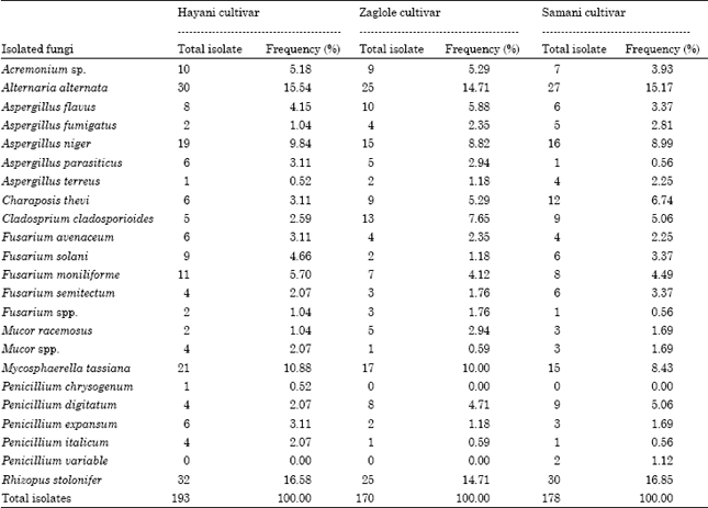
Isolation and identification of fungi associated with date fruits: Twenty-three species belonging to 10 genera were collected from three cultivars of date palm fruit. Results illustrated in (Table 1) revealed that Hayani cv. was the most susceptible cultivar comparing with Samani and Zaghlol cultivars.
Alternaria alternata was the most common fungi on Hayani, Zaghlol and Samani cultivars. The total numbers of isolates obtained from previous cultivars were 193, 170 and 178, respectively.
There are many spices of Fusarium which isolated from tested cultivars and there is no complete matching through the number of isolates and percentage of frequency between the cultivars. Fusarium moniliforme was the most predominant fungus among different Fusarium species in all tested cultivars. The isolation number for F. moniliforme in Hayani, Zaghlol and Samani were (11, 7 and 8) while percentage of frequency (5.70, 4.12 and 4.49), respectively.
Pathogenicity test: The pathogenic capabilities of twenty-six isolates of Fusarium moniliforme isolated from different date palm fruits were evaluated using Hayani cv. (the most susceptible cultivar under natural conditions).
| Table 1: | Occurrence and frequency (%) of fungi associated with date palm of three different cultivars using PDA medium at 27±2°C |
 | |
| LSD for fungusxCultivar interaction = 0.56 (p≤0.05) | |
All tested isolates of F. moniliforme were able to infect date palm fruits and able to induce all symptoms typically caused by F. moniliforme after 5 days from artificial inoculation at 27°C and varied in their percentage of infection. Eleven isolates were selected according to their pathogenic capability for further studies.
Mycotoxins determination: Results in Table 2 indicate that all the selected isolates of F. moniliforme were varied in their ability to produce different types of toxins. In general, all isolates produced low amounts of Zearalenone toxin compared with the toxin Fumonisin and T-2. Isolates No. 8, 1, 10 and 2 of F. moniliforme consider the highest toxin producers as compared with the other tested isolates. Isolate No. 7 was the lowest toxin producers meanwhile, the ability of isolate No. H-3 to produce any toxins was null. Isolates No. H-8 and H-3 were selected for further studies.
Biochemical changes
Determination of protein, total sugar, fat and fiber content: Results in Table 3 reveal that, in general, the infection of Hayani, Zaghlol and Samani date cultivars with H-8 and H-3 isolates of F. moniliforme resulted in a decrease of proteins, sugars, fats and fibers contents as compared with control. The lowest amount of these components was noticed in different infected cultivars with F. moniliforme isolate No. H-8 comparing with isolate No. H-3 which showed a little effect on the contents of date fruits.
| Table 2: | Mycotoxins concentration (ppm) produced by isolates of Fusarium moniliforme in tested date palm cultivars Hayani, Zaglole and Samani after 10 days of incubation at 25±2 °C |
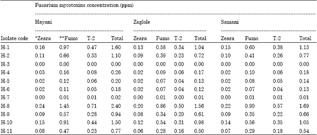 | |
| *Zeara: Zearalenone toxin, ** Fumo: Fumonisin toxin | |
| Table 3: | Effect of toxic and non-toxic isolates of Fusarium moniliforme on protein, total sugar, fat and fiber contents of date fruits cultivar Hayani, Zaghlol and Samani after 10 days of incubation at 27±2°C |
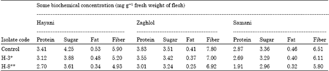 | |
| H-3*: Non-toxic isolate, H-8**: The highest toxic isolate: LSD for fungusxCultivar interaction at 5% for: Protein: 0.20, Sugar: 0.30, Fat: 0.04, Fiber: 0.42 | |
The lowest amount of protein content was record in the infected Samani cv. with isolate No. H-8 followed by Hayani and Zaghlol in comparison with their control. The analogous values were 1.91, 2.70 and 3.01 mg g-1 fresh weight of flesh, respectively. On the other hand, non toxic isolate of F. moniliforme (H-3) showed the lowest value of protein in Samani cv. followed by Hayani and Zaghlol (being 2.69, 3.12 and 3.55) comparing with their control.
Data presented in Table 3 show that total soluble sugar content was decreased in the inoculated samples compared with the control as the same trend with protein contents. The most toxigenic isolate of F. moniliforme, i.e., No. H-8 caused a higher reduction of this component and the corresponding values were 3.61, 3.24 and 2.96 mg g-1 fresh weight flesh in Hayani, Zaghlol and Samani cvs., respectively. While the non-toxic of F. moniliforme i.e., No. H-3 had a slight effect in decrease the total sugar content comparing with their control. The lowest value was noticed in Samani, Hayani and Zaghlol (being 3.42, 3.88 and 3.92 mg g-1 fresh weight flesh, respectively).
Data presented in Table 3 indicate that the infection with any of the tested isolates of F. moniliforme led to a decrease of fat content in date fruit if compared with the un-inoculated samples. Isolate No. H-8 (the highly toxigenic) caused a higher reduction, followed by isolates No. H-3 (non-toxigenic) compared with the control. The corresponding values of fat content in cultivar Hayani, Zaghlol and Samani which inoculated with toxigenic F. moniliforme isolate (H-8) were 0.34, 0.25 and 0.32 mg g-1 flesh fresh weight, respectively. On the other hand, the amount of fat content in the same tested cultivars inoculated with non-toxigenic isolate (H-3) was 0.48, 0.37 and 0.40 mg g-1 flesh fresh weight, respectively.
The crud fiber content was decreased in all tested cultivars of date fruit (Hayani, Zaghlol and Samani cvs.) after inoculated with any toxic and non toxic F. moniliforme isolates. Fiber decreasing ratio in tested cultivars (Hayani, Zaghlol and Samani cvs.) inoculated with toxic isolate No. H-8 was greater than in case of non-toxic isolate H-3. The analogues value was 4.93, 6.92, 5.80, 5.2, 7.0 and 6.11, respectively while the control value was 5.9, 7.8 and 6.51 mg g-1 flesh fresh weight, respectively. The effect of non-toxic isolate on fiber content was very slight comparing with toxic isolate as positive control and also negative control.
Fractionation of amino acids: Data presented in Table 4 revealed that there were twenty-one fractions of amino acids in the un-inoculated fruits (control) of Hayani cultivar while there were 20 and 19 only fractions in both Zaghlol and Samani, respectively. In general this number of fractions was reduced in the inoculated date palm with each of the tested isolates of F. moniliforme and this extent of the decrease was largely depend on fungal species and toxicity.
| Table 4: | Effect of toxic and non-toxic isolates of Fusarium moniliforme on amino acids contents of date fruits cultivar Hayani, Zaghlol and Samani after 10 days of incubation at 27±2°C |
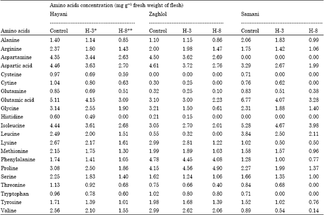 | |
| H-3*: Non-toxic isolate, H-8**: The highest toxic isolate, LSD for at 5%: Amino acids contentxCultivar interaction = 0.576, Amino acids contentxFungal isolates interaction = 0.761 | |
| Table 5: | Effect of toxic and non-toxic isolates of Fusarium moniliforme on water soluble vitamins (B1, B2 and C) and fat soluble vitamin (A, D and E) of date fruits cultivar Hayani, Zaghlol and Samani after 10 days of incubation at 27±2°C |
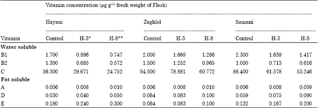 | |
| LSD for vitamin contentxCultivar interaction and/or isolates at 5% for: B1: 0.09, B2: 0.06, C: 9.7, A: 0.001, D: 0.01, E: 0.03 | |
For instance, date palm fruit inoculated with the most toxigenic isolate i.e., No. H-8 contained low level of most amino acids fractions comparing with non-toxigenic one No. H-3. The levels of amino acids fractions were decreased in number and amount in case of toxic isolate (H-8) comparing with non-toxic and control one. In Hayaini cultivar the fraction of Histidine was disappeared with toxic isolate (H-8) and also Cysteine, Cystine, Histidine and Leucine in Zaghlol cultivar while concentration of proline was increased. In Samani cultivar Cysteine, Cystine, Threonine and tryptophan were not found with toxic isolate.
Determination of vitamins content: Data obtained in Table 5 revealed that, inoculation with any tested isolate of F. moniliforme led to an increase of Fat soluble Vitamins A, D and E as compared with the control. The degree of increment was more pronounced for toxic isolates of F. moniliforme (H-8) than that in non-toxic (H-3). The higher level of Vitamin E was noticed in Hayani cv. followed by Samani and Zaghlol cvs. inoculated with F. moniliforme isolate No. H-8 (toxigenic), being 3.00, 2.00 and 1.00 μg g-1 fresh weight, respectively. The value in the control treatment was 0.180, 0.132 and 0.064 μg g-1 fresh weight. On contrast, all water soluble Vitamins (B1, B2 and C) had vice versa trend of fat soluble Vitamins and a less quantity of Vitamins was recorded in date palm fruits infected with the isolate No. H-8 (toxigenic) followed by No. H-3 (non-toxigenic). The highest value in all tested water soluble Vitamins was record for Vitamin C in date palm healthy fruit cv. Zaghlol followed by Samani and Hayani cvs. (being 94.5, 86.4 and 56.3 μg g-1 fresh weight of flesh, respectively) while in infected sample with toxigenic isolate was 60.772, 53.246 and 24.752 μg g-1 fresh weight of flesh, respectively. In Zaghlol, Samani and Hayani cultivars inoculated with non-toxigenic isolate the Vitamin C was 78.881, 61.578 and 29.671, respectively. Other soluble Vitamin (BI and B2) had the same trend of Vitamin C.
DISCUSSION
Mycotoxins are toxic secondary metabolites produced by fungi,. There are hundreds of mycotoxins types contaminate about 25% of the worldwide plants crops according to the estimation of FAO organization (Fandohan et al., 2003) Isolation trial reveal Fusarium is one of the major fungal genera associated with date palm fruits. There were differences between cultivars in the rate of fungal contamination which may be due to the nutritional content. Fusarium moniliforme was the most predominant fungus among different Fusarium species in all tested cultivars and this result was agreement with results of Bokhary (2010), Wan Norhasima et al. (2009) and Armengol et al. (2005) but was differed with the aforementioned isolates of F. moniliforme were screened for pathogenicity capability and their ability to produce different types of toxins. All the tested isolates of F. moniliforme produced low levels of zearalenone toxin compared with the toxin fumonisin and T-2, in addition there was one isolate of F. moniliforme hadn't ability to produce any toxins in the tested cultivar (H-3) while, the most excreted of different toxins was (H-8). Similar results were reported by Torres et al. (2010), Myung et al. (2009), Fatima et al. (2007), Abo-Elnaga and Ahmed (2007) and Hadiani et al. (2003).
Mazzani et al. (2001) and Logrieco et al. (2002) reported that Zearalenone and Fumonisin are the toxins that mostly produced by F. moniliforme and the commonly Fusarium toxins found were Trichothecenes, Zearalenone, Fumonisin and Moniliformin. The differences between these results and our results may be due to the fact that toxin production is influenced by some factors such as the physiological behavior of the tested isolates, the genetic capabilities of fungus and/or fungal substrate.
Infection of date palm by F. moniliforme isolates induced several biochemical changes. The rate of variation in the tissue composition was differed according to the tested isolate. There was a parallel relationship between mycotoxins formation and consequent biochemical changes (proteins, sugars, fats, fibers, amino acids and vitamins). This result was in agreements with Scarpari et al. (2005), Meinhardt et al. (2008) and Campos et al. (2009). In the present investigation, it was found that the protein content was reduced in inoculated samples by the tested isolates of F. moniliforme. The decline in protein content probably may be due to its decomposition by the tested fungal isolates.
Total soluble sugar content was decreased in infected sample may be due to its utilization by the infectd isolate in respiration, growth and/or mycotoxins formation after cellulose and pectin degradation in sample by the hydrolysis enzymes produced by fungi (Aly, 1982). It is possible, therefore, that isolates may play an active role in deterioration of fiber by hydrolysis enzymes which produced by toxigenic isolate more than non-toxigenic one (Ushamalini et al., 1998). The most toxigenic isolate of F. moniliforme i.e., No. H-8 was the most effective in reducing fat contents. There was direct correlation between Fusarium mycotoxins and reduction of fat content. The decrease in fat content may be attributed to the lipases production by the fungi invading which converting the fat into free fatty acids and glycerol. The obtained results are in agreement with those recorded by Shih and Zheng (1983). Neergaard (1977) report that the greatest reduction in oil content may have been caused by the lipolytic activity of the seed-borne fungi.The effect of Fusarium moniliforme isolates on the amino acids fractionation showed several changes in these fractions such as absence or reduction of certain components and decrease in the amount of other ones especially with toxigenic isolates comparing with non-toxigenic. The reduction in amino acids amount may be due to the fact that amino acids are important nutrients that are transferred from the plant to the fungus can function as nitrogen sources for the fungus (Scarpari et al., 2005).
An experiment was carried out to determine vitamins content in the inoculated sample by various isolates of Fusarium moniliforme. Data revealed that the inoculation by the toxic tested isolates led to a decrease content of some water-soluble Vitamins (B1, B2 and C). This reduction was increased with increasing of isolates toxicity. On contrary, the infection with toxigenic F. moniliforme isolate led to an increase of fat soluble Vitamins (A, D and E) and the degree of increment was more than that of non-toxigenic isolate. The increasing of fat soluble Vitamins may be due to degradation of sample fat with different lipase enzymes which produce by tested fungi to main component as fatty acids, glycerol and also fat soluble Vitamins. These results are in disagreement with those reported by Shaker and Darwish (2005) who found that there was a reversal relation among Vitamin E content and the infection with Fusarium spp. This variation among isolates may be attributed to some genetic factors which in turn control the enzyme system operating in the isolate. The obtained results are in agreement with those obtained by Jain et al. (2002) who mentioned that the content of fruits of Vitamin C was the highest in the un-inoculated control and the lowest in plants inoculated with each of Fusarium spp.
CONCLUSION
Through finding there was effect of some mycotoxin which produced by Fusarium moniliforme on nutrition value of date fruits. Although the risk of Fumonisin and zearalenone and t-2 toxin on the date fruits as very toxic substances but also had significant effect on biochemical change; sugar, fiber, Vitamins, protein and amino acids in quantity and quality.
REFERENCES
- Al-Shahib, W. and R.J. Marshall, 2003. The fruit of the date palm: Its possible use as the best food for the future? Int. J. Food. Sci. Nutr., 54: 247-259.
CrossRefPubMedDirect Link - Bradford, M.M., 1976. A rapid and sensitive method for the quantitation of microgram quantities of protein utilizing the principle of protein-dye binding. Anal. Biochem., 72: 248-254.
CrossRefPubMedDirect Link - Domsch, K.H., W. Gams and T.H. Anderson, 1980. Compendium of Soil Fungi. Academic Press, London, United Kingdom, ISBN: 9780122204012, Pages: 1264.
Direct Link - Jain, A., B.K. Goswami and L. Singh, 2002. Effect of Meloidogyne incognita and Fusarium oxysporum both alone and in combinations on ascorbic acid content of tomato fruits. Adv. Plant Sci., 15: 383-384.
Direct Link - Logrieco, A., G. Mule, A. Moretti and A. Bottalico, 2002. Toxigenic Fusarium species and mycotoxins associated with maize ear rot in Europe. Eur. J. Plant Pathol., 108: 597-609.
Direct Link - Martins, M.L., H.M. Martins and F. Bernardo, 2003. Fungal flora and mycotoxins detection in commercial pet food. Revista Portuguesa Ciencias Veterinarias, 98: 179-183.
Direct Link - Mazzani, C., O. Borges, O. Luzon, V. Barrientos and P. Quijada, 2001. Occurrence of Fusarium moniliforme and fumonisins in kernels of maize hybrids in venezuela. Brazilian J. Microbiol., 32: 345-349.
CrossRefDirect Link - Shaker, E.S. and I.M. Darwish, 2005. Antioxidative and antifungal activity of azadirachtin derivative on infected tomato seed germination. Bull. Faculty Agric. Univ. Cairo, 56: 483-500.
Direct Link - Torres, A.M., M.L. Ramirez and S.N. Chulze, 2010. Fusarium and fumonisins in maize in South America. Mycotoxins Food Feed Bioweapon, 2: 179-200.
CrossRef - Armengol, J., A. Moretti, G. Perrone, A. Vicent, J.A. Bengoechea and J. Garcıa-Jimenez, 2005. Identification, incidence and characterization of Fusarium proliferatum on ornamental palms in Spain. Eur. J. Plant Pathol., 112: 123-131.
CrossRef - Bokhary, H.A., 2010. Seed-borne fungi of date-palm, Phoenix dactylifera L. from Saudi Arabia. Saudi J. Biol. Sci., 17: 327-329.
CrossRef - Campos, A., G. Da Costa, A.V. Coelho and P. Fevereiro, 2009. Identification of bacterial protein markers and enolase as a plant response protein in the infection of Olea europaea subsp. europaea by Pseudomonas savastanoi pv. Savastanoi. Eur. J. Plant Pathol., 125: 603-616.
CrossRef - Dong, M., X.J. He, P. Tulayakul, J.Y. Li and K.S. Dong et al., 2010. The toxic effects and fate of intravenously administered zearalenone in goats. Toxicon, 55: 523-530.
CrossRef - Fandohan, P., K. Hell, W.F.O. Marasas and M.J. Wingfield, 2003. Infection of maize by Fusarium species and contamination with fumonisin in Africa. Afr. J. Biotechnol., 2: 570-579.
Direct Link - Fatima J., A. Meddich and I. El-Hadrami, 2007. Effectiveness of arbuscular mycorrhizal fungi in the protection of date palm (Phoenix dactylifera L.) against bayoud disease. Physiol. Mol. Plant Pathol., 71: 166-173.
CrossRef - Hadiani, M.R., H. Yazdanpanah, M. Ghazi-Khansariy, A.M. Cheraghaliz and M. Goodarziz, 2003. Survey of the natural occurrence of zearalenone in maize from northern Iran by thin-layer chromatography densitometry. Food Addit. Contam., 20: 380-385.
PubMed - Meinhardt, L.W., J. Rincones, B.A. Bailey, M.C. Aime, G.W. Griffith, D. Zhang and G.A.G. Pereira, 2008. Moniliophthora perniciosa, the causal agent of witches` broom disease of cacao: What`s new from this old foe. Mol. Plant Pathol., 9: 577-588.
CrossRef - Ortoneda, M., J. Capilla, F.J. Pastor, I. Pujol and J. Guarro, 2002. Efficacy of liposomal amphotericin B in treatment of systemic murine fusariosis. Antimiocrobial agents Chemother., 46: 2273-2273.
Direct Link - Myung, K., S. Li , R.A.E. Butchkom, M. Busman, R.H. Proctor, H.K. Abbas and A.M. Calvo, 2009. FvVE1 regulates biosynthesis of the mycotoxins fumonisins and fusarins in Fusarium verticillioides. J. Agric. Food Chem., 57: 5089-5094.
CrossRef - Scarpari, L.M., L.W. Meinhardt, P. Mazzafera, A.W.V. Pomella, M.A. Schiavinato, J.C.M. Cascardo and G.A.G. Pereira, 2005. Biochemical changes during the development of witches` broom: The most important disease of cocoa in Brazil caused by Crinipellis perniciosa. J. Exp. Bot., 56: 865-877.
CrossRef - Tirado, M.C., R. Clarke, L.A. Jaykus, A. McQuatters-Gollop and J.M. Frank, 2010. Climate change and food safety: A review. Food Res. Int., 43: 1745-1765.
CrossRef - Wan Norhasima, W.M., A.S. Abdulamir, F.A. Bakar, R. Son and A. Norhafniza, 2009. The health and toxic adverse effects of Fusarium fungal mycotoxin, fumonisins, on human population. Am. J. Infec. Dis., 5: 273-281.
CrossRefDirect Link - AOAC, 1990. Official Methods of Analysis. 15th Edn., Association of Official Analytical Chemists, Washington, DC., USA., pp: 200-210.
Direct Link - Martins, H.M., M.M. Guerra and F. Bernardo, 2006. Zearalenone, deoxynivalenol and fumonisins in mixed-feed for laying hens. Mycotoxin Res., 22: 206-210.
CrossRef - Cabassi, E., F. Miduri and A.M. Cantoni, 2005. Intoxication with fumonisin B1 (FB1) in piglets and supplementation with granulated activated carbon: Cellular-mediated immunoresponse. Vet. Res. Commun., 29: 225-227.
CrossRef - Abo-Elnaga, H.I.G. and N.G. Ahmed, 2007. Pathogenicity, toxicity and Gibberellic Acid Content of Fusarium moniliforme causing root rot and damping off of pepper. Plant Pathol. J., 6: 318-323.
CrossRefDirect Link