

P. Chattopadhyay
Birla Institute Technology and Sciences, Pilani-333031, Rajasthan, India
National Biotechnology Centre, Indian Veterinary Research Institute, Izatnagar-243122, UP, India
G. Sukla
Microbiology and Cellular Biology Laboratory, College of Pharmacy, IFTM, Lodhipur Rajput, Moradabad-24001, UP, India
R. Gautam
Microbiology and Cellular Biology Laboratory, College of Pharmacy, IFTM, Lodhipur Rajput, Moradabad-24001, UP, India
L. Singh
Microbiology and Cellular Biology Laboratory, College of Pharmacy, IFTM, Lodhipur Rajput, Moradabad-24001, UP, India
A. Verma
Microbiology and Cellular Biology Laboratory, College of Pharmacy, IFTM, Lodhipur Rajput, Moradabad-24001, UP, India
N. Verma
Microbiology and Cellular Biology Laboratory, College of Pharmacy, IFTM, Lodhipur Rajput, Moradabad-24001, UP, India
S. Kumar
Microbiology and Cellular Biology Laboratory, College of Pharmacy, IFTM, Lodhipur Rajput, Moradabad-24001, UP, India
A.K. Wahi
Microbiology and Cellular Biology Laboratory, College of Pharmacy, IFTM, Lodhipur Rajput, Moradabad-24001, UP, India
Asian Journal of Biochemistry
Year: 2007 | Volume: 2 | Issue: 6 | Page No.: 427-431





ABSTRACT
The internucleosomal cleavage of genomic DNA is a biochemical hallmark of apoptosis. DNase endonuclease has been suggested to be one of the apoptotic endonuclease, but its biochemical characteristic has not been fully elucidated in ischemic and reperfusion (IR) injury. The present study suggests that DNase-I is involved in the apoptosis of IR injured rat liver The finding suggests a possible biological basis of endonuclease associated apoptotic effects of DNA in ischemic and reperfused hepatocytes.
PDF Abstract XML References Citation
How to cite this article
P. Chattopadhyay, G. Sukla, R. Gautam, L. Singh, A. Verma, N. Verma, S. Kumar and A.K. Wahi, 2007. Isolation and evaluation of Endonucleases in apoptotic activity of DNA in Ischemic Injury and Reperfusion Injury of Rat Hepatocytes. Asian Journal of Biochemistry, 2: 427-431.
DOI: 10.3923/ajb.2007.427.431
URL: https://scialert.net/abstract/?doi=ajb.2007.427.431
DOI: 10.3923/ajb.2007.427.431
URL: https://scialert.net/abstract/?doi=ajb.2007.427.431
INTRODUCTION
Temporary clamping of the portal triad, i.e., inflow occlusion by the Pringle's maneuver, is a common strategy to minimize bleeding during hepatic resection. Increasing evidences suggests that oxygen derived free radicals and reintroduction of oxygen in ischemic tissue lead to ischemic and reperfusion injury (IR). Death of hepatocytes and other hepatic cell types is a characteristic feature of liver diseases as diverse as cholestasis, viral hepatitis, IR, liver preservation for transplantation and drug/toxicant-induced injury. Cell death typically follows one of two patterns: oncotic necrosis and apoptosis. Apoptosis plays a crucial role after IR in organ transplantation. Organ-specific endonuclease might play a role in apoptosis in laboratory rodent tissues. Liver damage is observed during various steps of liver transplantation (Clavien et al., 1992). Injury occurs before the removal of the graft from the donor (preservation injury), during the period of cold storage (preservation injury), during implantation in the recipient (rewiring injury) and after revisualization (reperfusion injury). Apoptosis is widely believed to play pivotal role in the progress of chronic hepatitis to cirrhosis. The signal that initiates atrophy and apoptosis in response to the ischemia are not certain deprivation of oxygen, nutrients, or growth factor may be important. Cell atrophy involve to auto-phagocytes of organelles induced by ischemia (Thompson, 1995). The signal that initiates atrophy and apoptosis in response to the ischemia are not certain, but presumably are due to deprivation of oxygen, nutrients, or growth factor may be important (Ericsson, 1969). Free radicals influence gene expression, regulate cellular response to cytokines as well as proliferate events of a cell. All these have been implicated as possible triggering mechanisms of apoptosis (Granger et al., 1986). A hall mark of apoptosis is the inter-nucleosomal DNA degradation for which a Ca+2, Mg+2 dependent endonuclease has been postulated (Peitsch et al., 1993). The inter-nucleosomal cleavage of genomic DNA is a biochemical hallmark of apoptosis. DNase gamma, an Mg2+/Ca2+ -dependent endonuclease, has been suggested to be one of the apoptotic endonucleases (Mizuta et al., 2006). Role of endonucleases in liver ischemia and reperfusion has not been fully elucidated. Therefore it was decided to study the effectiveness and DNase-I acting in apoptosis in IR liver.
MATERIALS AND METHODS
Animals
Swiss male albino rats weighing around 250 to 300 g procured from College of Pharmacy, IFTM, Moradabad were used in the present study. Animals had free access to food and water and were maintained under standard laboratory conditions with natural light and dark cycle. Food given to rat consisted of standard laboratory chaw (Hindustan Liver, Mumbai, India) and water ad libitum at all times. The animals were acclimatized for at least 5 days before behavioral experiments. Experiments were carried out between 09:00 and 15:00 h and the animals were starved for 18 h before laprotomy during the period of fasting. The rats were allowed to drink water ad libitum. Hepatic Ischemia was followed as per the procedure described by Hayashi et al. (1986). The rats were divided into two groups with 6 animals each and studies were made after 3h of reperfusion. Group I served as vehicle control, Group II served as only I/R injury. After 3 h, the left and median lobes of the liver were removed and used for the experiments.
Isolation of Hepatocytes
Hepatocytes were prepared by collagenase perfusion (Seglen, 1973). Liver was perfused with 0.05% collagenase. Reperfused livers were passed through 0.0125 mm gauze and resultant cell suspension was washed and counted using a haemocytometer. Viability was determined by trypan blue exclusion. Isolated cells viability were 70-90% after isolation.
Analysis of DNA Fragmentation
IR hepatocytes were lysed in buffer containing 50 mM Tris HCl (pH 7.8), 1mmM EDTA and 0.5% w/v sodium lauryl sulphate. DNA was extracted from hepatocytes as per method described by Herrmanam et al. (1994). Gel electrophoresis of DNA (10 μg/well) was performed at 100 mV for 1 h on 1% agarose gel containing 0.004% ethidium bromide in 10 mM Tris (hydroxymethyl) aminomethane (Tris)-1 mM EDTA (pH 8.0). The loading buffer consisted of 0.25% xylene cynole FF, 0.25% bromophenol blue, 40% sucrose and 1 mM ethidium bromide. The gels were visualized and photographed (Patel et al., 1994).
Extraction of Endonuclease
IR injured hepatocytes were washed with 10 volume of PBS buffer and then homogenized by 10 strokes in a teflon/glass homogenizer with 0.1% nonidet P-450. The homogenate was filtered through 200 μm stainless mess and the nuclei were collected by centrifugation at 4000 g for 5 min. Isolated nuclei were then re-suspended in 8 volume of buffer (10 m Tris HCl, pH 7.8, 3 mM Mg Cl2, 2 mM 2-mercaptoethanol, 3 mM PMSF, 0.5 M ammonium sulphate) and sonicated with sonicator. The mixtures were next stirred on ice for 30 min and the nuclear debris was removed by centrifugation at 20,000 x g for 60 min. The supernatant was taken as nuclear extract.
Endonuclease Assay
Endonuclease activity was measured as describe Shiokwa et al. (1994). A 1 μL aliquot of endonuclease fraction was added to reaction mixture containing 50 mM MOPS/NaOH, pH 7.2, 3 mM Cacl2, 3 mM MgCl2, 1 mM 2-meracaptoethanol, 0.1 mM PMSF and freshly prepared normal hepatocytes and incubated at 37°C for 60 min. In control hepatocytes were prepared in similar manner except 1 μL of buffer added instead endonuclease fraction. After incubation hepatocytes were centrifuged at 20, 000 x g for 10 min and obtained precipitated hepatocytes were lysed in buffer which contains 50 mM Tris HCl (pH 7.8), 1 mmM EDTA and 0.5% w/v sodium lauryl sulphate. DNA was isolated as described by Herrmann et al. (1994). The resulting DNA preparations were analyzed by electrophoresis 1% agarose gels contains 0.5 μg mL-1 ethidum bromide. The DNA fragment pattern was examined and photographed under UV illumumination.
RESULTS
Figure 1 shows the IR injured hepatocytes DNA was in apoptosis and produced laddering formation (Lane No. 2). Normal hepatocytes DNA not produced any laddering formation and intact
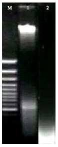 | |
| Fig. 1: | DNA ladder of ischemic and reperfused injury rat hepatocytes. Lane 1: Normal rats DNA isolated from hepatocytes. Lane 2: Apoptotic DNA of 3 h ischemic and reperfused rat isolated from hepatocytes |
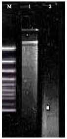 | |
| Fig. 2: | Measurement of endonuclease activity. Lane 1: DNA of normal hepatocytes and 1 μL endonuclease of 1 h induced and reperfused rat hepatocytes Lane 2: Apoptotic DNA of 3 h ischemic and reperfused rat isolated from hepatocytes |
DNA (Lane No. 1). Figure 2 shows the isolated endonuclease activity in normal hepatocytes. Normal hepatocytes undergo apoptosis in the presence of isolated endonuclease from IR hepatocytes and I/R injured hepatocytes causes inter-nucleosomal ladder-like pattern of DNA fragmentation (Lane 2).
DISCUSSION
Apoptosis represents a crucial mechanism of ischemia-reperfusion injury after liver transplantation. During ischemia and reperfusion of liver formed apoptotic cells and it may be one of the reasons of graft failure. Endonuclease is responsible for apoptosis of cells. DNase gamma, a member of the DNase I family, has been suggested to cause DNA fragmentation during apoptosis (Sunaga et al., 2006). In the present investigation endonuclease were separated from ischemic and reperfused liver. DNA fragmentation study showed endonuclease similar to DNase-I.So far, four distinct, but highly related, DNase I like genes have been identified in humans (Parrish et al., 1995; Sharks et al., 1990). Enzymes of the DNase I class similar to bovine pancreatic DNase-I with respect to molecular weight and ionic and pH requirements were found in various tissue of the rat. High levels of DNase I was found in digestive tissues, like parotid and sub maxillary, salivary glands and seminal vesicle. No activity was found in pancreatic extracts. However, no literature has reported about hepatic endonuclease. The present study shows that the hepatic endonuclease similar to DNase-I and cause apoptosis. The future drug discovery may be take attention to inhibitor of DNaseI in liver transplantation and related IR associated disease. Several evidences indicate the involvement of DNase γ in DNA fragmentation during apoptosis. DNase γ, a Mg+2/Ca+2 dependent endonuclease has been suggested to be one of the apoptotic endonucleases (Lacks, 1981; Mizuta et al., 2006). The present study showed that DNase is responsible for internuclosomal cleavage of chromatin during ischemic and reperfusion apoptosis. Further studies to elucidate and characterize endonuclease of hepatocytes and expression of DNase-I genes during ischemic and reperfusion injury needs to be conducted.
ACKNOWLEDGMENTS
The authors thank Prof. R.M. Dubey, Managing Director, IFTM, Moradabad and U.P., India for the provision of research bursary.
REFERENCES
- Clavien, P.A., P.R.C. Harvey and S.M. Strasburg, 1992. Preservation a reperfusion injuries in liver allograft, overview and synthesis of current studies. Transplantation, 53: 957-978.
Direct Link - Herrmann, M., M.H. Lorenz, R. Vell, M. Grunke, W. Woith and J.R. Kalde, 1994. A rapid and simple method for the isolation of apoptotic DNA fragments. Nucl. Acid. Res., 22: 5506-5507.
Direct Link - Mizuta, R., M. Mizuta, S. Araki, D. Shiokwa, S. Tanuma and D. Kitamura, 2006. Action of apoptotic endonuclease DNase gamma on naked DNA and chromatin substrates. Biochem. Biophys. Res. Commun., 345: 560-567.
Direct Link - Parrish, J.E., A. Uiccodicola, M. Wehhert G.F. Cox, E. Chen and D.L. Nelson, 1995. A muscle-specific DNase I-like gene in human X q 28. Hum. Mol. Gen., 4: 1557-1564.
Direct Link - Patel, T., S. Bronk and G.J. Gores, 1994. Increase of intracellular magnesium promotes glycodexycholate-induced apoptosis in rats hepatocytes. J. Clin. Invest., 94: 2183-2192.
Direct Link - Peitsch, M.C., B. Polzar, H. Stephan, T. Cromptron and H.R. McDonald, 1993. Characterization of the endogenous deoxyribonuclease involved in nuclear DNA degradation during apoptosis. EMBO J., 12: 371-377.
Direct Link - Sharks, S., D.J. Capon, R. Hellmis, S.A. Marsters and C.L. Baker, 1990. Recombinant human DNase I reduces the viscosity of cystic fibrosis sputum. Proc. Natl. Acad. Sci. USA., 87: 9188-9192.
Direct Link - Shiokwa, D., H. Ohyama, T. Yamda, T. Takahashi and S. Tanuma, 1994. Identification of a endonuclease responsible for apoptosis and rat thermocytes. Eur. J. Biochem., 226: 23-30.
Direct Link - Sunaga, S., A. Yoshimori, D. Shiokawa and S. Tanuma, 2006. Structure basis for the inhibitory mechanism of a novel Dnase gamma-specific inhibitor, DR396. Bioorg. Med. Chem., 14: 4217-4426.
Direct Link - Thompson, C.B., 1995. Apoptosis in the pathogenesis and treatment of disease. Science, 267: 1456-1462.
CrossRefPubMedDirect Link