
Maha A. Sabry
Department of Zoonoses, Faculty of Veterinary Medicine, Cairo University, Egypt
Journal of Biological Sciences
Year: 2007 | Volume: 7 | Issue: 5 | Page No.: 720-728


ABSTRACT
Fractionation of fertile Hydatid cyst fluid antigens (FHCFA) revealed 12 protein fractions at molecular weights (MW) of 105, 79, 62, 49, 38 28 24 21, 18, 8 and 2 kD bands over 105 kD. The bands at MW of 38, 36, 29 18, 16, 12 and 8 kD were reacted specifically with sera of Hydatid Crabbits as well as sera of surgically proved HC infected patients using enzyme linked immuno-transfer blot (EITB) technique. Evaluating the diagnostic efficacy of 3 eluted concentrated sub units of these antigens at MW range of 32-38, 16-18 and 8-12 kD, revealed that the fractions in the range of 8-12 kD appear as the most specific (96.66%) fractions. It didn`t cross reacted with anti bodies (Ab) in sera of patients infected by Fasciola, gastro-intestinal nematodes (G. I. N.) infected camel as well as Fasciola and Moneizia infected sheep. It clear mild level of cross reaction (10%) with Virus hepatitis C and Schistosoma manson infected patients sera. These fractions appear more sensitive (100%) for diagnosis of anti HC Ab in sera of surgically proved HC infected patients, HC infected camels and HC experimentally infected rabbits using ELISA technique. Fertile Hydatid cyst fluid antigen (FHCFA) of 32-38 kD appear less specific (93.33%) than the fraction of (8-12 kD), but this fraction still has absolute sensitivity (100%). The last groups of bands (16-18 kD) appear as the lowest specific (81.66%) and sensitive (92.0%) one. The protein fractions in the range of 8-12 kD showing marked diagnostic efficacy for hydatidosis infection in random samples of exposed people and pre-slaughtered animals. Diagnosis of infection by ELISA technique was closely related to diagnosis using sonographic examination in human and post mortem examination in animals. So that the former group of antigens identified as a good tool for diagnosis of the disease in human and investigation to infection status in living animals, the matter which is essential to improve the prognosis of the infected patients and breeding valuable animals.
PDF Abstract XML References Citation
How to cite this article
Maha A. Sabry, 2007. Advanced Concepts in Diagnosis of Hydatidosis in Human and Living Animals. Journal of Biological Sciences, 7: 720-728.
DOI: 10.3923/jbs.2007.720.728
URL: https://scialert.net/abstract/?doi=jbs.2007.720.728
DOI: 10.3923/jbs.2007.720.728
URL: https://scialert.net/abstract/?doi=jbs.2007.720.728
INTRODUCTION
Cystic Echinococcosis (CE) or Hydatid Disease (HD) is recognized as an important worldwide distributed disease from the clinical, economical and zoonotic point of view. The disease in man and other intermediate hosts is caused by ingestion of food or water contaminated with eggs of the dog tapeworm, Echinococcus granulosus (Devi and Parija, 2003).
Currently, E. granulosus is the only species of the genus echinococcus that is found in the region of the Mediterranean areas and the domestic dog serves as the only known reservoir for the adult tapeworm and therefore plays a recognized role as the main source of infection causing both health and veterinary problems (Lamar et al., 2001). Distribution of the disease is normally associated with underdeveloped countries especially in rural communities where man and other domestic animals are maintain in close contact with the dog (Ahmadi, 2004).
In animals, hydatid usually remains as asymptomatic disease producing no clinical symptoms and its course is slow. In domestic animals diagnosis is almost made during postmortem, large unilocular cysts are usually not diagnosed in young animals until middle life or later (Hassan, 1991).
El-Askalany (1981) recorded infection by Hydatid Cysts (H. C) in slaughtered camels reached to 31.26%, while Derbala and Zayed (1997) found H. C infection in camels, donkeys, horses, pigs and sheep as 40, 7.69, 6.25, 0.92 and 0.77%, respectively. They failed to detect. infection in slaughtered cows and buffaloes. Serologically El-Baz (1994) mentioned that the infection rates of hydatidosis in camels, donkeys, horses pigs and sheep was 40, 7.69, 6.25, 0.92 and 0.77%, respectively.
At the level of human in Egypt, the impression that hydatid disease was rare existed as accurate epidemiological information was lacking. This was attributed to the concern of medical authorities with the more important disease such as malaria, schistosomiasis and filariasis so that hydatidosis was not a notifiable disease. Also may be due to the slow clinical course of the disease which was not alarming and not contagious (Osman, 2006).
Lightowlers and Gottstein (1995) mentioned that, insensitive and non specific tests for diagnosis of hydatidosis include the casoni intradermal test, the complement fixation test, the indirect haemagglutination test and the latex agglutination test been replaced by the Enzyme Linked Immunosorbent Assay (ELISA), the indirect immunofluorescence antibody Test, Immuno Electro Phoresis (IEP) and Immuno Blotting (IB) in routine laboratory application.
There are Varity of antigens can be prepared from Hydatid Cysts (HC) include that of internal germinal layer, scoleces, or from cystic fluids either as sterile or Fertile Hydatid Cyst Antigen (FHCA) (Osman, 2006). Hydatid cystic fluid is a complex mixture of parasite-derived and host-derived molecules. It contains several antigens derived from the metabolism of the parasite together with many components from the host (Irabuena et al., 2000).
Fractionation of Hydatid fluid antigen using SDS-PAGE and identification to specific bands by immunoblotting resulted in identification of the arc-5 subunits, including two subunits with relative molecular mass between 37 and 38 kD and 20-22 to 24 kD (Poretti et al., 1999). Moreover, Lightowlers and Goltorti (1995) identified another thermostable lipoprotein called antigen B. The major components of this antigen resolved as 3 bands of 8-12 kD, 16 kD and 23-24 kD protein fraction.
Concerning the diagnostic values of these fractions, Poretti et al. (1999) cleared that FHCA of 8 kD and that of 29 and 34 kD bands exhibited high specificity than sensitivity, moreover 8-kD showing relatively frequent cross-reactions in tumor patients and in patients with other parasitic diseases but it high specific in differentiation between infection by cystic HC and alveolar one. At the same time, Ito et al. (1999) stated that this fraction (8 kD), revealed no cross-reactivity with any sera from patients with cysticercosis, other parasitic diseases, or with hepatomas, or from healthy controls as genus-specific for echinococcus. According to Dreweck et al. (1997). FHC protein fractions of 26, 18, 16 kD and 12 kD appear as more specific for detection of IgG antibodies of echinococcosis in general using ELISA technique. They considered as useful approach for post-treatment follow-up of patients at risk of developing recrudescent disease.
As mentioned by Babba et al. (1994) ELISA considered as one of the best sensitive serodiagnostic technique for diagnosis of Hydatidosis in man in comparison with latex agglutination, counter immuno electrophoresis using antigen of 38 kD specially if they associated with Echography and/or chest radiograph, They added that Immunoblotting, succeed in determination of 1-3 specific protein bands of 65, 12 and 8 kD. The sensitivity of the ELISA did not increase with either the size or type of cyst. (Coltorti et al., 1990).
The present study aimed to identify specific and sensitive protein fraction from fertile Hydatid cysts of camel lung via SDS-PAGE and EITB technique and estimate the value of these fractions in diagnosis of hydatidosis infection in suspected human and living animals using ELISA technique.
MATERIALS AND METHODS
Fertil Hydatid Cysts Fluid Antigen (FHCFA) preparation: Hydatid Cysts (HC) were collected from freshly slaughtered camel lungs (from Cairo slaughter abattoir). They examined from the aspect of viability as well as sample from their fluid was aspirated and microscopically examined for the presence of protoscolices.
These cysts were used for collection of fluid antigens and also for induction of experimental infection in young puppies for Echinococcus granulosus (Eg) Gravid Segment (GS) collection which used for induction of infection in rabbit for production of reference sera (versus HC) later on.
FHCFA was prepared according to Ito et al. (1999) where the fluid was collected from fertile cysts and clarified by centrifugation at 5000 rpm for 15 min at 4 °C, dialyzed against 5 mM Tris-H cl (pH 7.4) for 48 h at 4 °C, after determination their protein content by method of Lowery et al. (1951). The antigen was allocated into 1 mL vial and stored at -20 °C until use.
Fractionation of FHCFA using SDS-PAGE: The prepared antigens were resolved using 1.5 mm thickness, Sodium Dodecyl Sulfate-Polyacrylamide Gel Electrophoresis (SDS-PAGE) according to Laemmli (1970) in 12% polyacrylamide gel slabs in Tris-glycine buffer, pH 8.3 under reducing conditions. The stacking gel consisted of 5% acrylamide prepared in 12.5 mM Tris-HCL buffer (pH 6.7) (Sigma chemical Co.). Prestained low molecular weight (MW standard was employed (Sigma SDS-100B). The comb was adjusted as one small well for standard and one large for the sample.
Electrophoretic transfer of protein fractions onto nitrocellulose sheet: Electrophoresis transfer of fractionated proteins onto nitro cellulose sheet (NC) for Electro-Immuno Transfer Blot technique (EITB) was performed according to Towbin et al. (1979) using transfer buffer (25 mM tris-base, 192 mM glycine, 20% (v/v) methanol at pH 8.3). Transferring was carried out at 10V, 100 mA overnight at 4 °C. Longitudinal strip from the side of the NC membrane containing the MW protein standards and the whole fractionated antigens were cut off, washed by distilled water and stained with ponceau S stain (0.2% ponceau S in 3% trichloroacetic acid, Sigma).
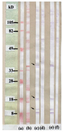 | |
| Fig. 1: | Recognition of Fertile Hydatid cysts antigen fractions using Ponceau S stain and EITB technique. Lane (a): Low molecular weight marker stained with Ponceau S solution, Lane (b): NC strip demonstrate the different antigenic fractions stained by Ponceau S solution, Lane (c): NC strip of the fractionated antigen reacted with positive rabbit sera. Lane (d): NC strip of the fractionated antigen reacted with negative rabbit sera. Lane (e): NC strip of the fractionated antigen reacted with hydatidodis infected patient sera, Lane (f): NC strip of the fractionated antigen reacted with negative human sera |
The excess dye was washed off by distilled water. The relative MW of the visualized bands was estimated from the reference MW standard curve (Fig. 1).
Determination of FHCFA specific protein fractions using EITB: According to Towbin et al. (1979) EITB technique was applied in order to determine the specific diagnostic fractions of FHCFA longitudinal NC strips (15 x0.5 cm) were cut out and allowed to react versus hydatid experimentally infected rabbits (3 samples) and 5 samples of Hydatid surgically proved infected human as well as negative control samples at (1:100 dilution), 0.5 mL of sera/strip. The used conjugate was horseradish peroxidase conjugated goat anti-human and anti-rabbit IgG (Sigma Immunochemicals) at 1:1000 in 3% BSA/PBS. The used substrate is 4-chloro-1-naphthol. Fractions that react versus reference positive sera and in the same time did not react versus negative control one, considered as specific protein fractions (Fig. 1).
These strips of NC were returned back to its original position on NC sheet for determination to the sit of the specific reacted kD protein fractions on the whole NC sheet.
Isolation of selected protein fractions from NC by electro elution: Three transverse pieces of NC membrane corresponding to the protein fractions of 32-38, 16-18 and 8-12 kD were cut out horizontally across the whole NC. Each transverse NC strip was transferred separately to elution tube membrane 4-6 MW cut off (Spectrum Medical Inc., Los Angeles, CA 900060).The tube were filled with PBS (pH 7.4) and kept in Bio-Rad elution unit at 10 V, 100 mA over night at 4 °C. NC materials were removed and the volume was reduced using polyethyleneglycol in molecular porous membrane tubing 4-6 MW cut off according to Katrak et al. (1992). The protein contents of the eluted concentrated materials was determined and kept in 1 mL vial at -20 °C till use for coating of the ELISA plate.
Indirect ELISA technique: ELISA test was done as described by Gottstein et al. (1993). Each plate was coated with antigen at its optimal concentration 2 ug mL -1 coating buffer (adjusted after checkerboard titration). Peroxidase-conjugated protein A (Sigma) was used at 1: 2000 dilution. Ortho-phenylenediamidine-OPD was added at a concentration of 340 ug mL -1 substrate buffer. Absorbance was read at 492 nm using a Titerteck multiskan ELISA reader which calculate the positive and negative value automatically.
The test was applied for determination specificity of 3 fractions (32-38, 16-18 and 8-12 kD) versus control and tested sera at 1:100 serum dilution. Specificity of the tested fraction was evaluated according to (Abdel-Rahman et al. (1998), as the ability of the tested fraction to detect its target Ab from different antibodies of other parasites using the following equation:
Specificity % = (T-P)/T x100/100 |
Where T = No. of tested sample P = No. of+Ve samples. This at standard serum dilution, while sensitivity is the ability of the tested antigenic fractions to detect its target Ab in samples infected by the parasite at standard serum dilutions (% of positive sera among the total number of the positive samples).
Estimation to the diagnostic efficacy of the selected fractions using ELISA technique
Collected samples: In order to estimate the diagnostic efficacy of the selected eluted concentrated FHCFA protein fractions, 3 experiments were designed, one for testing the sensitivity, another for evaluating their specificity and the 3rd one as application for diagnostic efficacy of these fractions.
To fulfill the requirement of these 3 experiments, special serum samples of known parasitic status were selected from large number of examined samples as the following:
Human samples: A number of 110 stool and blood samples were collected from out-patient clinic in Kasr El Any Medical hospital, Tropical Medicine and Hygiene Dept. (Kasr El-Any street) as well as private clinics from patients complained by digestive disturbances, fever and abdominal pain. Some of these patients were exposed to Sonographic diagnosis of hydatidosis on chest and liver.
Animal samples: A number of 145 sheep and 140 camel fecal and blood samples were collected from these animals at time of slaughtering in Cairo abattoir. Post mortem data of most of these animals could be obtained after evisceration concerning presence of HC and different other tissue parasites.
Fecal samples were examined parasitologically in order to determine the parasitic infection in each case, by one or more method include direct smear method according to the technique of WHO (1983) Concentration floatation technique using saturated sodium chloride solution according to Wattal et al. (1986), while the large eggs were diagnosed via two successive sieve system (Fluke finder, Moscow, ID) according to Welch et al. (1987). The collected blood samples were used for separation of the required serum.
From the previous samples and according to parasitic infection in stool or PM and Sonographic examination the following serum samples were selected according to the requirements of each experiment:
Selected human sera
| • | Ten samples of patients had Schistosoma mansoni eggs only in their stool. |
| • | Five serum samples from healthy people (as negative control). |
Selected animal sera
| • | Ten samples of camels harbor Gastro-Intestinal Parasites (GIP) eggs only in their feces. |
| • | Ten samples of sheep had Fasciola eggs only in their faeces. |
| • | Five samples of sheep feces contain Moneizia eggs only. |
| • | Ten sample from hydatid infected Cairo abattoir slaughtered camels (3-15 cysts in lung) |
| • | Five samples from young camels has parasite free faecal and PM (as negative control). |
| • | Five samples from sheep of parasite free feces and PM (as negative control). |
Experimental control samples
| • | Sera of Hydatid cysts experimentally infected rabbits: Three rabbits were experimentally infected by E. granulosus Gravid Segment (GS) prepared in other related work (Osman, 2006) under supervision of the author. The used GS were collected after scarification of one dog 60 days post experimentally infected by fertile Hydatid cysts. The infected rabbits were scarified 60 days post infection where high anti-Hydatid cysts antibodies could be detected in their sera and used as positive control. |
| • | Three samples of non-infected rabbits. |
Samples via personal communications
| • | Five Fasciola and 10 of virus hepatitis (C) infected patients were obtained from Institute of Tropical Medicine and Hygiene, Kasr El-Any street. |
| • | Five surgical proved HC infected human sera (Dept of Parasitology Fac. of Medicine, Cairo University) |
For testing the diagnostic efficacy of the selected fractions
| • | Forty serum samples were collected from HC suspected camel at the quarantine pre slaughtering and they were confirming post slaughtering. |
| • | Forty five serum samples were collected from suspected sheep at the quarantine pre slaughtering and they were confirm post slaughtering |
| • | Thirty five serum samples from people exposed to infection (veterinary clinic workers) have available Sonographic data. |
These samples were included in sero-diagnosis for anti-HC antibodies by ELISA techniques and their data was confirmed by PM examination for slaughtered animals or sonographic findings for human to confirm the results.
RESULTS AND DISCUSSION
Distribution of hydatidosis is related to underdeveloped countries especially in rural communities where man remains in close contact with the dog (definitive host) and the various domestic animals act as intermediate hosts. Dogs were infected by eating infected carcasses containing the hydatid cyst. The parasite was transmitted to man by ingestion of food or water contaminated with dog faeces containing the infective eggs (Ahmadi, 2004).
In Egypt, El-Askalany (1981) mentioned that, incidence of human infection call for three existing factors: an existing sheep raising district, the presence of large dog population which has access to the offal of dead or slaughter animals and sanitation that permits a close association between these dogs and human being. All these factors were present in the Egyptian villages where dogs were allowed into houses. Animals shed eggs in stool and children were allowed to play with them.
In human, diagnosis of hydatidosis infection based mainly on imaging studies and immunodiagnostic procedures, Caremani et al. (1997), while the condition was differ in animals, the infection was diagnosed during PM examination of the slaughtered carcasses mainly and numbers from these cysts can reach to dogs by different ways or even pass with the meat for consumers. Presence of an accurate mean for diagnosis of infection in living animals can prefer a good tool for control the spread of this disease.
As mentioned by De Morilla et al. (1983) and Irabuena e t al. (2000), serological diagnosis of Hydatidosis, could be considered as beneficial tool in this respect, but cross-reaction between different parasites is still as a questionable point making some difficulties in the accurate evaluation of the infection status of suspect animals, especially under the level of field collected polyclonal sera. They added that, accuracy of these tests was affected markedly by degree of purity and specificity of the used antigens. For this reason Identification of special hydatid fraction could be improve the accuracy of the used diagnostic technique.
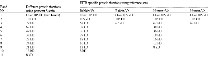
The present study was focused on identification of FHCFA specific diagnostic protein fractions using EITB technique. The results in Table 1 and Fig. 1 demonstrate that Ponceau-s stained longitudinal strips, blotted with fractionated FHCFA antigen revealed 12 protein fractions were identified using the molecular weight standard curve as 105, 79, 62, 49, 38, 28, 24, 21, 18, 8 and 2 kD bands over 105 kD.
Treatment of corresponding NC strips using HC experimentally infected rabbit and surgically proved hydatid infected human sera via EITB test revealed 10 fractions of these bands react versus the above sera, three from these bands were react non-specifically versus negative rabbit and human sera (that over 105 kD, 105 kD and 62 kD), while the other 7 bands could be considered as specific recognized by specific anti-bodies only as in le and Fig. (1). These specific bands were corresponded to the MW standard of 38, 36, 29, 18,16, 12 and 8 kD, with different amount of protein content as in Fig. 1.
The fraction of 29 kD could not be detected using human HC infected serum and in the same time, didn ’t react with the negative control sera. These data was agreed with that mentioned by Ito et al. (1999) and Poretti et al. (1999) as FHCF antigens revealed several fractions in the range of 8 kD to over than 105 kD. Failure of 29 kD fraction to react versus human sera may be due to the origin of this antigen as it camel non human origin.
EITB is one of the most specific sero-diagnostic techniques but it considered non-practical for current field application in comparison with ELISA technique (Ibarra et al., 1998). De Morilla et al. (1983) mentioned that ELISA technique was a sensitive serological test, able to analyze many samples simultaneously.
| Table 1: | Specific and non specific protein fractions of FHCA using SDS-PAGE and Western blot versus reference positive control sera |
 | |
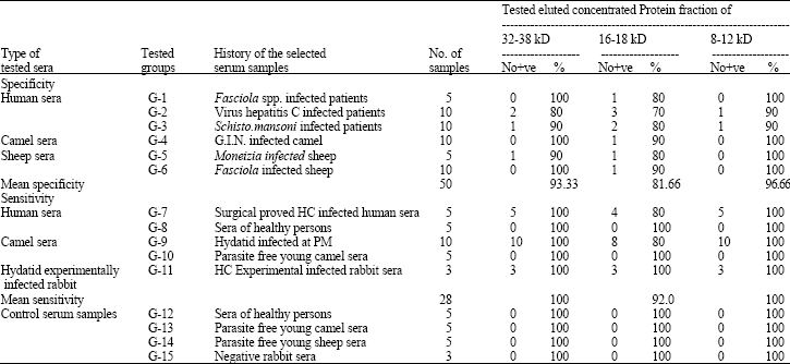
| Table 2: | Specificity and sensitivity of different eluted FHC protein fractions in diagnosis of infection in selected serum samples using ELISA technique (1:100 serum dilution) |
 | |
Moreover, Babba et al (1994), identify (ELISA) as one of the best sensitive sero-diagnostic technique for diagnosis of hydatidosis in man in comparison with latex agglutination and counter immunoelectrophoresis Collectively these protein fractions occur among 3 main subunits in the MW zones of 32-38, 16-18 and 8-12 kD. The sites of these 3 groups of bands were cut transversely from the whole NC sheet. The protein contents of these bands were eluted and concentrated and used for more characterization.
Estimation to the diagnostic efficacy of these 3 sub-unites (32-38, 16-18 and 8-12kD) via ELISA technique as in Table (2), cleared that the fraction of 8-12 kD appear as the most specific (96.66%) and sensitive (100%) one for diagnosis of hydatid anti-bodies in different serum samples. This group of fractions did not cross react with anti-Fasciola antibodies in infected patients or animals, did not cross react with antibodies produced from GIN infections in camels and Moneizia infection in sheep. They cross react with one sample (10%) of S. mansoni and Virus hepatitis C infected patient. These data came in agreement with Ito et al. (1999) and Babba et al.(1994) as the fraction of 8 kD and that of 12 kD did not cross react with different parasites and liver affections via ELISA and EITB techniques. On the contrary with these authors, these sub-units have low specificity (90%) versus S. mansoni infected patients.
The sub unites antigens in the range of 32-38 kD appear of lower specificity (93.33%) and of good sensitivity (100%). The marked diagnostic value of these 2 sub-units fractions was previously mentioned by Babba et al. (1994) and Wen and Craig (1994) using ELISA techniques, as antibodies in sera of HC infected patients were react specifically versus protein fractions of 20, 16, 12 and 38-kD subunit of antigen 5 from FHCA. In the contrary with Dreweck et al. (1997) the band fractions of 16-18 appear lower in specificity (81.66%) and sensitivity (92.0%) than the other tested fractions this may be due to the difference in the strain of the used hydatid fluid antigens. Also with d'Amelio et al. (1985) where they identify 4 fractions from FHCFA of human origin at MW in the range of 32-13, 67 and 52 kD, as they specifically react versus sera of infected patients.
Hydatidosis is worldwide problem, WHO (1999) showed that number of yearly infected people with E. granulosus was 2.7 millions. Human cystic echinococcosis, is recognized globally as an increasing major zoonotic disease and remains as significant health problem in Arab countries (Dar and Al-Karmi, 1997). In order to this, extraction of special protein fraction which able to induce accurate diagnosis for hydatid infected animals during their life could be facilitate control of the disease as mentioned by El-Askalany (1981) as the prevalence of the disease in man is conditioned by the degree of infection in sheep, cattle and camel.
The marked specificity and sensitivity of 8-12 kD protein fractions could be demonstrate this antigen as a high valuable purified antigen help in accurate diagnosis of hydatid infected animals in Egypt during breeding and before slaughtering without interest to the other parasitic infections which could be diagnosed in these animals.
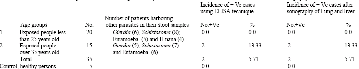
Diagnosis of hydatidosis infection in group of exposed people using this fraction as demonstrated in Table 3 revealed that the serological incidence of infection using this fraction was (5.71%) which is exactly the same figure by sonographic examination (5.71%).
| Table 3: | Value of 8-12 kD FHC protein fraction in Diagnosis of hydatidosis in human and confirmation of the result after sonography on the chest and liver |
 | |
| Table 4: | Value of 8-12 kd FHC protein fraction in Diagnosis of hydatidosis in living animals and confirmation of the data post slaughtering |
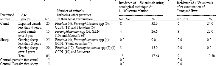 | |
The infection was recorded in suspected older patients (over 35 years old) than in young individuals. Presence of different enteric parasites in both groups of patients (Giardia, S. mansoni, Entamoeba histolytica and hymenolepis nana) did not produce cross reactions in diagnosis by these protein fractions. This identifies the value of this fraction for current diagnosis of suspected patients using ELISA technique. Concerning to pre-slaughtered camels and sheep (Table 4) the incidence of infection was higher by ELISA technique (17.64%) in comparison with the data of P.M. examination of lung and liver (10.58%). This was agreed with El-baz (1994) as the difference between sero-diagnosis and PM diagnosis of hydatidosis may be related to presence of hydatid infection in small size which could not be demonstrated by visual examination or the infections in other parts of the body did not accurately investigated. Enteric parasits diagnosed in fecal examination of these animals (Fasciola, Paramphistomum spp., GIN, Moneizia eggs and coccidian oocysts), previously proved hat they did not cross reacted with these protein fractions.
CONCLUSION
FHCF fraction of 8-12 kD (camels origin) could be considered as a good tool for early diagnosis of the disease in human and investigation to infection status in living animals. This way facilitated early diagnosis of infection in human and animals the matter which is essential to improve the prognosis of the infected patients as mentioned by Fujimoto et al. (2005). Identification of infected animals during their life could be facilitate slaughtering them under special control measures which ensure total condemnations of their infected tissues and eliminate the random arrival of the cysts to dogs the matter which play a role in minimize the infection in dogs and wide spread of the disease. Moreover it is necessary to mention that, control measures of hydatidosis must be carried on paralleled to that of human beings, also against the definitive host and the other intermediate ones.
REFERENCES
- Ahmadi, N.A., 2004. sing morphometry of the larval rostellar hooks to distinguish Iranian strains of Echinococcus granulosus. Ann. Trop. Med. Parasitol., 98: 211-220.
CrossRefDirect Link - Babba, H., A. Messedi, S. Masmoudi, M. Zribi and R. Grillot et al., 1994. Diagnosis of human hydatidosis: Comparison between imagery and six serologic techniques. Am. J. Trop. Med. Hyg., 50: 64-68.
Direct Link - Caremani, M., A. Benci, R. Maestrini, A. Accorsi, D. Caremani and L. Lapini, 1997. Ultrasound imaging in cystic echinococcosis. Proposal of a new sonographic classification. Acta Trop., 67: 91-105.
Direct Link - Coltorti, E.A., E. Fernandez, E.R. Marguet, J.D. Scozzina, E.A. Guarnera, 1990. Detection of asymptomatic carriers of hydatid cysts: Specificity increase of the immunoenzyme assay. Rev. Inst. Med. Trop. Sao Paulo, 32: 275-284.
Direct Link - Dar, F.K. and T. Alkarmi, 1997. Public health aspects of cystic echinococcosis in the Arab countries. Acta Tropica, 67: 125-132.
CrossRefDirect Link - Devi, C. and S.C. Parija, 2003. Latex Agglutination Test (LAT) for antigen detection in the cystic fluid for the diagnosis of cystic echinococcosis. Diag. Microbiol. Infect. Dis., 45: 123-126.
Direct Link - Dreweck, C.M., C.G. Luder, P.T. Soboslay and P. Kern, 1997. Subclass-specific serological reactivity and IgG4-specific antigen recognition in human echinococcosis. Trop. Med. Int. Health, 2: 779-787.
CrossRefPubMedDirect Link - Fujimoto, Y., A. Ito, Y. Ishikawa, M. Inoue, Y. Suzuki, M. Ohhira, T. Ohtake and Y. Kohgo, 2005. Usefulness of recombinant Em18-ELISA to evaluate efficacy of treatment in patients with alveolar echinococcosis. J. Gastroenterol., 40: 426-431.
Direct Link - Gottstein, B., P. Jacquier, S. Bresson-Hadni and J. Eckert, 1993. Improved primary immunodiagnosis of alveolar echinoccosis in human by ELISA using the Em2plus antigen. J.Clin. Microbiol, 31: 373-376.
Direct Link - Ibarra, F., N. Montenegro, Y. Vera, C. Boulard, J. Flore and P. Ochoa, 1998. Comparison of three ELISA tests for seroepidemiology of bovine fascioliasis. Vet. Parasitol., 77: 229-236.
Direct Link - Ito, A., L. Ma, P.M. Schantz, B. Gottstein and Y.H. Liu et al., 1999. Differential serodiagnosis for cystic and alveolar echinococcosis using fractions of Echinococcus granulosus cyst fluid (antigen B) and E. multilocularis protoscolex (EM18). Am. J. Trop. Med. Hyg., 60: 188-192.
Direct Link - Katrak, K., B.P. Mahon, W.C. Jones, S. Brautigam and K.H. Mills, 1992. Preparative separation of foreign antigens for highly efficient presentation of T cells in vitro. J. Immunol. Meth., 156: 247-254.
CrossRef - Laemmli, U.K., 1970. Cleavage of structural proteins during the assembly of the head of bacteriophage T4. Nature, 227: 680-685.
CrossRefDirect Link - Lamar, S., M. Kilani and P. Torgerson, 2001. Frequency distributions of Echinococcus granulosus and other helminths in stray dogs in Tunisia. Ann. Trop. Med. Parasitol., 95: 69-76.
PubMedDirect Link - Poretti, D., E. Felleisen, F. Grimm, M. Pfister and F. Teuscher et al., 1999. Differential immunodiagnosis between cystic hydatid disease and other cross-reactive pathologies. Am. J. Trop. Med. Hyg., 60: 193-198.
Direct Link - Towbin, H., T. Staehelin and J. Gordon, 1979. Electrophoretic transfer of proteins from polyacrylamide gels to nitrocellulose sheets: Procedure and some applications. Proc. Natl. Acad. Sci. USA., 76: 4350-4354.
PubMedDirect Link - Welch, R.D., P.H. Smith, J.B. Malone, R.A. Holmes and J.P. Geaghan, 1987. Herd evaluation of Fasciola hepatica infection in Louisiana cattle by an ELISA. Am. J. Vet. Res., 48: 345-347.
PubMed - Wen, H. and P.S. Craig, 1994. Immunoglobulin G subclasses in human cystic and alveolar echinococcosis. Am. J. Trop. Med. Hyg., 51: 741-748.
Direct Link