

Nasreen Akhter
Faculty of Animal Husbandry and Veterinary Sciences,Sindh Agriculture University, Tandojam, Pakistan
I. R. Mohammad
Faculty of Animal Husbandry and Veterinary Sciences,Sindh Agriculture University, Tandojam, Pakistan
K. B. Mirbahar
Faculty of Animal Husbandry and Veterinary Sciences,Sindh Agriculture University, Tandojam, Pakistan
M. I. Memon
Faculty of Animal Husbandry and Veterinary Sciences,Sindh Agriculture University, Tandojam, Pakistan
S. A. Soomro
Faculty of Animal Husbandry and Veterinary Sciences,Sindh Agriculture University, Tandojam, Pakistan
Journal of Biological Sciences
Year: 2001 | Volume: 1 | Issue: 3 | Page No.: 158-159


ABSTRACT
The prevalence of helminthiasis was found to be 15.2 % in buffaloes. Out of 500 samples examined, 9.2 % and 5.4 % were infected with nematodes and trematodes respectively. Mixed infection was observed in 0.6 %. The chief helminths identified were: Toxocara vitulorum (3.6 %), Fasciola gigantica (3.2 %) Oesophagostomum radiatum (3.0 %) Strongyloides papillosus (2.4 %), Fasciola hepatica (2.2 %), Ostertagia ostertagi (1.0 %), Paramphistomum cervi, (0.8 %) and Trichuris Spp. (0.2 %).
PDF Abstract XML References Citation
How to cite this article
Nasreen Akhter, I. R. Mohammad, K. B. Mirbahar, M. I. Memon and S. A. Soomro, 2001. Prevalence of Helminthiasis in Buffaloes in Cattle Colony Hyderabad. Journal of Biological Sciences, 1: 158-159.
DOI: 10.3923/jbs.2001.158.159
URL: https://scialert.net/abstract/?doi=jbs.2001.158.159
DOI: 10.3923/jbs.2001.158.159
URL: https://scialert.net/abstract/?doi=jbs.2001.158.159
INTRODUCTION
Buffalo is considered as leading species amongst the livestock for its high milk yield. It shares about 72% of the total milk produced in the country (Mahadevan, 1978). Buffaloes are susceptible to a majority of important parasitic diseases. According to field reports over 275 thousand buffaloes are lost annually due to different diseases (Junejo and Qureshi, 1993). Gastrointestinal helminthiasis syndrome is caused by a mixture of many species of helminthic parasites in the alimentary tract (Chaudhry et al., 1984).
In Pakistan, with lower hygienic standards, the parasites are responsible for huge economic losses in livestock industry. These parasites not only affect the health of the animal but also adversely affect their productivity, decrease resistance to other diseases and cause mortality leading to heavy economic losses (Irfan, 1984).
It is difficult to assess the exact economic losses due to parasitism. It is estimated that millions of rupees are lost every year due to rejection of meat, edible offals, depreciation of wool, skins and hides. Keeping in view the great economic importance of parasitic infections, this study was carried out to identify the species of Gastro-intestinal parasites and to determine their prevalence in buffaloes.
MATERIALS AND METHODS
Five hundred faecal samples of buffaloes were collected randomly from 30 different commercial dairy farms from cattle colony Hyderabad. Faecal samples were examined by Direct Smear Method and Sedimentation Method.
Direct Smear Method: The direct smear method as described by Urquhart et al. (1988) was adopted. A drop of water plus equivalent amount of faeces were thoroughly mixed on a microscopic slide to obtain a relatively homogenous and sufficiently transparent preparation. A cover slip was placed on the fluid and the preparation was allowed to settle for few minutes. The preparation was then systematically examined under low power microscope for the presence of eggs or larvae.
Sedimentation Method: The sedimentation method as described by Lynne and Lawrence (1979) was used for the identification of eggs. Approximately 1.0 g of faeces was taken into test tube containing 10-12 ml of normal saline. The faecal material was emulsified thoroughly in the saline by glass rod. The emulsion was filtered through the mesh sieve into the centrifuge tube and centrifuged at 2000 rpm for 2-3 minutes. The supernatant was discarded and the process was repeated twice. 7 ml of 10 percent formalin solution was then added to the sediments and shaken vigorously. 2-3 ml ether was added to the test tube and shaken vigorously to get a homogenous mixture. The mixture was centrifuged again at 2000 rpm for five minutes. The contents of the tube were separated into four layers i.e., sediment, formalin layer, debris plug and ether layer. The supernatant was discarded all at once until only the sediment was left. This was diluted with little formalin ( 2-3 drops ) and mixed homogeneously. With a pasteur pipette, a few drops of sediment were removed to object glass and examined under low magnification power of microscope.
RESULTS AND DISCUSSION
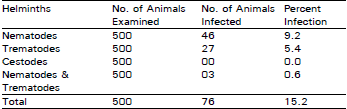
Present study was carried out to determine the prevalence of Helminthiasis in buffaloes at Cattle Colony, Hyderabad. Five hundred faecal samples were collected and examined. The prevalence of helminthiasis was calculated as 15.2% (Table 1). The prevalence of nematodes, trematodes and the mixed infection was 9.2, 5.4 and 0.6% respectively. The cestodes were, however, not found. The over all prevalence of 15.2% is very low when compared to that reported by Ishtiaq (1995). He reported a prevalence of 56.99%. The difference may be due to the climatic conditions, or difference in the age of animals, and the number of samples examined. An other important factor which may have accounted for the lower prevalence of the helminths recorded in this study, may be the fact that the samples were obtained from cattle colony Hyderabad, where animals kept for commercial purposes are reasonably well managed, and are regularly dewormed.
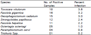
The helminths observed in the present study have also been identified by many workers from different parts of the world. Toxocara vitulorum was found with the highest prevalence i.e., 3.6% (Table 2) followed by Fasciola gigentica, Oesophagostomum radiatum, Strongiloides papillosus and Fasciola hepatica. The prevalence rate of T. vitulorum is comparable to the 4.8 % recorded by Iqbal (1987) in buffaloes at Faisalabad. Ishtiaq (1995) has observed higher percentage of T.vitolurum in buffalo calves. He reported, 100 percent. The variation may be incriminated to management practices and the prevalence of infection in that area. All animals infected with T. vitolurum were young calves i.e less than six months of age which is in agreement with the observations of Urquhart et al. (1988). Fasciola gigantica was the second commonest helminth and prevailed at a rate of 3.2% which is lower than that reported by Bilqees and Alam (1988). They detected prevalence of F. gigantica at a rate of 8.5% in buffaloes in Karachi. The variation may be due to the different source of samples and the number of animals examined.
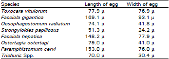
Helminths identified on the basis of morphological characteristics and the of size of the eggs were; T. vitulorum, F. gigantica, O. radiatum, S. papillosus, F. hepatica, O. ostertagi, P. cervi and Trichuris Spp. (Table 3).Several workers have reported the presence of different species of helminths in buffaloes and other ruminants in Pakistan and other parts of the world.
| Table 1: | Prevalence of helminthiasis in buffaloes |
 | |
| Table 2: | Helminth species identified in buffaloes. |
 | |
| Table 3: | Measurement of eggs of various helminth Species found in buffaloes |
 | |
Hingra (1991) identified, Neoascaris vitulorum, Haemonchus, Cooperia, Ostertagia and Trichostrongoides spp. in buffaloes at Lahore. While the prominent helminths identified in Faisalbad area include O. radiatum, Trichuris spp. T. ovis, Bunostomum phlebatumum, N. vitulorum, Moniezia benedeni, Haemonchus placei, S. papillosus and Ostertagia (Ahmed 1991). The results of above workers are not in complete agreement with the findings of the present study. The complete absence of Trematodes reported by above workers may be due to the age of the animals studied, management practices and intermediate host population in that area.
Shahid et al. (1993) conducted studies to assess the prevalence of endo-parasites in buffaloes and cattle in okara. They reported, O. radiatum, Mecistocirrus digitatus, B. Phlebotomum, Strongyloides spp. and Haemonchus contortus. These findings are in contrast to the findings of present study except O. radiatum.
Tarigan et al. (1987) identified the Paramphistomum, Fasciola and Nematodes as major species in Indonesian buffalo.
Similarly, Mourad et al. (1985) studied the gastro-intestinal parasitism of buffaloes in Egypt and reported, the presence of Trichostrongylus, Moniezia, Fasciola and paramphistomes. The findings of the present study are in general agreement with these workers. However, a complete absence of Moniesia spp. and the presence of T. vitulorum and Trichuris spp. in the present study may be attributed to the environmental conditions and the presence of intermediate host.