
Souravh Bais
Department of Pharmacology, Rayat Institute of Pharmacy, Railmajra, District SBS Nagar, Punjab, 144533, India
Yash Prashar
Department of Pharmacology, Rayat Institute of Pharmacy, Railmajra, District SBS Nagar, Punjab, 144533, India
Research Journal of Phytochemistry
Year: 2015 | Volume: 9 | Issue: 2 | Page No.: 41-55


ABSTRACT
The aim of the present study was to investigate the protective effects of amentoflavone (AF), isolated from six species of Juniperus (AF1-AF6) (Cupressaceae) Juniperus communis L. (AF1), Juniperus wallichiana L. (AF2), Juniperus indica L. (AF3), Juniperus macropoda L. (AF4), juniperus recurva L. (AF5) and Juniperus polycarpos L. (AF6)] against H2O2 induced oxidative damage in human erythrocytes and leucocytes. The experiment was set and designed as per method available in literature. Amentoflavone was isolated from aerial parts of Juniperus species (above six) and authenticated by TLC, IR, HPLC and physicochemical parameters. The two concentration of isolated AF (A = 250 μL, B = 500 μL) were used to assessed its protective effects against H2O2 induced oxidative damage in human erythrocytes and leucocytes. The measurements of different antioxidant parameters like, CAT, SOD and GPx, LPO and GSH levels were performed under each set extracts. Juniperus communis L. (AF1B) (500 μL) and Juniperus indica L. (AF3B) (500 μL) were the most significant one on CAT (28.35±2.980 and 27.73±1.580 AU g–1 Hb), GPx (128.53±3.10 and 41.7±1.420 μg g–1 Hb) and SOD (1193.5±19.7 and 1532.6±17.60 U g–1 Hb) enzyme systems of erythrocytes. Among plant extracts, Juniperus communis L. (AF1B) showed the highest activities on CAT (13.79±1.832 AU mg–1 protein) and SOD (820±14.50 U mg–1 protein) while Juniperus indica L. (AF3B) showed significant effects on Gpx (152.72±3.70 U mg–1 protein), GSH (25.93±1.560 μg mg–1 protein) and LPO (10.54±2.90 nmol mg–1 protein) enzyme systems of leucocytes. The study concludes that isolated fractions of AF from Juniperus species (among six species), has a potential source of natural antioxidants for treatment and prevention of diseases in which oxidative stress takes place.
PDF Abstract XML References Citation
Received: May 10, 2015;
Accepted: July 09, 2015;
Published: August 19, 2015
How to cite this article
Souravh Bais and Yash Prashar, 2015. Identification and Characterization of Amentoflavone from Six Species of Juniperus Against H2O2 Induced Oxidative Damage in Human Erythrocytes and Leucocytes. Research Journal of Phytochemistry, 9: 41-55.
URL: https://scialert.net/abstract/?doi=rjphyto.2015.41.55
URL: https://scialert.net/abstract/?doi=rjphyto.2015.41.55
INTRODUCTION
Reactive Oxygen Species (ROS) such as O2- and H2O2 play an important role in normal cellular function and cellular signaling. At lower concentrations they participate in various cellular physiological reactions. The increase in ROS results in increased leukocyte and platelet activation and increased leukocyte recruitment (Schopfer et al., 2001; Patel et al., 2000; Cooper et al., 2002; Stokes et al., 2002a, b). The modification of cellular phenotype and increased levels of ROS is associated with oxidative stress and vascular disease formation and progression. Increased pro-oxidants are associated with vascular diseases and are thought to be an important step in vascular disease development, including atherosclerosis and hypertension (Landmesser et al., 2003). Multiple pro-oxidants and anti-oxidants participate in the normal physiologic balance that is lost with oxidative stress. The herbal plants are valuable sources of antioxidants, as their chemical constituents help to maintain the balance between prooxidants and antioxidants which increase the quality lifespan.
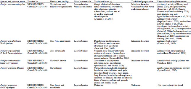
About 60-70 species of aromatic evergreen trees or shrubs constituting the genus Juniperus of the cypress family (Cupressaceae), is distributed throughout the Northern Hemisphere. Juniper is found in Himachal Pradesh at an altitude of 3000-4200 m. It is mainly distributed in Manimahesh in Chamba, Kullu, Churdhar in Sirmour, Chhota and Bara Bhanghal in Kangra, Kinnaur and Pattan valley in Lahaul-Spiti districts. The plant also grows in Europe south-western Asia and North America (Sharma and Lal, 2005). The juvenile leaves of juniper are needle like. Mature leaves are awl-shaped, spreading and arranged in pairs or in whorls of three. Some species have small, scale like leaves, often bearing an oil gland, which are pressed closely to the rounded or four-angled branchlets. Male and female reproductive structures usually are borne on separate plants. The reddish brown or bluish cones are fleshy and berry like and often have a grayish, waxy covering. They mature in 1-3 seasons and contain 1-12 seeds (Adams, 2014). Juniperus used traditionally, in the treatment of various disease and disorders (Table 1). The leaves of Juniperus contain enriched amounts of phenols and flavonoids (Tavares et al., 2012). Amentoflavone is a dimer of apigenin, found in several plants and reported as antiviral (Lin et al., 1999), antioxidant, antidepressant, anti-inflammatory and analgesic activity (Cholbi et al., 1991; Baureithel et al., 1997; Kim et al., 1998; Da Silva et al., 2000).
The aim of the present study was to investigate the protective effects of amentoflavone isolated from six wild Juniperus species found in Himalaya region, used in traditional indian medicine, including Juniperus communis L., Juniperus wallichiana L., Juniperus indica L., Juniperus macropoda L., Juniperus recurva L. and Juniperus turbinate L. against H2O2 induced oxidative damage in human erythrocytes and leucocytes.
MATERIALS AND METHODS
Chemicals: GPx and SOD activity were determined using commercial available enzyme kits such as Ransel, RANDOX/RS 504 and Ransod, RANDOX/SD 125, RANDOX Laboratories, U.K. Hydrogen peroxide, 3% (Max Laboratories Pvt. Ltd.), pyrogallol (High media), amentoflavone (Highmedia) gallic acid (LOBA Chem) butylated hydroxyl anisole, potassium ferricyanide, nitro blue tetrazolium, thiobarbituric acid (TBA), trichloroacetic acid ethylene diamine tetra acetic acid, ammonium thiocyanate, potassium persulfate, ferrous chloride, quercetin and ascorbic acid were obtained from Sigma Chemicals, USA and SD. LOBA-Chem Ltd., Baddi (H.P). All other chemicals and reagents used for experimental work were of analytical grade (Fig. 1).
Plant material collection and authentication: All the species of Juniperus were collected in the month of November-December from various locations of Punjab and Himachal Pradesh, frozen and freeze dried. Plants were identified by the chief scientist of NISCAIR and kept in the Herbarium (NISCAIR/RHMD/Consult/2014/2424-03) NISCAIR New Delhi.
Preparation of extract: Five hundred gram of air dried powdered leaves of Juniperus species were extracted with methanol.
| Table 1: | Common names, part or material, habitat and locality, traditional preparation(s), reported activities and traditional uses of Juniperus species from Himalaya |
 | |
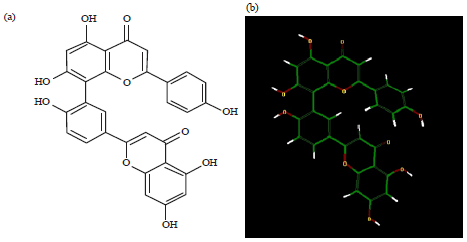 | |
| Fig. 1(a-b): | Structural diagram of amentoflavone |
After the extraction, excess solvent was completely removed by using a rotatory flash evaporator to get concentrated, then completely dried in freeze drier and all the extracts were preserved in airtight container under refrigeration. The extracts were used for the estimation of total polyphenols, flavonoids contents.
Chemical characterization
Total phenol quatification: Determination of total phenolic content of freeze-dried extracts of Juniperus species was performed by the Folin-Cio calteau method (Makkar, 2003). The absorbance was recorded at 725 nm against the reagent blank with a double beam UV/Visible spectrophotometer (EI model No. 5512, Japan). The amount of total phenols was calculated as pyrogallol equivalents (even though pyrogallol was used as a standard in this work, it is not a natural constituent of Juniperus in general) from the calibration curve by linear regression.
Estimation of total flavonoid: The aluminium chloride colorimetric technique was used for estimation of flavonoids (Woisky and Salatino, 1998). The ME fraction (0.5 mL each) was taken (100 mg mL–1 of ethanol) in test tube and mixed with 1.5 mL of methanol, 0.1 mL of 10% aluminium chloride, 0.1 mL of 1 M potassium acetate and 2.8 mL of distilled water. The reaction proceeded at room temperature for 30 min and the absorbance was subsequently measured at 415 nm. The calibration curve was plotted by preparing quercetin standard solution across a range of 10-70 ppm in methanol. The amount of flavonoid was calculated from the standard quercetin graph (Siddique et al., 2010).
Extraction and isolation of amentoflavone: The dried extract (10 g) of six different species was suspended in water separately followed by successive partitioning with chloroform, ethanol, n-butanol, respectively. The aqueous/n-butanol fraction (2 g) was chromatographed on silica gel column, eluted with a gradient of chloroform: methanol (100:00:100). Fractions with similar Rf were pooled together affording five subtractions of individual species. The most active fractions were re chromatographed with addition of 5% water and selected for structural confirmation (Ishola et al., 2012).
Authentication of amentoflavone
Preparation of stock solution: Amentoflavone (standard and isolated), 10 mg was dissolved in 5 mL of dimethyl sulfoxide (DMSO), diluted with 20 mL of methanol and filtered through Whatman filter paper to recover an amentoflavone solution of 500 μg mL–1.
Thin layer chromatography: Freshly coated with silica gel G254 plates were allowed to air dry at room temperature, transferred to an oven for activation and kept at 110°C for 30 min. Commonly used solvent system for identification and quantification of flavonoids, benzene: pyridine: Formic acid (36:9:5) (BPF) was used for co-thin layer chromatograph to compare Rf values of standard and isolated amentoflavone (Ali et al., 2011). Stock solution (20 μL) was spotted; plates were placed into the developing chamber and allowed to run until it reached a height of about 10 cm from the point of spotting. Spots were viewed under U.V light and as developed in iodine vapour showed the spot with different fluorescence. After development, the plates were kept in an oven maintained at 110°C in order to achieve optimal color development.
Identification of amentoflavone: Melting point was determined with the help of a digital melting point apparatus (Microteknik, India). All selected fractions of six species were identified by spectral data (IR), which was in accordance with those previously described (Ishola et al., 2012; Markham et al., 1987; Zheng et al., 2011). The HPLC was used with C18 Column (250×4.6 mm, 5 μm) column. The mobile phase consisted of methanol and phosphate buffer. The gradient condition was 0-25 min with methanol 40-48% and 20-45 min with methanol 68-80%. The column temperature was 40°C. The flow rate was 1.0 mL min–1 and the detection wavelength at 338 nm.
Calculation of the other parameters: All the physicochemical properties viz. MR (Molecular Refractivity), MV (Molecular Volume), Pc (Parachor), η (Index of refraction), ST (Surface Tension), D (density) and Pol (Polarizability) were calculated by ACD lab freeware (Chemsketch 5.0). All the topological parameters and other descriptors were calculated by dragon 5.0 and some non conventional parameter Viz. ASA (Approximate Surface Area) and SAG (Surface Area Grid) are calculated by Hyperchem 6 (demo version).
Isolation of leucocytes: Human leukocytes were isolated from freshly sampled venous blood (12-15 mL) of healthy volunteers by using dextran (Macrodex: 6% dextran in 0.9% NaCl solution) and heparin tubes (25,000 IU mL–1).
Isolation of erythrocytes: Fresh blood samples from healthy volunteers (12-15 mL) were collected and centrifuged at 3000 rpm for 15 min and plasma and buffy coats were removed. Red cells were washed with Phosphate Buffer Solution (PBS) (pH 7.00, containing 140 mM NaCl) 3 times and erythrocytes were haemolyzed with ice-cold distilled water.
Preparation of incubations with Juniperus species (Isolated AF fractions) For erythrocytes:
| • | Group I: | Control contains erythrocyte haemolysate 750 μL, PBS 1000 μL and distilled water 250 μL |
| • | Group II: | H2O2, erythrocyte haemolysate 750 μL, H2O2 (10 mM) 50 μL, PBS 950 μL and distilled water 250 μL |
| • | Group III: |
| • | AF1A group; erythrocytes hemolysate 750 μL, H2O2 (10 mM) 50 μL, AF 1A 250 μL and PBS 950 μL | |
| • | AF1B group; erythrocytes hemolysate 750 μL, H2O2 (10 mM) 50 μL, AF 1B 500 μL and PBS 950 μL | |
| • | AF2A group; erythrocytes hemolysate 750 μL, H2O2 (10 mM) 50 μL, AF2A 250 μL and PBS 950 μL | |
| • | AF2B group; erythrocytes hemolysate 750 μL, H2O2 (10 mM) 50 μL, AF2B 500 μL and PBS 950 μL | |
| • | AF3A group; erythrocytes hemolysate 750 μL, H2O2 (10 mM) 50 μL, AF3A 250 μL and PBS 950 μL | |
| • | AF3B group; erythrocytes hemolysate 750 μL, H2O2 (10 mM) 50 μL, AF3B 500 μL and PBS 950 μL | |
| • | AF4A group; erythrocytes hemolysate 750 μL, H2O2 (10 mM) 50 μL, AF4A 250 μL and PBS 950 μL | |
| • | AF4B group; erythrocytes hemolysate 750 μL, H2O2 (10 mM) 50 μL, AF4B 500 μL and PBS 950 μL | |
| • | AF5A group; erythrocytes hemolysate 750 μL, H2O2 (10 mM) 50 μL, AF5A 250 μL and PBS 950 μL | |
| • | AF5B group; erythrocytes hemolysate 750 μL, H2O2 (10 mM) 50 μL, AF5B 500 μL and PBS 950 μL | |
| • | AF6A group; erythrocytes hemolysate 750 μL, H2O2 (10 mM) 50 μL, AF6A 250 μL and PBS 950 μL | |
| • | AF6B group; erythrocytes hemolysate 750 μL, H2O2 (10 mM) 50 μL, AF6B 500 μL and PBS 950 μL |
For leucocytes:
| • | Group I: | Control contains leucocytes haemolysate 750 μL, PBS 1000 μL and distilled water 250 μL |
| • | Group II: | H2O2, leucocytes haemolysate 750 μL, H2O2 (10 mM) 50 μL, PBS 950 μL and distilled water 250 μL |
| • | AF1A group; leucocytes hemolysate 750 μL, H2O2 (10 mM) 50 μL, AF1A 250 μL and PBS 950 μL |
| • | AF1B group; leucocytes hemolysate 750 μL, H2O2 (10 mM) 50 μL, AF1B 500 μL and PBS 950 μL |
| • | AF2A group; leucocytes hemolysate 750 μL, H2O2 (10 mM) 50 μL, AF2A 250 μL and PBS 950 μL |
| • | AF2B group; leucocytes hemolysate 750 μL, H2O2 (10 mM) 50 μL, AF2B 500 μL and PBS 950 μL |
| • | AF3A group; leucocytes hemolysate 750 μL, H2O2 (10 mM) 50 μL, AF3A 250 μL and PBS 950 μL |
| • | AF3B group; leucocytes hemolysate 750 μL, H2O2 (10 mM) 50 μL, AF3B 500 μL and PBS 950 μL. |
| • | AF4A group; leucocytes hemolysate 750 μL, H2O2 (10 mM) 50 μL, AF4A 250 μL and PBS 950 μL |
| • | AF4B group; leucocytes hemolysate 750 μL, H2O2 (10 mM) 50 μL, AF4B 500 μL and PBS 950 μL |
| • | AF5A group; leucocytes hemolysate 750 μL, H2O2 (10 mM) 50 μL, AF5A 250 μL and PBS 950 μL |
| • | AF5B group; leucocytes hemolysate 750 μL, H2O2 (10 mM) 50 μL, AF5B 500 μL and PBS 950 μL |
| • | AF6A group; leucocytes hemolysate 750 μL, H2O2 (10 mM) 50 μL, AF6A 250 μL and PBS 950 μL |
| • | AF6B group; leucocytes hemolysate 750 μL, H2O2 (10 mM) 50 μL, AF6B 500 μL and PBS 950 μL |
Assays of antioxidant activity
Assays of catalase activity: The reaction mixture consisted of 1 mL PBS (50 mM, pH 7.00) and 2 mL diluted erythrocytes or leucocytes homogenate. The mixture was incubated at 25°C for 3 min and the reaction started by the addition of 1 mL of 30 mM H2O2. The decomposition of H2O2 was followed directly by the decrease in absorbance at 240 nm at 25°C in a double beam spectrophotometer (EI model No. 7612, Japan) (Aebi, 1984). The results were expressed for erythrocytes as AU g–1 hemoglobin (Hb) and for the leucocytes as U mg–1 protein
Assays of SOD (Superoxide dismutase) activity: The SOD activity was determined using the RANDOX Ransod enzyme kit. This method employs xanthine and xanthine oxidase (XOD) generated superoxide radicals, which react with 2-(4-iodophenyl)-3-(4-nitropheno)-5- phenyltetrazolium-chloride to form the red formazon dye. The SOD activity was measured by the degree of inhibition of this reaction (Konyalioglu and Karamenderes, 2005). The results were expressed for erythrocytes as U g–1 Hb and for leucocytes as U mg–1 protein.
Assays of GPx (Glutathion peroxidase) activity: The GPx activity was determined using the RANDOX Ransel enzyme kit. In this method, GPx catalyses the oxidation of GSH by hydrogen peroxide. In the presence of GSH reductase and reduced nicotinamide adenine dinucleotide phosphate (NADPH), the oxidised glutathione (GSSG) was immediately converted to the reduced form with a concomitant oxidation of NADPH to NADP+ (oxide form). The decrease in absorbances at 340 nm was measured (Konyalioglu and Karamenderes, 2005). The results were expressed for erythrocytes as U g–1 Hb and for leucocytes as U mg–1 protein.
Determination of GSH (reduced glutathion) content in leucocytes and erythrocytes : The GSH was determined by using 5, 5’ di-thio-bis-2-nitro benzoic acid (DTNB). In this method, molecule of DTNB was reduced to 2-nitro-5 mercapto benzoic acid (NMBA) by GSH. The NMBA was deep yellow and this colour was used to measure SH groups by spectrophotometrically at 412 nm erythrocyte and leucocyte homogenate samples (1 mL) were taken. Following this, i.e., 4 mL of 5% trichloroacetic acid (TCA) was added in centrifuge tubes. This mixture was centrifuged at 1000 rpm for 15 min. The PBS (50 mM, pH 8.00) 2 mL and 5 μM DTNB 250 μL were mixed with each of 200 μL erythrocyte and leucocyte supernatants. The absorbance of the mixture was measured against blank tube (added 200 μL distilled water instead of supernatant) at 412 nm (Duh and Yen, 1997). The results were expressed for erythrocytes as μg g–1 Hb and for leucocytes as μg mg–1 protein.
Determination of LPO (lipid peroxidase): The LPO was measured by TBA method (Satoh, 1978; Yagi, 1984), this method evaluates oxidative stress by measuring MDA, the last product of lipid breakdown caused by oxidative stress. All experimental groups of erythrocyte and leucocyte homogenate samples were used. Test solutions (samples and standards) of 0.5 mL were added to 4.0 mL of N/12 H2 SO4 followed by the addition of 0.5 mL of 10% phosphotungustic acid and allowed to stand at room temperature for 5 min and then centrifuged for 10 min at 3,000 rpm and supernatant was discarded. The 2.5 mL N/12 H2SO4 and 0.2% TBA was added to these tubes and allowed to stand at boiling water bath for 60 min. After being cooled with tap water, 3 mL of the mixture of n butanol and HCl (15:1, v/v) was added and the mixture was shaken vigorously and absorbance of the organic layer (upper layer) was measured at 532 nm (Duh and Yen, 1997). The results were expressed for erythrocytes as nmol g–1 Hb and for leucocytes as nmol mg–1 protein.
Determination of total protein concentration in erythrocyte: The Hb concentration was determined by Drabkin method in erythrocyte hemolysate (Lowry et al., 1951).
Determination of Hb concentration in leukocytes: Total protein concentration in leucocyte haemolysate was evaluated by using bovine serum albumin as standard (Bauer et al., 1974).
RESULTS
The results of the preliminary phytochemical screening of methanolic extract of Juniperus showed the presence of alkaloids, tannins, flavonoids and terpenoids and steroids. The total phenolic contents of juniperus species were estimated by folin catechu method in terms pyrogallol equiv L–1. The contents of phenolic compounds were highest in Juniperus communis (2.14±0.21 pyrogallol equiv L–1) and the total flavonoid contents were highest in Juniperus indica (0.94±0.14 quercetin equiv L–1) (Table 2). Melting points of isolated AF fractions were shown in Table 3 which were matched with in a range of standard amentoflavone (218-220°C). The AF fractions (AF1-AF7) gave Rf value 0.17 compared to standard amentoflavone 0.17 in solvent system of benzene: pyridine: formic acid (36:9:5) (BPF). The UV absorption maxima of isolated AF fractions were 338 nm. The IR spectrum of isolated AF from different species of JC, JW, JP, JM, JI and JR showed a strong bond of 3351, 1632, 1498 and 1452 cm–1 suggests presence of hydrogen OH stretching, chelated carbonyl C = O stretching and aromatic ring functionalities (C = C aromatic at 1608 cm–1) similar to standard amentoflavone.
Effects on neurochemical parameters: All the results indicated that all six extracts of Juniperus species were found to be significant (p<0.001) on antioxidant enzyme systems of erythrocytes and leucocytes compared with the H2O2 group. Juniperus communis (AF1) and Juniperus indica (AF3) were the most effective one on CAT, GPx and SOD enzyme systems of erythrocytes (Table 4). Among plant extracts, (AF1) (500 μL) and (AF3) (500 μL) showed the highest significant activities on CAT, Juniperus communis (500 μL) on SOD, GPx and on LPO enzyme system of leucocytes (Table 5). All extracts (six) of Juniperus sp. were found protective (250 and 500 μL) on GSH levels of erythrocytes and leucocytes against H2O2 induced oxidative stress. AF6 (24.54±1.210, 45.6±1.770 at (500 μL) and AF4 (25.96±1.760, 41.5±3.100 at (500 μL) have the significant activity on GSH levels of erythrocytes and leucocytes, respectively.
| Table 2: | Total phenolic contents from different species of Juniperus |
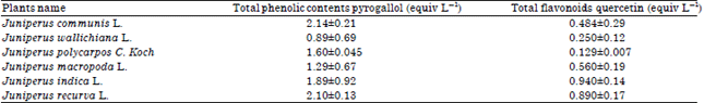 | |
| All values expressed as Mean±SEM (n = 4) | |
| Table 3: | Various identification characteristics of amentoflavone |
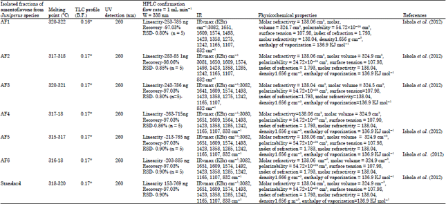 | |
| AF: Amentoflavone, RSD: Relative standard deviation, TLC: Thin layer chromatography, HPLC: High performance liquid choromatography, UV: Ultraviolet, IR: Infra red | |
| Table 4: | Effects of Juniperus species on erythrocytes antioxidant enzyme activity, glutathione and lipid peroxidase levels* |
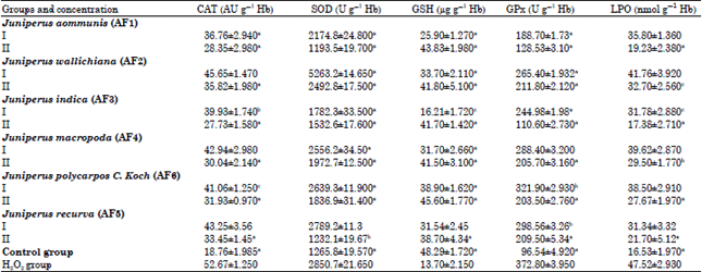 | |
| *All the value expressed as Mean±Sem, (n = 3) at ap<0.001, bp<0.01, cp<0.05 when compared with H2O2 group (one- way ANOVA followed by Dunnets test), SOD: Superoxide dismutase, Gpx: Glutathion peroxidase, LPO Lipid peroxidase, CAT: Catalase, GSH: Glutathione | |
| Table 5: | Effects of Juniperus species on leucocytes antioxidant enzyme activity, Glutathione and lipid peroxidase levels* |
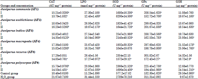 | |
| *All the value expressed as Mean±Sem, (n = 3), at ap<0.001, bp<0.01, cp<0.05 when compared with H2O2 group (one- way ANOVA followed by dunnets test), SOD: Superoxide dismutase, Gpx: Glutathion peroxidase, LPO Lipid peroxidase, CAT: Catalase, GSH: Glutathione | |
DISCUSSION
The protective effect of methanolic extracts from different species of juniperus in different concentration (250 and 500 μL) has been seen against H2O2 induced oxidative damage in human erythrocytes and leucocytes. Juniperus species was used traditionally, in the treatment of various diseases and disorder like cough, abdominal disorders, renal suppression, leucorrhoea, skin affections, infantile tuberculosis, asthma and also used in acute and chronic cystitis Juniper berries are used commercially for the preparation essential oil (flavoring agents), gin and as a spice (Marse, 1991). According to traditional usage of Juniperus species, we evaluated methanolic extracts of these plants against H2O2 induced oxidative damage in human erythrocytes and leucocytes. Juniperus species are reported for presence of numerous chemicals i.e. volatile oils (Vichi et al., 2007), coumarines, bicyclic diterpenes (Teresa et al., 1980), flavonoids and phenolic compounds (Lamer-Zarawska, 1975; Hiermann et al., 1996; Ilyas and Ilyas, 1990; Khare, 2007; Bais et al., 2015). The phenolic compounds obtained from four species of Juniperus have been reported as neuroprotective (Tavares et al., 2012; Bais and Mali, 2013). Present study also focused on the effect of junipers against oxidative stress model. Human blood cells are the key fighters against various oxidative stress diseases and remain the primary target for natural products and plants. These cells are continuously exposed to radicals and are unable to replace damaged components, especially polyunsaturated fatty acids chains from peroxidation (Bukowska, 2003).
During the reduction of molecular oxygen to water through acceptance of four electrons, active oxygen species, such as superoxide anion radicals, H2O2 and OH• radicals are generated. Against such active oxygen’s, cells have some defensive mechanisms including superoxide dismutase, catalase, several peroxidases and antioxidants, such as ascorbate, tocopherol, uric acid, β-carotene and GSH (Izawa et al., 1995; Yen et al., 2003).
In response to oxidative stress, GSH protect the thiol groups on membrane surface. The decreased availability of GSH indicates an increased demand (GSH) in erythrocytes to regain the storage mediated oxidative stress (Ault and Lawrence, 2003; Konyalioglu and Karamenderes, 2005). In present study, all extracts had significant (p<0.001) protective effect on GSH level as compared to the hydrogen peroxide group. Reactive oxygen species degrade polyunsaturated lipids, forming malondialdehyde (Schopfer et al., 2001). This is an end product of erythrocyte membrane lipid peroxidation. In various clinical conditions in humans and in vitro studies in animal erythrocytes and leucocytes, MDA a highly reactive, bifunctional molecule has been shown to cross-link erythrocyte and leucocyte phospholipids and proteins to impair a variety of the membrane-related functions and ultimately leading to diminished erythrocytes and leucocytes survival (Chiu et al., 1989; Sugihara et al., 1991; Ault and Lawrence, 2003). All extracts of juniperus species significantly decreased the level of LPO in dose dependent manner when compared with H2O2 group and this showed the protective effect against oxidative damage. Juniperus communis (500 μL) and Juniperus indica (500 μL) have the lowest LPO levels on erythrocytes (Table 4). Juniperus polycarpos C. Koch (500 μL) and Juniperus indica (500 μL) showed highest protective effects on leucocytes (Table 5). As a conclusion, Juniperus species extracts are found to have protective effect on antioxidant enzyme systems, GSH and LPO levels of erythrocytes and leucocytes against H2O2 Induced oxidative damage. Observed activity can be attributed to the phenol contents of these plants. The present results demonstrate that extracts of Juniperus communis, Juniperus wallichiana, Juniperus indica, Juniperus macropoda, Juniperus recurva and Juniperus polycarpos are potential sources of natural antioxidants. In future, further in vitro and in vivo studies are needed to assess the effects of these species on antioxidant system of the body.
ACKNOWLEDGMENTS
Authors are thankful to the Principal, Rayat Institute of Pharmacy, Punjab for providing the necessary facilities during this research work. Authors also wish to thanks Pinnacle Biomedical Research Institute, Bhopal.
REFERENCES
- Schopfer, P., C. Plachy and G. Frahry, 2001. Release of reactive oxygen intermediates (superoxide radicals, hydrogen peroxide and hydroxyl radicals) and peroxidase in germinating radish seeds controlled by light, gibberellin and abscisic acid. Plant Physiol., 125: 1591-1602.
Direct Link - Patel, R.P., D. Moellering, J. Murphy-Ullrich, H. Jo, J.S. Beckman and V.M. Darley-Usmar, 2000. Cell signaling by reactive nitrogen and oxygen species in atherosclerosis. Free Radical Biol. Med., 28: 1780-1794.
CrossRefDirect Link - Cooper, D., K.Y. Stokes, A. Tailor and D.N. Granger, 2002. Oxidative stress promotes blood cell-endothelial cell interactions in the microcirculation. Cardiovascular Toxicol., 2: 165-180.
CrossRefDirect Link - Stokes, K.Y., E.C. Clanton, K.S. Bowles, J.W. Fuseler and D. Chervenak et al., 2002. The role of T-lymphocytes in hypercholesterolemia-induced leukocyte-endothelial interactions. Microcirculation, 9: 407-417.
CrossRefDirect Link - Stokes, K.Y., D. Cooper, A. Tailor and D.N. Granger, 2002. Hypercholesterolemia promotes inflammation and microvascular dysfunction: Role of nitric oxide and superoxide. Free Radical Biol. Med., 33: 1026-1036.
CrossRefDirect Link - Landmesser, U., S. Dikalov, S.R. Price, L. McCann and T. Fukai et al., 2003. Oxidation of tetrahydrobiopterin leads to uncoupling of endothelial cell nitric oxide synthase in hypertension. J. Clin. Invest., 111: 1201-1209.
CrossRefDirect Link - Sharma, P.K. and B. Lal, 2005. Ethnobotanical notes on some medicinal and aromatic plants of Himachal Pradesh. Indian J. Trad. Knowl., 4: 424-428.
Direct Link - Tavares, L., G.J. McDougall, S. Fortalezas, D. Stewart, R.B. Ferreira and C.N. Santos, 2012. The neuroprotective potential of phenolic-enriched fractions from four Juniperus species found in Portugal. Food Chem., 135: 562-570.
CrossRefDirect Link - Lin, Y.M., M.T. Flavin, R. Schure, F.C. Chen and R. Sidwell et al., 1999. Antiviral activities of biflavonoids. Planta Med., 65: 120-125.
CrossRefDirect Link - Cholbi, M.R., M. Paya and M.J. Alcaraz, 1991. Inhibitory effects of phenolic compounds on CCl4-induced microsomal lipid peroxidation. Cell. Mol. Life Sci., 47: 195-199.
CrossRefDirect Link - Baureithel, K.H., K.B. Buter, A. Engesser, W. Burkard and W. Schaffner, 1997. Inhibition of benzodiazepine binding in vitro by amentoflavone, a constituent of various species of Hypericum. Pharmaceutica Acta Helvetiae, 72: 153-157.
CrossRefDirect Link - Kim, H.K., K.H. Son, H.W. Chang, S.S. Kang and H.P. Kim, 1998. Amentoflavone, a plant biflavone: A new potential anti-inflammatory agent. Arch. Pharm. Res., 21: 406-410.
CrossRefDirect Link - Da Silva, K.L., A.R. dos Santos, P.E. Mattos, R.A. Yunes, F. Delle-Monache and V. Cechinel-Filho, 2000. Chemical composition and analgesic activity of Calophyllum brasiliense leaves. Therapie, 56: 431-434.
PubMedDirect Link - Siddique, N.A., M. Mujeeb, A.K. Najmi and M. Akram, 2010. Evaluation of antioxidant activity, quantitative estimation of phenols and flavonoids in different parts of Aegle marmelos. Afr. J. Plant Sci., 4: 1-5.
Direct Link - Ishola, I.O., M. Chatterjee, S. Tota, N. Tadigopulla, O.O. Adeyemi, G. Palit and R. Shukla, 2012. Antidepressant and anxiolytic effects of amentoflavone isolated from Cnestis ferruginea in mice. Pharmacol. Biochem. Behav., 103: 322-331.
CrossRefDirect Link - Ali, S.A., O.R. Abdulraheem, T.E. Alemika, I.M. Sule and M. Ilyas et al., 2011. Phytochemical screening and identification of compounds in the leaves of Callitris glauca (Cupresseceae). Res. J. Pharma. Biol. Chem. Sci., 2: 624-635.
Direct Link - Markham, K.R., C. Sheppard and H. Geiger, 1987. 13C NMR studies of some naturally occurring amentoflavone and hinokiflavone biflavonoids. Phytochemistry, 26: 3335-3337.
CrossRefDirect Link - Zheng, J.X., Y. Zheng, H. Zhi, Y. Dai and N.L. Wang et al., 2011. New 3′, 8′′-linked biflavonoids from Selaginella uncinata displaying protective effect against anoxia. Molecules, 16: 6206-6214.
CrossRefDirect Link - Konyalioglu, S. and C. Karamenderes, 2005. The protective effects of Achillea L. species native in Turkey against H2O2-induced oxidative damage in human erythrocytes and leucocytes. J. Ethnopharmacol., 102: 221-227.
CrossRefDirect Link - Duh, P.D. and G.C. Yen, 1997. Antioxidative activity of three herbal water extracts. Food Chem., 60: 639-645.
CrossRefDirect Link - Satoh, K., 1978. Serum lipid peroxide in cerebrovascular disorders determined by a new colorimetric method. Clin. Chim. Acta, 90: 37-43.
CrossRefPubMedDirect Link - Yagi, K., 1984. Assay for blood plasma or serum. Methods Enzymol., 105: 328-331.
CrossRefPubMedDirect Link - Lowry, O.H., N.J. Rosebrough, A.L. Farr and R.J. Randall, 1951. Protein measurement with the folin phenol reagent. J. Biol. Chem., 193: 265-275.
CrossRefPubMedDirect Link - Abbassy, M.A. and G.I. Marei, 2013. Antifungal and chemical composition of essential oils of Juniperus communis L. and Thymus vulgaris L. against two phytopathogenic fungi. J. Applied Sci. Res., 9: 4584-4588.
Direct Link - Banerjee, S., A. Mukherjee and T.K. Chatterjee, 2012. Evaluation of analgesic activities of methanolic extract of medicinal plant Juniperus communis Linn. Int. J. Pharm. Pharmaceutical Sci., 4: 547-550.
Direct Link - Manvi and G.P. Garg, 2010. Screening and evaluation of pharmacognostic, phytochemical and hepatoprotective activity of Juniperus communis Linn. stems. Int. J. Pharma. Bio Sci., 1: 1-11.
Direct Link - Banerjee, S., H. Singh and T.K. Chatterjee, 2013. Evaluation of anti-diabetic and anti-hyperlipidemic potential of methanolic extract of Juniperus communis (L.) in streptozotocinnicotinamide induced diabetic rats. Int. J. Pharm. Bio Sci., 4: 10-17.
Direct Link - Hoferl, M., I. Stoilova, E. Schmidt, J. Wanner and L. Jirovetz et al., 2014. Chemical composition and antioxidant properties of juniper berry (Juniperus communis L.) essential oil. action of the essential oil on the antioxidant protection of Saccharomyces cerevisiae model organism. Antioxidants, 3: 81-98.
CrossRefDirect Link - Akdogan, M., A. Koyu, M. Ciris and K. Yildiz, 2012. Anti-hypercholesterolemic activity of Juniperus communis Lynn Oil in rats: A biochemical and histopathological investigation. Biomed. Res., 23: 321-328.
Direct Link - Sati, S.C. and S. Joshi, 2010. Antibacterial potential of leaf extracts of Juniperus communis L. from Kumaun Himalaya. Afr. J. Microbiol. Res., 4: 1291-1294.
Direct Link - Tunon, H., C. Olavsdotter and L. Bohlin, 1995. Evaluation of anti-inflammatory activity of some Swedish medicinal plants. Inhibition of prostaglandin biosynthesis and PAF-induced exocytosis. J. Ethnopharmacol., 48: 61-76.
CrossRefDirect Link - Haider, S.Z., M. Mohan, S. Sah and R. Seth, 2013. Comparative study on composition and antimicrobial activity of Juniperus wallichiana essential oils against clinical isolates. Med. Plants-Int. J. Phytomedicines Related Ind., 5: 90-95.
CrossRefDirect Link - Moein, M.R., Y. Ghasemi, S. Moein and M. Nejati, 2010. Analysis of antimicrobial, antifungal and antioxidant activities of Juniperus excelsa M. B subsp. Polycarpos (K. Koch) Takhtajan essential oil. Pharmacognosy Res., 2: 128-131.
CrossRefDirect Link - Gyawali, R., M. Binita and T.M. Shrestha, 2012. Antibacterial and cytotoxic activity of juniperus indica bertol from nepalese Himalaya. Int. J. Pharma. Sci. Res., 3: 1104-1107.
Direct Link - Vichi, S., M. Riu-Aumatell, M. Mora-Pons, J.M. Guadayol, S. Buxaderas and E. Lopez-Tamames, 2007. HS-SPME coupled to GC/MS for quality control of Juniperus communis L. berries used for gin aromatization. Food Chem., 105: 1748-1754.
CrossRefDirect Link - Teresa, J.D.P., A.F. Barrero, L. Muriel, A.S. Feliciano and M. Grande, 1980. New natural diterpene acids from Juniperus communis. Phytochemistry, 19: 1153-1156.
CrossRefDirect Link - Lamer-Zarawska, E., 1975. Biflavonoids in Juniperus L. sp (Cupressaceae). Polish J. Pharmacol. Pharm., 27: 81-87.
PubMedDirect Link - Bukowska, B., 2003. Effects of 2,4-D and its metabolite 2,4-dichlorophenol on antioxidant enzymes and level of glutathione in human erythrocytes. Comp. Biochem. Physiol. C Toxicol. Pharmacol., 135: 435-441.
CrossRefPubMedDirect Link - Izawa, S., Y. Inoue and A. Kimura, 1995. Oxidative stress response in yeast: Effect of glutathione on adaptation to hydrogen peroxide stress in Saccharomyces cerevisiae. FEBS Lett., 368: 73-76.
CrossRefDirect Link - Yen, G.C., H.C. Chiang, C.H. Wu and C.T. Yeh, 2003. The protective effects of Aspergillus candidus metabolites against hydrogen peroxide-induced oxidative damage to Int 407 cells. Food Chem. Toxicol., 41: 1561-1567.
CrossRefDirect Link - Ault, J.G. and D.A. Lawrence, 2003. Glutathione distribution in normal and oxidatively stressed cells. Exp. Cell Res., 285: 9-14.
CrossRefDirect Link - Chiu, D., F. Kuypers and B. Lubin, 1989. Lipid peroxidation in human red cells. Semin. Hematol., 26: 257-276.
PubMedDirect Link - Sugihara, T., W. Rawicz, E.A. Evans and R.P. Hebbel, 1991. Lipid hydroperoxides permit deformation-dependent leak of monovalent cation from erythrocytes. Blood, 77: 2757-2763.
PubMedDirect Link - Bais, S.S. and P.Y. Mali, 2013. Protective effect of Amorphophallus campanulatus tuber extracts against H2O2 induced oxidative damage in human erythrocytes and leucocytes. Int. J. Green Pharm., 7: 111-116.
Direct Link - Bais, S., N.S. Gill, N. Rana and S. Shandil, 2014. A phytopharmacological review on a medicinal plant: Juniperus communis. Int. Scholarly Res. Notices, Vol. 2014.
CrossRefDirect Link - Bais, S., N.S. Gill and N. Rana, 2014. Effect of Juniperus communis extract on reserpine induced catalepsy. Inventi Impact: Ethnopharmacol., 2014: 117-120.
Direct Link - Bais, S., N.S. Gill and N. Kumar, 2015. Neuroprotective effect of Juniperus communis on chlorpromazine induced Parkinson disease in animal model. Chin. J. Biol., Vol. 2015.
CrossRefDirect Link - Woisky, R.G. and A. Salatino, 1998. Analysis of propolis: Some parameters and procedures for chemical quality control. J. Apicult. Res., 37: 99-105.
CrossRefDirect Link - Aebi, H., 1984. Catalase in vitro. In: Methods in Enzymology, Packer, L., Academic Press, Cambridge, Massachusetts, United States, ISBN: 9780121820053, pp: 121-126.
CrossRefDirect Link - Gumral, N., D.D. Kumbul, F. Aylak, M. Saygin and E. Savik, 2015. Juniperus communis Linn oil decreases oxidative stress and increases antioxidant enzymes in the heart of rats administered a diet rich in cholesterol. Toxicol. Ind. Health, 31: 85-91.
CrossRefDirect Link - Mishra, P. and C.S. Chauhan, 1984. Antimicrobial studies of the essential oil of the berries of Juniperus macropoda Boiss. Hindustan Antibiotics Bull., 26: 38-40.
PubMedDirect Link