
Nobuko Mori
Department of Veterinary Science, School of Veterinary Medicine, Nippon Veterinary and Life Science University, Tokyo, Japan
Peter Lee
Department of Veterinary Science, School of Veterinary Medicine, Nippon Veterinary and Life Science University, Tokyo, Japan
Ichiro Yamamoto
Department of Veterinary Science, School of Veterinary Medicine, Nippon Veterinary and Life Science University, Tokyo, Japan
Satoshi Nozawa
Department of Veterinary Nursing, School of Veterinary Medicine, Nippon Veterinary and Life Science University, Tokyo, Japan
Toshiro Arai
Department of Veterinary Science, School of Veterinary Medicine, Nippon Veterinary and Life Science University, Tokyo, Japan
Asian Journal of Animal and Veterinary Advances
Year: 2011 | Volume: 6 | Issue: 8 | Page No.: 844-850


ABSTRACT
The aim of this study was to determine what insulin-induced daily changes occur with adiponectin, Tumor Necrosis Factor-alpha (TNF-α) and other relevant lipid metabolic parameters such as Free Fatty Acid (FFA), Total Cholesterol (T-Cho) and Triglyceride (TG) levels in dogs suffering from type 1 Diabetes Mellitus (T1DM). Six dogs were treated with insulin injections at the Nippon Veterinary and Life Science University Animal Clinical Center for T1DM. They were clinically diagnosed as T1DM by signs of polydipsia, polyuria, glycosuria and high fasting plasma glucose (over 250 mg dL-1). Blood samples of dogs were collected twice a day, before breakfast and after 6 hours of insulin injection. As results, post-insulin treatment value of TG, BUN, creatinin and chyromicron significantly increased, whereas ALT, ALP, protein and FFA significantly decreased post treatment. Significant reductions in total cholesterol, Very Low- Density Lipoprotein-cholesterol (VLDL) and Low-Density Lipoprotein-cholesterol (LDL) concentrations were also observed. Phospholipid Transfer Protein (PLTP) activity increased post-insulin treatment. The increase in Cholesteryl Ester Transfer Protein (CETP) activity was significant. Remarkably, insulin treatment reduced plasma adiponectin levels in T1DM dogs. Insulin resistance results in an increased risk of atherosclerosis on a long term basis as evidenced by atherogenicity markers of the plasma lipoprotein profile such as increased CETP and PLTP activities. Under obese condition adiponectin has been shown to negatively correlate with several inflammatory mediators (IL-1β, IL-6, IL-8, TNF-α). Low-grade chronic systemic inflammation has been associated with obesity potentially leading to insulin resistance.
PDF Abstract XML References Citation
Received: February 28, 2011;
Accepted: May 25, 2011;
Published: June 29, 2011
How to cite this article
Nobuko Mori, Peter Lee, Ichiro Yamamoto, Satoshi Nozawa and Toshiro Arai, 2011. Insulin Treatment-Induced Daily Changes to Plasma Adiponectin and TNF-α Level and Lipid Metabolism Parameters in Dogs Suffering from Type 1 Diabetes Mellitus. Asian Journal of Animal and Veterinary Advances, 6: 844-850.
DOI: 10.3923/ajava.2011.844.850
URL: https://scialert.net/abstract/?doi=ajava.2011.844.850
DOI: 10.3923/ajava.2011.844.850
URL: https://scialert.net/abstract/?doi=ajava.2011.844.850
INTRODUCTION
Plasma glucose and lipid metabolite concentrations reflect changes in energy metabolic and physical conditions of animal suffering from various metabolic disorders (Arai et al., 2003; Hsiao et al., 2007). It is known that plasma insulin concentration decreases whereas Free Fatty Acid (FFA) and glucose concentrations significantly increase in Type 1 Diabetes Mellitus (T1DM) dogs. Plasma lipoprotein profiles are possible to characterize different species by fraction patterns (Terpstra et al., 1982). As such, animals are generally classified into 2 main lipoprotein groups: HDL and LDL dominant mammals. Dogs are classified with HDL dominant mammals as cats, horses, mice and rats; whereas the LDL dominant group includes humans, hamsters, guinea pigs, pigs and rabbits (Chapman, 1986). The dog is a species with predominant HDL and very few VLDL (Mahley and Weisgraber, 1974). Hence, in this species, HDL is the main plasma carriers of cholesterol, with 2 HDL sub fractions being identifiable (Bauer, 2004): small dense particles called HDL2 and 3 and large buoyant particles called HDL1. HDL1 is unique to dogs and is enriched in both cholesterol and apolipoprotein (apo) E, tending to be prevalent in dogs fed on high amounts of cholesterol and or, saturated fat. They are also referred to as HDLc (Watson, 1996). Coincidentally, adiponectin concentrations also markedly decrease in plasma of dogs suffering from obesity. Adiponectin is an adipokine secreted by mature adipocytes and is commonly found in systemic blood circulation (Ishioka et al., 2006). Its characterized effected functions include: enhancement of insulin sensitivity, anti-inflammatory properties and inhibiting atherosclerosis in humans (Whitehead et al., 2006). Moreover, it is known that tumor necrosis factor-alpha (TNF-α) inhibits adiponectin secretion while stimulating FFA release. Studies have shown that obesity promotes a pro-inflammatory state thereby potentially increasing circulating TNF-α level in plasma. Studies have previously reported on insulin-induced predicted changes and alterations to glucose and energy metabolism in T1DM dogs, by comparing pre and post insulin metabolic values (Mori et al., 2007). However, it is unknown how and whether insulin treatment has any effect on lipid metabolism. Therefore, the aim of this study was to determine what insulin-induced daily changes occur with plasma levels of adiponectin, TNF-α and other relevant lipid metabolic parameters in dogs suffering from Type 1 DM.
MATERIALS AND METHODS
This study started 26th February 2010. Six dogs (age: 1-10 years; sex: 5 female and 1 male) housed and treated for diabetes mellitus at the Nippon Veterinary and Life Science University Animal Clinical Center were selected for this research. They were clinically diagnosed to be suffering from T1DM by signs of polydipsia, polyuria, glycosuria and high fasting plasma glucose (over 250 mg dL-1) and T1DM classification specifically based on anamnesis, clinical signs and clinical chemistry, respectively. Diabetic dogs were treated with 0.5 to 3.0 IU kg-1 body weight of insulin injections, Detemir insulin or NPH insulin (Both insulin; Novo Nordisk, Tokyo), twice daily (8 am and 8 pm) depending on their diabetic states. Fasted pre-insulin treatment blood samples were obtained from the jugular vein and collected into heparinized plastic tubes prior to 8 am breakfast. Animals were given 5-15 min to finish their breakfast, before they received appropriate amounts of insulin treatment depending on each individual animal’s status. Six hours post insulin injection, post-insulin treatment blood samples were collected again from the same dogs. Plasma was recovered by centrifugation at 1700 g for 10 min at 4°C and subsequently stored at -25°C.
Plasma Albumin (ALB), Alkaline Phosphatase (ALP), Alanine Aminotransferase (ALT), Aspartate Aminotransferase (AST) and Lactate Dehydrogenase (LDH) activities and Blood Urea Nitrogen (BUN), Creatinin (CRE), Glucose (GLU), Total-Cholesterol (T-Cho) and TG concentrations were measured using an Olympus AU680 auto analyzer (Olympus Corporation, Tokyo, Japan) with the manufacturer’s reagents. Adiponectin, CETP, FFA, insulin, PLTP and TNF-α were determined with the following commercial kits, respectively: Mouse/Rat adiponectin ELISA kit (Otsuka Pharmaceutical Co., Tokyo, Japan), CETP Activity Assay Kit and PLTP Activity Assay Kit (BioVision Reseach Products, Mountain View, CA), NEFA-C test Wako(Wako Pure Chemical Industries, Inc., Tokyo, Japan), Lbis Insulin kit (SHIBAYAGI Co., Gunma, Japan) and Canine TNF-α/TNF SF1A Immunoassay(R and D Systems, Inc., Minneapolis, MN).
Plasma cholesterol lipoprotein fractions were determined by biphasic agarose gel electrophoresis method and analyzed using an Epalyzer 2 Electrophoresis (Helena Laboratories, Saitama, Japan) according to the manufacturer’s protocol. High-density lipoprotein 1, 2 and 3 (HDL1, HDL2 and 3), low-density lipoprotein and very low-density lipoprotein (LDL and VLDL) and chylomicron fractions were assessed and analyzed using Edbank III analysis software (Helena Laboratories).
Statistical analysis: Results are presented as Means±95% C.I. The Student’s t-test for paired groups was performed on the various data sets in order to determine significance using Graph Pad Prism software version 4.0 (Graph Pad Soft ware, San Diego, CA). Differences were considered as significant when the p-value was <0.05.
RESULTS AND DISCUSSION
Table 1 shows that pre- treatment TG concentrations were low, whereas ALT, AST activities and FFA concentrations were high which are common characteristics of DM (Radin et al., 2009).
| Table 1: | Comparison of plasma metabolites, hormones, TNF-α concentrations and enzyme activities in type 1 diabetes mellitus dogs pre/post insulin treatment |
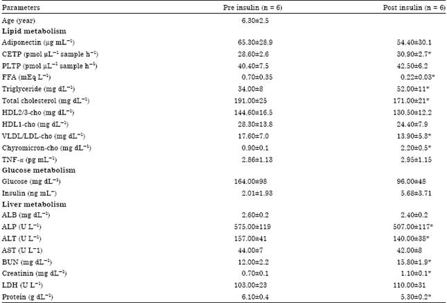 | |
| Values are presented as Means±95%C.I. *indicates significance (paired t-test, p<0.05) when compared against corresponding pre insulin shot group. Cho: Cholesterol, HDL: High density lipoprotein, LDL: Low density lipoprotein, VLDL: Very low density lipoprotein FFA: Free fatty acid, CETP: Cholesteryl ester transfer protein, PLTP: Phospholipid transfer protein TNF-α: Tumor necrosis factor alpha | |
TG concentrations significantly increased, whereas ALT and FFA levels significantly decreased post treatment. Significant reduction in concentrations of T-Cho and its sub-fractions (HDL1, HDL2 and 3, VLDL and LDL-cho) was also observed. PLTP activity also increased post-insulin treatment; however unlike CETP, the increase in CETP activity was significant. Lastly, insulin treatment reduced adiponectin concentrations in T1DM dogs.
In this study, significant reduction in plasma levels of FFA and ALT were observed following insulin treatment. ALT is a sensitive liver-specific indicator of damage in dogs (Raja et al., 2011) and insulin treatment seemed to improve liver condition. FFA level has been shown to induce a pro-inflammatory response in pancreatic islets via the IL-1 receptor (Boni-Schnetzler et al., 2009), therefore, a reduction in circulating FFA level would benefit T1DM dogs. Surprisingly, FFA level positively correlated with levels of VLDL and LDL (pre-insulin: r = 0.55; post-insulin: r = 0.68) and significant reductions in T-Cho, HDL1, HDL2 and 3, VLDL and LDL-cho levels were also observed in the T1DM. HDL is the predominant type of lipoprotein in canine plasma and plays a major role in cholesterol-transport. Canine HDL is composed of two sub fractions: HDL1 (large, buoyant particles which overlap with LDL) and HDL2 and 3 (small, dense particles equivalent to human HDL). In addition to its cholesterol-transport role, HDL can also reverse transport cholesterol that is excessive to tissue requirements back to the liver for storage, excretion or redistribution. We investigated the activity levels of two distinct lipid transfer proteins: CETP and PLTP, to see whether they may have any role to play in the resultant changes. It is known that lipoprotein particles can exchange lipid species (cholesterylesters, phospholipids, TG) between one another (Tall, 1995). Moreover, both CETP and PLTP may constitute two determinants of the atherogenicity of the plasma lipoprotein profile. Interestingly, animals with well-documented atherogenesis susceptibility (chicken, pig, rabbit and humans) display significantly higher mean CETP activity, but lower mean PLTP activity than known 'resistant' animals (cat, dog, mouse and rat). CETP is an important protein in regulating HDL-cho concentration and it catalyzes the transfer of cholesterol esters from HDL to VLDL and LDL. It is believed that CETP mainly participates in the reverse cholesterol transport pathway. In this study, CETP activity under post-insulin treatment increased significantly. CETP positively correlated with T-Cho (r = 0.78) and specifically with HDL1 (r = 0.50) and HDL2 and 3 (r = 0.58) sub fractions under pre-insulin conditions. CETP correlation under post-insulin strongly increased for T-Cho (r = 0.85), HDL1 (r = 0.76) and HDL2 and 3 (r = 0.66), while also positively correlating with TG, (r = 0.54), VLDL (r = 0.59) and chylomicron (r = 0.66). Lagrost et al. (1999) reported that levels of CETP, TG and non-high-density lipoprotein cholesterol increased as CETP activity increased in humans (Zeller et al., 2007). CETP also positively correlated with ALT (pre: r = 0.82; post: r = 0.84) and AST (pre: r = 0.91; post: r = 0.86) as markers for liver failure. However, CETP activity tends to be low or undetectable in dogs as compared to humans and other animals. Alternatively, regardless of insulin treatment, PLTP appeared to be more active than CETP (33% increase) which is in agreement with what has been reported using radioimmunoassay measurement (Guyard-Dangremont et al., 1998). PLTP regulates the size of HDL particles by transferring phospholipids from triglyceride-rich lipoproteins to HDL. In addition, PLTP levels positively correlated with TG (r = 0.61) and VLDL and LDL (r = 0.87) under pre-insulin conditions. However, post-insulin treatment resulted in lipid metabolic changes whereby PLTP correlated positively with T- cho (r = 0.49), HDL2 and 3 (r = 0.42), LDL and VLDL (r = 0.48) and chylomicron (r = 0.89). The correlation strength with LDL and VLDL was significantly reduced. It is known that PLTP increases hepatic VLDL secretion and decreases plasma HDL levels, resulting in a more atherogenic lipoprotein profile. Furthermore, PLTP unfavorably affects the anti-inflammatory and anti-oxidative properties of HDL particles (Moerland et al., 2008).
Plasma TNF-α concentration was almost not changed after insulin treatment and level of TNF-α would corroborate with the reduced anti-inflammatory and anti-oxidative effect of HDL in plasma due to reduced HDL. However, TNF-α positively correlated with adiponectin (pre: r = 0.99; post: r = 0.78) which would seem to contradict with what has been reported, thus indicating that TNF-α may not function as a trigger for decreasing adiponectin levels in dogs. Precious evidence that adiponectin plays a role in increasing insulin sensitivity and decreasing of circulating adiponectin levels play causative role in the development of insulin resistance (Assal et al., 2007). There was some evidence that the primary mechanism by which adiponectin enhance insulin sensitivity appears to be through fatty acid oxidation and inhibition of hepatic glucose production (El-Ghaffar and El-Said, 2006). Adiponectin has also been reported to negatively correlate with TG and positively correlate with HDL in humans from T2DM (Hotta et al., 2000). Interestingly, adiponectin levels in this study also negatively correlated with TG (pre: r = -0.89; post: r = -0.77), while positively correlating with HDL1 (pre: r = 0.58; post: r = 0.54), HDL2 and 3 (pre: r = 0.82; post: r = 0.89) and chylomicron (pre: r = 0.75; post: r = 0.63), although the animals were suffering from T1DM. On the other hand, adiponecitin negatively correlated with ALT (pre: r = -0.87; post: r = -0.91) and AST (pre: r = -0.60; post: r = -0.99) indicating that sustained reduced adiponectin levels could have a long term impact on liver stress. In addition, insulin treatment in T1DM dogs reduces adiponectin levels which would indicate that adiponectin alone is not the sole factor for enhancing insulin sensitivity. Instead, other factors may also contribute and have a synergistic effect with insulin. The results are in agreement with the previous reported on the inverse relationship between adiponectin and insulin. Moreover, euglycemic- hyperinsulinemic clamp tests in both humans and rats have indicated that insulin injection leads to reduced circulating adiponectin levels, partially because insulin exerts an acute effect on adipocytes to decrease production and/or secretion of this adipocytokine (Yu et al., 2002). Therefore, adipocyte insulin signal transduction may be more relevant to understanding adiponectin levels post-insulin treatment rather than the absolute amount of insulin being given as treatment. Generally adponectin has been shown to negatively correlate with several inflammatory mediators (IL-1β, IL-6, IL-8, TNF-α) (Roth et al., 2010). Low-grade chronic systemic inflammation has been associated with obesity potentially leading to insulin resistance.
CONCLUSION
Insulin treatment in dogs suffering from T1DM appeared to stimulate lipid metabolism and improve liver condition as indicated by reduced ALT levels. In addition, exogenous insulin reduced plasma adiponectin level which has been shown to positively correlate with TNF-α in T1DM dogs. Although insulin treatment improve glucose metabolism in T1DM dogs, it may appear to bring an increased risk for atherosclerosis on a long term basis.
ACKNOWLEDGMENTS
This work was supported in part by the Strategic Research Base Development Program for Private Universities from the Ministry of Education, Culture, Sports, Science and Technology of Japan (MEXT), 2008-2012 and Grant-in Aid for Scientific Research (No. 21380195 to T. Arai) from the MEXT.
REFERENCES
- Assal, H.S., M. Fath-Allah and A. Elsherbiny, 2007. Serum leptin and adiponectin in obese diabetic and non-diabetic. J. Med. Sci., 7: 865-869.
CrossRefDirect Link - Arai, T., M. Nakamura, E. Magori, H. Fukuda and T. Sako, 2003. Decrease in malate dehydrogenase activities in peripheral leucocytes of type 1 diabetic dogs. Res. Vet. Sci., 74: 183-185.
CrossRefDirect Link - Bauer, J.E., 2004. Lipoprotein-mediated transport of dietary and synthesized lipids and lipid abnormalities of dogs and cats. J. Am. Vet. Med. Assoc., 224: 668-675.
PubMed - Boni-Schnetzler, M., S. Boller, S. Debray, K. Bouzakri and D.T. Meier et al., 2009. Free fatty acids induce a proinflammatory response in islets via the abundantly expressed interleukin-1 receptor I. Endocrinology, 150: 5218-5229.
CrossRefDirect Link - Abd El-Ghaffar, N. and N.H. El-Said,, 2006. Hypoadiponectinaemia in Egyptian patients with type II diabetes mellitus with vascular complications. J. Med. Sci., 6: 626-630.
CrossRefDirect Link - Guyard-Dangremont, V., C. Desrumaux, P. Gambert, C. Lallemant and L. Lagrost, 1998. Phospholipid and cholesteryl ester transfer activities in plasma from 14 vertebrate species. Relation to atherogenesis susceptibility. Comp. Biochem. Physiol. B. Biochem. Mol. Biol., 120: 517-525.
PubMed - Hsiao, P.J., K.K. Kuo, S.J. Shin, Y.H. Yang and W.Y. Lin et al., 2007. Significant correlations between severe fatty liver and risk factors for metabolic syndrome. J. Gastroenterol. Hepatol., 22: 2118-2123.
PubMed - Hotta, K., T. Funahashi, Y. Arita, M. Takahashi and M. Matsuda et al., 2000. Plasma concentrations of a novel, adipose-specific protein, adiponectin, in type 2 diabetic patients. Arterioscler. Thromb. Vasc. Biol., 20: 1595-1599.
CrossRefDirect Link - Ishioka, K., A. Omachi, M. Sagawa, H. Shibata, T. Honjoh, K. Kimura and M. Saito, 2006. Canine adiponectin: CDNA structure, mRNA expression in adipose tissues and reduced plasma level in obesity. Res. Vet. Sci., 80: 127-132.
CrossRef - Lagrost, L., A. Athias, N. Lemort, J.L. Richard and C. Desrumaux et al., 1999. Plasma lipoprotein distribution and lipid transfer activities in patients with type II b hyperlipidemia treated with simvastatin. Atherosclerosis, 143: 415-425.
Direct Link - Mahley, R.W. and K.H. Weisgraber, 1974. Canine lipoproteins and atherosclerosis. I. Isolation and characterization of plasma lipoproteins from control dogs. Circ. Res., 35: 713-721.
PubMed - Moerland, M., H. Samyn, T. van Gent, R. van Haperen and G. Dallinga-Thie et al., 2008. Acute elevation of plasma PLTP activity strongly increases pre-existing atherosclerosis. Arterioscler. Thromb. Vasc. Biol., 28: 1277-1282.
Direct Link - Mori, A., P. Lee, T. Sako, H. Mizutani and T. Arai, 2007. Successful intensive insulin treatment of type 1 diabetic dogs leads to restoration of peripheral leukocyte insulin signaling gene expression and enzyme activities. J. Vet. Med. Sci., 71: 1017-1026.
PubMed - Radin, M.J., L.C. Sharkey and B.J. Holycross, 2009. Adipokines: A review of biologicalandanalytical principles and an update in dogs, cats and horses. Vet. Clin. Pathol., 38: 136-156.
CrossRef - Raja, M.M.M., A. Raja, M.M. Imran, A.M.I. Santha and K. Devasena, 2011. Enzymes application in diagnostic prospects. Biotechnology, 10: 51-59.
CrossRefDirect Link - Roth, C.L., M. Kratz, M.M. Ralston and T. Reinehr, 2010. Changes in adipose-derived inflammatory cytokines and chemokines after successful lifestyle intervention in obese children. Metabolism.
CrossRef - Terpstra, A.H., F.J. Sanchez-Muniz, C.E. West and C.J. Woodward, 1982. The density profile and cholesterol concentration of serum lipoproteins in domestic and laboratory animals. Comp. Biochem. Physiol. B, 71: 669-673.
PubMed - Watson, T.D.G., 1996. Lipoprotein metabolism in dogs and cats. Comp. Hematol. Int., 6: 17-23.
CrossRef - Whitehead, J.P., A.A. Richards, I.J. Hickman, G.A. Macdonald and J.B. Prins, 2006. Adiponectin: A key adipokine in the metabolic syndrome. Diabetes. Obes. Metab., 8: 264-280.
CrossRef - Yu, J.G., S. Javorschi, A.L. Hevener, Y.T. Kruszynska, R.A. Norman, M. Sinha and J.M. Olefsky, 2002. The effect of thiazolidinediones on plasma adiponectin levels in normal, obese and type 2 diabetic subjects. Diabetes, 51: 2968-2974.
CrossRef - Zeller, M., D. Masson, M. Farnier, L. Lorgis and V. Deckert et al., 2007. High serum cholesteryl ester transfer rates and small high-density lipoproteins are associated with young age in patients with acute myocardial infarction. J. Am. Coll. Cardiol., 50: 1948-1955.
CrossRef