
Mohammad M. Fatthy Ramadan
Departments of Otorhinolaryngology, Faculty of Medicine, Al-Azhar University, Egypt
Ahmed Abd Elrahman Mahmoud
Departments of Otorhinolaryngology, Faculty of Medicine, Al-Azhar University, Egypt
Ahmed M. Abdelhady Elsheikh
Departments of Otorhinolaryngology, Faculty of Medicine, Al-Azhar University, Egypt
Hatem Salah Eldin Elhabashy
Departments of Otorhinolaryngology, Faculty of Medicine, Al-Azhar University, Egypt
Trends in Medical Research
Year: 2013 | Volume: 8 | Issue: 1 | Page No.: 16-26


ABSTRACT
The aim of this study is to evaluate safety and feasibility of endoscopic surgery for management of nasal Inverted Papilloma (IP). The study estimated tissue extract Epidermal Growth Factor Receptor (EGFR) and serum Squamous Cell Carcinoma Antigen (SCCA) levels to determine their applicability for differentiation between different pathological entities of IP. All patients with IP underwent full history taking, general and local examination and CT imaging. Endoscopic biopsy was taken and then endoscopic excision was conducted under general anesthesia. Freshly excised IP were divided into two parts; the first for pathological examination and the second part was used for estimation of tissue extract level of EGFR. Venous blood samples were obtained before and after surgical resection for enzyme-linked immunosorbent assay (ELISA) estimation of serum SCCA. The study also included 10 healthy volunteers as a control group. The study included 19 IP patients; 9 patients had do novo IP, 6 patients had recurrent IP and 4 had IP with Squamous Cell Carcinoma (SCC) changes. All surgeries were conducted safely without complications within a mean operative time of 74.5±13.5 min, mean blood loss of 158.2±27.8 mL and mean postoperative duration of hospital stay of 5.4±1.3 days. Mean preoperative serum SCCA levels were significantly higher in IP patients compared to controls and in patients had IP with SCC compared to those had benign IP with non-significantly higher levels detected in sera of patients had recurrent IP compared to those had de novo IP. Postoperative serum levels of SCCA were significantly lower in all patients compared to their respective preoperative levels and non-significantly higher levels in patients had IP with SCC compared to those had benign IP. Mean tissue extract levels of EGFR were significantly higher in tissues of IP with SCC compared to tissues of benign IP with significantly higher tissue extract levels in recurrent IP compared to tissues of de novo IP. Functional endoscopic surgery could be considered as safe and effective line of management of IP. Estimation of EGFR tissue expression level could be used for differentiation between benign IP and IP with malignant transformation even in early phases of dysplasia and serum SCCA could be used as a marker for complete operative excision of IP irrespective of its pathological diagnosis.
PDF Abstract XML References Citation
Received: March 01, 2013;
Accepted: April 20, 2013;
Published: June 17, 2013
How to cite this article
Mohammad M. Fatthy Ramadan, Ahmed Abd Elrahman Mahmoud, Ahmed M. Abdelhady Elsheikh and Hatem Salah Eldin Elhabashy, 2013. Applicability of Biomarkers for Differentiation of Inverted Papilloma Assigned for Endoscopic Surgery. Trends in Medical Research, 8: 16-26.
URL: https://scialert.net/abstract/?doi=tmr.2013.16.26
URL: https://scialert.net/abstract/?doi=tmr.2013.16.26
INTRODUCTION
The lining epithelium of the respiratory tract is basically composed of psudostratified squamous type (respiratory epithelium with columnar cells), whereas, stratified squamous epithelium is present in the pharynx and part of the larynx. This coexistence of two different types of epithelia creates squamo-columnar junctions at different sites in the respiratory tract. It is thought to be a prerequisite for the spread of human papilloma virus infections at such junctions. A vast variety of benign and malignant neoplasms arises in the respiratory tract. This is may be due to this divergent histology of the epithelial lining (Sarioglu, 2007; Fitzhugh and Mirani, 2008; Kirdar et al., 2009).
Epidermal Growth Factor Receptor (EGFR) is made of extracellular domains, including a ligand-binding domain which is a hydrophobic trans-membrane region and a tyrosine kinase-containing cytoplasmic region. Stimulation of the EGFR by endogenous ligands, EGF or Tissue-growth Factor-alpha (TGF-α) results in a conformational change in the receptor, permitting its dimerization and subsequent activation of intracellular tyrosine kinase, protein phosphorylation and stimulation of various cell signaling pathways that mediate gene transcription and cell cycle progression (Penault-Llorca et al., 2003). EGFR is present in normal human cells particularly in injured tissues to help wound healing (Avissar et al., 2000). It has been shown that higher levels of expression of the receptor is correlated with malignancy in a variety of tumors. The EGFR activation participates in oncogenesis by inducing cell proliferation, cell mobility and angiogenesis and inhibiting apoptosis. This activation might be due to numerous abnormalities, including increased expression of its ligand (Psyrri et al., 2005; Walker et al., 2009).
The Squamous Cell Carcinoma Antigen (SCCA) is a group of glycoproteins belongs to the family of serine/cysteine-protease inhibitors (Suminami et al., 1991). SCCA first was isolated biochemically from Squamous Cell Carcinoma (SCC) tissue of the uterine cervix and consists of at least 10 sub-fractions differing in iso-electric point (Kato and Torigoe, 1977). Molecular studies have showed that SCC antigen is transcribed by two nearly identical genes; SCCA1 and SCCA2 (Schneider et al., 1995). Both genes are protease inhibitors that map to the serine protease inhibitor (serpin) cluster at 18q21. These gene products are expressed in SCC tissues as well as normal squamous epithelium (Hamada et al., 2001).
The exophytic and inverted pattern of growth found microscopically in Inverted Papilloma (IP) is often confused with sinonasal adenocarcinoma which contain single-layered epithelium only. Clinically, its behavior is similar to that of inverted papilloma because of local recurrence and malignancy coexistence. The factors responsible for associated malignancy are not well known. It is also still unknown whether carcinomas in inverted papilloma arise meta-or synchronous. (Mirza et al., 2007; Mendenhall et al., 2007; Knopf et al., 2008; Wang et al., 2011). Owing to its liability for recurrence, postoperative long-term follow-up of IP patients is recommended, so, the establishment of a biologic marker reflecting the extent of disease can be of great help to the clinician (Liu et al., 2011; Koo et al., 2011).
The current selective prospective study aimed to evaluate the safety and feasibility of endoscopic surgery as a therapeutic modality for IP. It also aimed to estimate the excised specimen EGFR tissue extract level and serum SCCA levels in patients with IP. This study is a trial to determine the role of EGFR and SCCA in pathogenesis and/or their applicability for differentiation between different pathological entities of IP.
MATERIALS AND METHODS
The current prospective 5 year study was conducted at Otorhinolaryngology departments, Al Azhar University Hospitals, Egypt, so as to include all patients with sinonasal inverted papilloma, irrespective of its pathological state. The study also included 10 healthy volunteers chosen from those attending Hospital Blood Bank for blood donation to give blood samples as control group. After obtaining fully informed patients’ consent, all patients underwent full history taking, general and local examination. CT imaging was conducted and its results were verified according to Lund and Lloyd (1984).
Prior to surgery, a biopsy was performed endoscopically, usually under local anesthesia. General anesthesia was used during the resection. Injection of epinephrine at a concentration of 1:100,000 was used to reduce the hemorrhage. Endoscopic en bloc resection was attempted in small tumors. However, when the origin of the growth was unclear and the tumor filled the nasal cavity, parts of the tumor are not involving the nasal cavity; mucosa were debulked to anatomically delineate the origin of the tumor’s growth and then, an en bloc excision of the tumor with as wide a margin as possible was performed. Secured margins were confirmed by frozen section.
Freshly excised IP or taken biopsy tissues were divided into two parts; the first was examined pathologically using hematoxylin and eosin (H and E) stain, according to the World Health Organization classification (Shanmugaratnam and Sobin, 1991). The second part was immediately kept frozen at -70°C until processed for estimation of tissue extract level of EGFR.
Tissue processing and estimation of tissue extract level of EGFR: Tumor specimens were finely minced and homogenized in 5 volumes of ice-cold buffer consisting of 25 mM Tris, 1.5 mM EDTA, 5 mM NaN3, 0.1% monothioglycerol and 20% glycerol by applying several intermittent bursts of an Ultra-turrax homogenizer. The crude homogenate was centrifuged at 7000 rpm for 20 min at 0°C and the supernatant was further centrifuged at 105 rpm for 75 min at 0°C to obtain a membrane fraction for EGFR assay. The membrane pellet was resuspended in 25 mM Tris, 1.5 mM EDTA, 5 mM NaN3, 20% glycerol and 10 mM MgCl2. Aliquots of the suspension (100 μL containing 300-500 μg of protein) were incubated with 125I-EGF (2.6 nM; 800,000 Ci/mmol; NEN, DuPont, Wilmington, DE) in the presence or absence of unlabeled EGF (1 μg) for 12-16 h at room temperature in a final volume of 400 μL. Binding was blocked by the addition of 3 mL of ice-cold 25 mM Tris, 20% glycerol, 5 mM NaN3 and 0.1% Bovine Serum Albumin (BSA). After centrifugation at 2000 rpm for 20 min at 0°C, the supernatant was carefully aspirated, pellets were counted in a gamma counter and levels were expressed as fmol/mg protein (Battaglia et al., 1988).
Venous blood samples were obtained prior to and after surgical resection and allowed to clot then serum was separated by centrifugation at 3000 rpm for 10 min. Serum was removed and stored at -70°C until Enzyme-linked Immunosorbent Assay (ELISA) assayed for estimation of serum SCCA (Fujirebio Diagnostics, Inc, Majnabbeterminalen, Göteborg, Sweden) (Roijer et al., 2003).
Statistical analysis: Obtained data were presented as Mean±SD, ranges, numbers and ratios. Results were analyzed using Wilcoxon (Z-test) test. Statistical analysis was conducted using the SPSS (Version 15, 2006) for Windows statistical package. p value <0.05 was considered statistically significant.
RESULTS
The study included 19 patients; 15 males and 4 females, with mean age of 65.1±4.7, range: 56-77 years. Histopathological examination of excised specimens defined 4 cases had IP with SCC changes; while the remaining 15 cases had benign IP, 9 patients had do novo IP and 6 patients had recurrent IP. Patients had recurrent IP had a mean recurrence rate of 1.8±0.7; range: 1-3 recurrence times with a mean duration of the last endoscopic removal of 6.9±1.5; range: 5-9 months. All patients presented by nasal obstruction as the main presenting symptom, epistaxis was reported in 11 patients, headache in 9 patients and foul discharge in 7 patients, (Table 1). Additionally, one of patients with SCC changes in an IP presented by unilateral facial swelling. Two of cases had IP showing SCC changes presented by a left nasal mass extending into the ipsilateral maxillary sinus, the 3rd patient had a right nasal mass but sinuses were still free and the 4th patient had right side large soft tissue mass occupying the middle meatus of the nasal cavity with involvement of both the maxillary antrum and the ethmoidal sinuses, (Fig. 1).
Macroscopically benign IP appeared in the form of multiple polypoidal; firm mass with undulant or papillary surfaces, (Fig. 2). Microscopically, benign IP were characterized by focally soughed stratified squameous epithelial covering with a mildly exophytic surface, (Fig. 3a) and multiple invaginations into the underlying focally edematous, vascular and focally fibrotic stroma that showed moderate to marked non-specific inflammatory cellular infiltration, (Fig. 3b).
| Table 1: | Patients’ characters: number, ages, genders and presenting symptoms of patients with benign IP and with IP with SCC |
 | |
| Data are presented as Mean±SD and numbers; ranges and percentages are in parenthesis | |
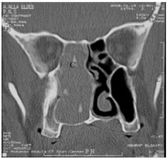 | |
| Fig. 1: | CT imaging, coronal view, showing a right side large soft tissue mass occupying the middle meatus of the nasal cavity with involvement of both the maxillary antrum and the ethmoidal sinuses. Areas of calcification could be seen within the mass (C) |
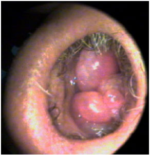 | |
| Fig. 2: | A photograph of benign IP appeared macroscopically in the form of multiple polypoidal mass with undulant surfaces |
 | |
| Fig. 3(a-b): | (a) A photograph of endoscopically excised IP showing focally sloughed squameous epithelial covering (Sq) with multiple invaginations (I) and mildly exophytic surface (Ex), (H and E x100) and (b) A photograph of endoscopically excised IP showing focally fibrotic stroma (F) and moderate to marked inflammatory cellular infiltrations (IC), (H and E x200) |
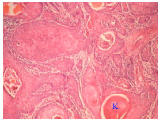 | |
| Fig. 4: | A photograph of endoscopically excised IP showed an ulcerating, infiltrating neoplastic squameous epithelial growth of nasal tissue, generally disposed in nests with central keratin (K) pearls surrounded by desmoplastic stroma. Focal high-grade dysplastic changes and few areas showing moderate anaplasia were also seen, (H and E x160) |
| Table 2: | Mean serum SCCA and tissue extract level of EGFR estimated in studied patients categorized according to histopathological data |
 | |
| Data are presented as Mean±SD; ranges are in parenthesis, *Significant versus controls, **Significant versus de novo cases, †Significant versus benign papilloma, ‡Significant versus preoperative levels | |
Microscopic examination of IP with SCC revealed an evidence of nasal tissue involvement by an ulcerating, infiltrating neoplastic squameous epithelial growth, generally disposed in nests with central keratin pearls surrounded by desmoplastic stroma showing chronic non-specific inflammatory cellular infiltration. Focal high-grade dysplastic changes up to focal in-situ changes and few areas showing moderate anaplasia were also seen, (Fig. 4).
All surgeries were conducted safely without intraoperative or postoperative complications within a mean operative time of 74.5±13.5; range: 50-100 min and a mean blood loss of 158.2±27.8; range: 120-210 mL. All patients were discharged after an uneventful postoperative course after a mean postoperative duration of hospital stay of 5.4±1.3; range: 3-7 days.
Mean preoperative serum SCCA levels were significantly (p<0.05) higher in patients with IP compared to control subjects. Mean preoperative serum SCCA levels detected in patients with IP associated with SCC were significantly (p<0.05) higher when compared to levels detected in patients with benign IP. These levels were non-significantly (p>0.05) higher in sera of patients with recurrent IP compared to those with de novo IP. Surgical resection significantly (p<0.05) reduced serum levels of SCCA in all patients compared to their respective preoperative levels and non-significantly (p>0.05) higher levels in patients had IP with SCC compared to those had benign IP (Table 2). Mean tissue extract levels of EGFR were significantly (p<0.05) higher in tissues of IP with SCC compared to tissues of benign IP with significantly (p<0.05) higher tissue extract levels in recurrent IP compared to tissues of de novo IP (Table 2).
DISCUSSION
The current selective study aimed to collection of IP cases; throughout the study period only 19 cases were collected and 4 of which were confirmed to have SCC changes. The presenting features are mosaic but nasal obstruction was predominant in association with headache, insomnia, epistaxis and/or foul discharge. These data supported that previously documented by Tanvetyanon et al. (2009) who retrospectively reviewed the medical records of patients with IP and SCC treated at their institution during 1999-2007 and identified 6 patients with SCC arising from IP. Also, Lathi et al. (2011) examined the clinico-pathological profile of sinonasal polypoid masses in a series of 112 patients and reported that the majority of patients were males. They found that IP represented 36.8% of benign lesions and nasal obstruction was the most common presenting complaint (97.3%), followed by rhinorrhoea, hyposmia, intermittent epistaxis, headache, facial swelling and eye-related symptoms (10.7%).
CT imaging showed areas of calcification within a large soft tissue mass occupying the middle meatus of the nasal cavity with sinus involvement and localized bone thickening. In agreement with such CT picture Lee et al. (2007) detected two patterns of localized bone thickening in associations with sinonasal IP. The first pattern is plaque-like bone thickening which was seen mainly when focal hyperostosis involved the lateral wall of the nasal cavity. The second pattern is cone-shaped bone thickening that was seen only in the walls of the paranasal sinuses or the bony septum.
All surgeries were conducted endoscopically without intraoperative or postoperative complications within a mean operative time of 74.5 min, mean blood loss of 158.2 mL and mean postoperative duration of hospital stay of 5.4 days. These data indicated safety and feasibility of functional endoscopic surgery for management of such lesions characterized by high liability of bleeding. In support of these data, Sun et al. (2011) and Dragonetti et al. (2011) reported that nasal endoscopic and endoscopy-assisted surgery can be used in the treatment for the malignant transformation of sinunasal IP, with the advantages of minimally invasion, fewer complications and the well-preserved nasal functions.
Intraoperatively, when the origin of the growth was unclear and the tumor filled the nasal cavity, mucosa were debulked to anatomically delineate the origin of the tumor’s growth and then surgery was completed. In line with such plane, Carta et al. (2011) documented that planning of the surgical strategy is based on the comparison of CT and MRI, with the aim of targeting the insertion of the tumor. With the help of expanded surgical procedures, endoscopic surgery has become the gold standard for the treatment of the vast majority of IP.
Tissue extract levels of EGFR where significantly elevated in IP with SCC compared to benign IP and in recurrent IP compared to de novo IP, a finding indicating increased local expression of growth factors in tissues of IP and illustrated the role played by growth factors for pathogenesis and progression of IP.
These findings go in hand with Katori et al. (2005) who immunohistochemically detected a significant increase of EGFR and tissue growth factor-alpha (TGF-α) in IP with severe dysplasia, IP with carcinoma and invasive SCC compared to IP with mild dysplasia and control nasal mucosa. They concluded that pre-cancerous lesions of IP exhibited elevated levels of EGFR and TGF-α and these expression may be associated with early events in IP carcinogenesis. Sauter et al., 2007 stated that an increased EGFR and TGF-alpha expression is associated with early changes in IP carcinogenesis.
Also, Li et al. (2007) found the expression of EGFR protein may be activated in the early stage of pathogenesis of SCC of nasal cavity and paranasal sinuses. Chao and Fang (2008) found that the positive rate of immunostaining, positive intensity of immunostaining for EGFR protein and EGFR mRNA were significantly up-regulated in the IP and synchronous SCC with IP compared with polyp and normal mucosa. They concluded that EGFR’s role in malignant transformation from IP to SCC of the nasal cavity is suggested.
In parallel to tissue expression levels of EGFR, preoperative serum SCCA levels were in patients with IP when compared to control. These levels are also significantly higher in IP patients with SCC when compared to levels estimated in controls and patients with benign IP whether de novo or recurrent. These levels are higher in sera of patients had recurrent IP compared to those had de novo IP. These data indicated a synchronous increased serum levels of SCCA with progression of IP either in the form of repeat growth or malignant transformation. Postoperative serum SCCA levels were significantly lower in all IP cases compared to their respective preoperative measures with non-significantly higher levels compared to control levels and among studied groups. These data indicated a fact that the source of SCCA estimated in serum was the papillomatous growth and the more the change the more the serum level.
In line with these data multiple studies detected a relation between both tissue expression of EGFR and/or serum levels of SCCA and papillomatous lesions anywhere in the body and with their progression or changed behavior. Huang et al. (2006) indicated that SCCA may be of great potential as the biomarker of tongue cancer and as the potential therapeutic target for gene therapy. Iwata et al. (2007) reported a case of IP causing high serum levels of SCCA and Carcinoembryonic Antigen (CEA), postoperatively, the serum levels of CEA and SCCA significantly decreased within 3 months. Lin et al. (2011) demonstrated that preoperative SCCA is a good marker of pathologic lymph node metastasis, an advanced tumor stage and a higher rate of distant metastasis in patients had oral carcinoma.
In support of the pathogenic role of EGFR in development and progression of IP, expression of different variants of EGFR was documented in airway papillomatous and/or malignant lesions; Yang et al. (2009) confirmed the expression of EGFR in laryngeal carcinoma and documented that it is tumor-specific and tends to be more frequent in EGFR-over expressing tumor tissues and poorly differentiated ones which may in part contribute to the malignant phenotype. Kourelis et al. (2009) documented that EGFR was upregulated significantly along the epithelial deterioration toward neoplasia. Also, Wu et al. (2010) reported that respiratory papillomas overexpress the EGFR.
The obtained results and review of literature allowed concluding that functional endoscopic surgery could be considered as safe and effective line of management of IP. Estimation of EGFR tissue expression level could be used for differentiation between benign and malignant IP even in early phases of dysplasia and serum SCCA could be used as a marker for complete operative excision of IP irrespective of its pathological diagnosis. However, further studies with larger series are needed to support these results and to clarify rationales.
CONCLUSION
Functional endoscopic surgery could be considered as safe and effective line of management of IP. Estimation of EGFR tissue expression level could be used for differentiation between benign IP and IP with malignant transformation even in early phases of dysplasia and serum SCCA could be used as a marker for complete operative excision of IP irrespective of its pathological diagnosis.
REFERENCES
- Avissar, N.E., H.T. Wang, J.H. Miller, P. Iannoli and H.C. Sax, 2000. Epidermal growth factor receptor is increased in rabbit intestinal brush border membrane after small bowel resection. Digestive Dis. Sci., 45: 1145-1152.
CrossRef - Battaglia, F., G. Polizzi, G. Scambia, S. Rossi and P. Benedetti Panici et al., 1988. Receptors for epidermal growth factor and steroid hormones in human breast cancer. Oncology, 45: 424-427.
CrossRef - Chao, J.C. and S.Y. Fang, 2008. Expression of epidermal growth factor receptor in the inverted papilloma and squamous cell carcinoma of nasal cavity. Eur. Arch. Oto-Rhino-Laryngol., 265: 917-922.
CrossRef - Dragonetti, A., R. Gera, A. Sciuto, A. Scotti, A. Bigoni, E. Barbaro and A. Minni, 2011. Sinonasal inverted papilloma: 84 patients treated by endoscopy and proposal for a new classification. Rhinology, 49: 207-213.
PubMedDirect Link - Fitzhugh, V.A. and N. Mirani, 2008. Respiratory epithelial adenomatoid hamartoma: A review. Head Neck Pathol., 2: 203-208.
CrossRef - Hamada, K., Y. Hanakawa, K. Hashimoto, M. Iwamoto and T. Kihana et al., 2001. Gene expression of human squamous cell carcinoma antigens 1 and 2 in human cell lines. Oncol. Rep., 8: 347-354.
PubMedDirect Link - Huang, X., Y. Wei, L. Li, Y. Wen and J. Yang et al., 2006. Serum proteomics study of the squamous cell carcinoma antigen 1 in tongue cancer. Oral Oncol., 42: 25-30.
CrossRef - Iwata, T., K. Inoue, N. Nishiyama, N. Izumi and S. Mizuguchi et al., 2007. Pulmonary inverted schneiderian papilloma causing high serum levels of carcinoembryonic antigen and squamous cell carcinoma-associated antigen: Report of a case. Surg. Today, 37: 790-793.
CrossRef - Kato, H. and T. Torigoe, 1977. Radioimmunoassay for tumor antigen of human cervical squamous cell carcinoma. Cancer, 40: 1621-1628.
Direct Link - Katori, H., A. Nozawa and M. Tsukuda, 2005. Markers of malignant transformation of sinonasal inverted papilloma. Eur. J. Surg. Oncol., 31: 905-911.
CrossRef - Kirdar, S., S. Basak, O. Odobasi, F.K. Doger and G. Erpek, 2009. Human papillomavirus in rare unilateral benign intranasal tumours. Rhinology, 47: 349-353.
Direct Link - Koo, B.S., B.J. Jung, S.G. Kim, Z.L. Liang, M.K. Yeong and K.S. Rha, 2011. Altered expression of E-cadherin and β-catenin in malignant transformation of sinonasal inverted papillomas. Rhinology, 49: 479-485.
PubMedDirect Link - Kourelis, K., G. Vandoros, T. Kourelis, P. Goumas and G. Sotiropoulou-Bonikou, 2009. Retinoid X receptor overexpression desensitizes laryngeal epithelium to carcinogenic effects associated with epidermal growth factor receptor upregulation. J. Otolaryngol. Head Neck Surg., 38: 233-239.
PubMedDirect Link - Lathi, A., M.M. Syed, P. Kalakoti, D. Qutub and S.P. Kishve, 2011. Clinico-pathological profile of sinonasal masses: A study from a tertiary care hospital of India. Acta Otorhinolaryngol. Ital., 31: 372-377.
PubMedDirect Link - Lee, D.K., S.K. Chung, H.J. Dhong, H.Y. Kim, H.J. Kim and K.H. Bok, 2007. Focal hyperostosis on CT of sinonasal inverted papilloma as a predictor of tumor origin. Am. J. Neuroradiol., 28: 618-621.
Direct Link - Li, J., B. Wang and L. Guo, 2007. Expressions and significances of EGFR, C-erbB2 and Ki-67 in nasal squamous cell carcinomas. Lin Chung Er Bi Yan Hou Tou Jing Wai Ke Za Zhi, 21: 501-503.
PubMedDirect Link - Lin, W.H., I.H. Chen, F.C. Wei, J.J. Huang and C.J. Kang et al., 2011. Clinical significance of preoperative squamous cell carcinoma antigen in oral-cavity squamous cell carcinoma. Laryngoscope, 121: 971-977.
CrossRef - Liu, W., Z. Li, Q. Luo, Y. Lai and J. Zhang et al., 2011. The elevated expression of osteopontin and vascular endothelial growth factor in sinonasal inverted papilloma and its relationship with clinical severity. Am. J. Rhinol. Allergy, 25: 313-317.
CrossRefDirect Link - Lund, V.J. and G.A.S. Lloyd, 1984. Radiological changes associated with inverted papilloma of the nose and paranasal sinuses. Br. J. Radiol., 57: 455-461.
CrossRefDirect Link - Mendenhall, W.M., R.W. Hinerman, R.S. Malyapa, J.W. Werning, R.J. Amdur, D.B. Villaret and N.P. Mendenhall, 2007. Inverted papilloma of the nasal cavity and paranasal sinuses. Am. J. Clin. Oncol., 30: 560-563.
CrossRefDirect Link - Mirza, S., P.J. Bradley, A. Acharya, M. Stacey and N.S. Jones, 2007. Sinonasal inverted papillomas: Recurrence and synchronous and metachronous malignancy. J. Laryngol. Otol., 121: 857-864.
CrossRef - Penault-Llorca, F., X. Durando and J.O. Bay, 2003. Prognostic value of epidermal growth factor receptor. Bull. Cancer, 90: S192-S196.
PubMedDirect Link - Psyrri, A., Z. Yu, P.M. Weinberger, C. Sasaki and B. Haffty et al., 2005. Quantitative determination of nuclear and cytoplasmic epidermal growth factor receptor expression in oropharyngeal squamous cell cancer by using automated quantitative analysis. Clin. Cancer Res., 11: 5856-5862.
CrossRefDirect Link - Sarioglu, S., 2007. Update on inverted epithelial lesions of the sinonasal and nasopharyngeal regions. Head Neck Pathol., 1: 44-49.
CrossRef - Sauter, A., R. Matharu, K. Hormann and R. Naim, 2007. Current advances in the basic research and clinical management of sinonasal inverted papilloma (review). Oncol. Rep., 17: 495-504.
PubMedDirect Link - Schneider, S.S., C. Schick, K.E. Fish, E. Miller and J.C. Pena et al., 1995. A serine proteinase inhibitor locus at 18q21.3 contains a tandem duplication of the human squamous cell carcinoma antigen gene. Proc. Natl. Acad. Sci. USA., 92: 3147-3151.
CrossRefDirect Link - Suminami, Y., F. Kishi, K. Sekiguchi and H. Kato, 1991. Squamous cell carcinoma antigen is a new member of the serine protease inhibitors. Biochem. Biophys. Res. Commun., 181: 51-58.
CrossRef - Sun, W.Y., N. Zhao, R.H. Zhai and Z.J. Ma, 2011. Endoscopic surgery and endoscopy-assisted surgery for the malignant transformation of sinonasal inverted papilloma. Zhonghua Er Bi Yan Hou Tou Jing Wai Ke Za Zhi, 46: 1036-1039.
PubMedDirect Link - Tanvetyanon, T., D. Qin, T. Padhya, R. Kapoor, J. McCaffrey and A. Trotti, 2009. Survival outcomes of squamous cell carcinoma arising from sinonasal inverted papilloma: report of 6 cases with systematic review and pooled analysis. Am. J. Otolaryngology, 30: 38-43.
CrossRef - Walker, F., L. Abramowitz, D. Benabderrahmane, X. Duval and V. Descatoire et al., 2009. Growth factor receptor expression in anal squamous lesions: modifications associated with oncogenic human papillomavirus and human immunodeficiency virus. Hum. Pathol., 40: 1517-1527.
CrossRef - Wang, X., G. Shi, Y. Liu, H. Ji, M. He, J. Li and H. Wang, 2011. Analysis of the clinical and pathological characteristics of sinonasal neoplasms. Lin Chung Er Bi Yan Hou Tou Jing Wai Ke Za Zhi, 25: 1071-1075.
PubMedDirect Link - Wu, R., A.L. Abramson, M.H. Symons and B.M. Steinberg, 2010. Pak1 and Pak2 are activated in recurrent respiratory papillomas, contributing to one pathway of Rac1-mediated COX-2 expression. Int. J. Cancer, 127: 2230-2237.
CrossRef - Yang, B., J. Chen, X. Zhang and J. Cao, 2009. Expression of Epidermal Growth Factor Receptor variant III in laryngeal carcinoma tissues. Auris Nasus Larynx, 36: 682-687.
CrossRef