
J.O. Ofeimun
Department of Pharmacognosy, Faculty of Pharmacy, University of Benin, Benin, Nigeria
B.A. Ayinde
Department of Pharmacognosy, Faculty of Pharmacy, University of Benin, Benin, Nigeria
I. Igbe
Department of Pharmacology and Toxicology, Faculty of Pharmacy, University of Benin, Benin, Nigeria
M. Aderogba
International Center for Chemical and Biological Sciences, University of Karachi, Karachi, Pakistan
A. Adhikari
International Center for Chemical and Biological Sciences, University of Karachi, Karachi, Pakistan
H. Amjad
International Center for Chemical and Biological Sciences, University of Karachi, Karachi, Pakistan
M.C. Iqbal
International Center for Chemical and Biological Sciences, University of Karachi, Karachi, Pakistan
Research Journal of Phytochemistry
Year: 2014 | Volume: 8 | Issue: 3 | Page No.: 127-132


ABSTRACT
Rhaphiostylis beninensis is a medicinal plant and a seasoning agent. Some biological and pharmacological reports have been made on the plant root bark extracts. There is a dearth of information on the phytochemical constituents of the plant, this study focused on the isolation and characterization of its components. After defatting with petroleum ether, the plant material was extracted with methanol. The extract obtained was partitioned into chloroform and aqueous phases followed by VLC of the chloroform fraction using hexane with increasing concentration of chloroform and ethyl acetate. Repeated column chromatography of combined fractions (3-5) and further purification on Sephadex LH-20 and preparative thin layer chromatography afforded compound 1. It was characterized using spectroscopic techniques (NMR and MS) and identified to be N, N-di (4-methoxybenzyl) thiourea. In the inhibition of rat-paw edema induced by carragenan, the compound (20 mg kg-1) slightly produced higher inhibitory effect than indomethacine (10 mg kg-1). It’s occurrence in this plant and its anti inflammatory potential are reported for the first time.
PDF Abstract XML References Citation
Received: January 08, 2014;
Accepted: March 22, 2014;
Published: June 19, 2014
How to cite this article
J.O. Ofeimun, B.A. Ayinde, I. Igbe, M. Aderogba, A. Adhikari, H. Amjad and M.C. Iqbal, 2014. Anti-inflammatory Constituent from the Root of Rhaphiostylis beninensis(Icacinaceae). Research Journal of Phytochemistry, 8: 127-132.
URL: https://scialert.net/abstract/?doi=rjphyto.2014.127.132
URL: https://scialert.net/abstract/?doi=rjphyto.2014.127.132
INTRODUCTION
Rahphiostylis beninensis is a woody climber found in the South and Eastern parts of Nigeria and other West African countries (Keay, 1989). It is known by different local names in Nigeria such as “Kpolokoto” by Igbos, “Usuende” by Binis and “Umeni” by the Urhobos (Lasisi et al., 2011). The plant is used in ethnomedicine in the treatment of fever, rheumatism, constipation, mental disorder, painful conditions and eye problem (Odugbemi, 2008; Bouquet and Debray, 1974).
Mosquito repellent activity of the aqueous leaf extract of the plant was reported by Adjanohoun and Ake Assi (1979). Oil obtained from the root and various extracts of the bark and fruit exhibited anti-microbial activity against Gram-positive and Gram-negative bacteria as well as fungi (Edema et al., 2009; Adebayo-Tayo et al., 2010). Furthermore, Lasisi et al. (2011) demonstrated the cytotoxic activity of the plant against brine shrimp while Ofeimun and Onwukeame (2006) reported the analgesic and anti-inflammatory effects of the root extract. Although, the plant was reported to test positive to the presence of anthraquinones, cardiac glycosides, flavonoids and triterpenes (Ofeimun and Onwukeame, 2006), there is a dearth of information on the isolation and identification of any compound from the plant. Therefore, this research study was aimed at isolation and identification of the root bark constituents.
MATERIALS AND METHODS
Collection and preparation of plant material: The root of R. beninensis was collected in Obayantor village, Edo State, Nigeria and it was identified and authenticated by Dr. Olufemi Shasanya of the Forestry Research Institution of Nigeria (FRIN) Ibadan Nigeria, where a herbarium specimen was deposited with the voucher number FHI-10068. The plant material was chopped into pieces and dried in the laboratory for 5 days followed by drying in the oven maintained at 40°C. The dried material was ground into powder form using an electric mill.
General experimental procedure: Mass spectrum data was acquired on a biospectrometry finnigan instrument while ID and 2D-NMR spectra were obtained using a JEOL ECA 500 spectrometer. Purification was monitored on TLC plates silica gel G-60 F254 using chloroform-ethyl acetate (9:1) as solvent mixture and detection with Dragendorf’s spray reagent.
Extraction and isolation: Powdered root material (850) was defatted with petroleum ether (40-60°C; 1.5 L) in a Soxhlet apparatus for 6 h. The marc was air dried and re-extracted with methanol (2 L) for 6 h. The extract obtained was concentrated under vacuum using a rotary evaporator maintained at 40°C. The methanol extract (40 g) was partitioned with chloroform (300 mLx3). The chloroform fraction was subjected to vacuum liquid chromatography using increasing concentration of chloroform in hexane, up to 100%, followed in turn by increasing concentration of ethylacetate and methanol. Out of 10 fractions from the VLC (F1-F10) obtained, fractions F3-F5 were combined and chromatographed on a silica gel (Mesh 60-120) column, eluted with increasing concentration of hexane in chloroform and latter, mixtures of chloroform and methanol. Test tube fractions 6-15 (2.29 g) were combined and chromatographed on a silica gel (Mesh 60-120) column and eluted with hexane and chloroform in a ratio of 95:5, 90:10 up to 5:95 and 100% chloroform. This was followed by increasing gradient of methanol in chloroform up to 20%. Of the 129 test tube fractions obtained, test tubes 40-54 were concentrated and subjected to further purification on a Sephadex LH-20 column using ethyl acetate and methanol (4:1) as solvent system. This afforded 17 test tube fractions with test tubes 11-14 combined and subjected to preparative thin layer chromatography using chloroform-ethylacetate (9:1) as solvent system. The broad band obtained was dissolved in chloroform and recrystalized in acetone to obtain an off-white crystalline compound 1 (34 mg). Structural elucidation of compound 1 was carried out based on data obtained from mass spectrometry and NMR (ID and 2D) analyses.
Anti-inflammaory evaluation of compound 1
Source of laboratory animals: The animals were obtained from the animal house of the School of Medicine, Ambrose Alli University Ekpoma, Edo state, Nigeria. Prior to use, they were kept in the animal house of the Department of Pharmacology, Faculty of Pharmacy, University of Benin, for two weeks to acclimatize. They were fed with grower’s mash (Life Flour Mills, Ibadan) and had unrestricted access to clean drinking water.
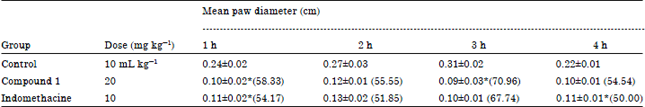
Male and female wistar rats (180-220 g) were used to test for the anti-inflammatory evaluations using the method of Winter et al. (1962) as modified by Neimeeger et al. (1964). The rats were divided into three groups of five animals each. Group 1 animals received 10 mL kg-1 5% tragacanth solution (control) while groups 2 and 3 were given 20 mg kg-1 of compound 1 and 10 mg kg-1 indomethacine. One hour after the administration, 0.1 mL of freshly prepared 1% w/v carrageenan suspension in normal saline was injected into the sub-plantar region of the left hind paw of each animal. The paw diameter was measured with the aid of a vernier caliper at 0, 1, 2, 3 and 4 h following the injection of carrageenan. The percentage inhibition was calculated from the following equation:
Where:
| Do | = | Paw diameter of the rat just before the administration of carrageenan (0 h) |
| Dt | = | Paw edema of the rat after the administration of carrageenan at different time interval |
Percentage inhibition of edema is proportional to anti-inflammatory activity.
Ethical approval was obtained from the Faculty’s ethical review committee on the use of laboratory animals.
Statistical analysis: Results obtained are presented as Mean±SEM. Statistical analyses were performed with the one-way analysis of variance (ANOVA) followed by the Duncan multiple range test. p<0.05 was considered significant.
RESULTS AND DISCUSSION
Compound 1 (Fig. 1) was obtained as an off-white crystalline substance (34 mg) and had an Rf of 0.69 (Chloroform-ethyl acetate; 9:1). The 1H-NMR (CDCl3, 500 MHz); (δ ppm): 7.2 (1H,singlet) 7.1 (2H, d, 8.5 Hz, H-2 and H-2'), 6.8 (2H, d, 8.5 Hz, H-3 and H-3'), 6.8 (2H, d, 8.5 Hz, H-5 and H-5'), 7.1 (1H, d, 8.5 Hz, H-6 and H-6'), 4.5 (2H singlet) 3.8 (3H, singlet, -OCH3). 13C-NMR (CDCl3, 125 MHz): 114 (C-1), 114.27 (C-2), 128.99 (C-3), 159.35 (C-4), 114.27 (C-5), 128.99 (C-6), 48.15 (C-7), 189 (C-8) 55.31 (-OCH3), 114 (C-1'), 114.27 (C-2'), 128.99 (C-3'), 159.35 (C-4'), 114.27 (C-5'), 128.99 (C-6'), 48.15 (C-7'), 55.13 (-OCH3). HR-EI-MS m/z: 316. 1201 (calculated for C17H20N2O2S, 316.2310).
The proton NMR showed that all the aromatic protons appeared as doublet while the singlet signals were attributable to the protons of the attached methoxy groups. The 13CNMR revealed the presence of seventeen carbon signals classified as two methyls, two methylenes, eight methines and five quaternary carbons. The two protons doublets each with double integrations in the aromatic region resonating at δH 7.1 and δH 6.8 were inferred from the HMBC interactions of H-2 and H-3 with C-7 at δC 48.15 and with methoxy carbon at δC 55.13, respectively. The same interactions were observed for H-2' and H-3' with C-7'. The double integrations of all types of protons suggested a dimeric structure for this compound. with a chiral centre at carbon position 8. The two methoxy groups with δH 3.8 were confirmed at C-4 and C-4'. The two aromatic quaternaries were confirmed at 1 and 1' through their HMBC interactions with NH protons.
Based on the NMR and mass spectrometry data, it can be concluded that compound 1 is a thiourea compound identified as N, N, di (4-methoxybenzyl) thiourea.
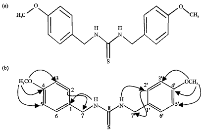 | |
| Fig. 1(a-b): | Structure of N, N-di (4-methoybenzyl) thiourea |
| Table 1: | Anti-inflammatory activity of methanol extract of root of Rhaphiostylis beninensis |
 | |
| Each value represent Mean±SEM of 5 rats. Values in parenthesis represent % inhibition of inflammation. *Significant at p<0.05 | |
| Table 2: | Anti-inflammatory effect of N, N, di (4-methoxybenzyl) thiourea |
 | |
| Each value represent Mean±SEM of 5 rats. Values in parenthesis represent% inhibition of inflammation. *Significant at p<0.05 | |
Compound 1 was observed to elicit remarkable anti-inflammatory activity at a dose of 20 mg kg-1 (Table 1). At 1 h the mean paw diameter observed in the control animals was 0.24±0.02 cm, whereas, the animals treated with 20 mg kg-1 of compound 1 had a mean paw diameter of 0.10±0.02 implying a 58.33% inhibition. At 3 h, the control animals had a mean paw diameter of 0.31±0.02 while the animals treated with the compound had a mean paw diameter of 0.09±0.03 cm indicating a 70.96% inhibition of inflammation. The activity of the compound was observed to be a little higher than that of indomethacine at 10 mg kg-1 (Table 2).
Carragenan is a standard phlogistic agent of choice for testing for the anti-inflammatory activity of drugs because it is considered non-toxic and devoid of apparent systemic effect (Chakraborty et al., 2004). It is also considered as a good example of an in vivo model as the procedure involves various mediators of inflammation mechanisms. Smith et al. (1974) postulated that carragenan induced edema shows three distinct phases with each corresponding to the involvement of particular mediators. Phase one is characterized by the release of histamine and 5- hydroxyltryptamine which occurs between 1-2 h. This is followed by the second phase that is characterized by the release of kinins and this occurs in the 3rd h of the inflammatory response. The last phase occurs between the 4th and 5th h and involves the release of prostaglandin. The fact that compound 1 significantly reduced the magnitude of the edema formed in the 1st h (compared to the control) showed that it effectively reduced the concentrations of histamine and 5-hydroxyltryptamine produced. The compound also significantly inhibited the release of kinins and further inhibited the inflammatory effects of the carragenan by blocking the release of prostaglandin. It can be inferred that the compound exhibited the highest effect at the 3rd h which corresponded to the release of kinin. The inhibition of inflammation observed with the compound (20 mg kg-1) was found to be slightly higher than that obtained with indomethacine (10 mg kg-1). The occurrence of the compound in nature was first reported in Pentadiplendra brazzeana (El-Migirab et al., 1977) and its occurrence and anti-inflammatory properties in this plant are reported for the first time.
Thioureas have been reported to possess anti-oxidant activity (Kajimoto and Murakami, 1998). A high level of correlation has been reported for anti-oxidant and anti-inflammatory activity and this is believed to be due to the fact that anti-oxidants exact their action by scavenging for free radicals and some of these free radicals such as nitric oxide have been implicated as pro inflammatory agents (Kulkarni et al., 2008). Although, the crude extract of the plant and compound 1 are yet to be evaluated for probable antioxidant properties, compound 1 can be said to one of the major anti-inflammatory agents in the root of R. beninensis.
ACKNOWLEDGMENT
The authors particularly, Ayinde B.A acknowledge the fellowship offered to him by NAM-ICCBS and also ICCBS, University of Karachi, Pakistan for the spectroscopic analyses.
REFERENCES
- Chakraborty, A., R.K.B. Devi, S. Rita, K. Sharatchandra and T.I. Singh, 2004. Preliminary studies on antiinflammatory and analgesic activities of Spilanthes acmella in experimental animal models. Indian J. Pharmacol., 36: 148-150.
Direct Link - El-Migirab, S., Y. Berger and J. Jadot, 1977. Isothiocyanates, thiourees et thiocarbamates isoles de Pentadiplandra brazzeana. Phytochemistry, 16: 1719-1721.
CrossRefDirect Link - Kulkarni, R.R., A.D. Virkar and P. D'mello, 2008. Antioxidant and antiinflammatory activity of Vitex negundo. Indian J. Pharm. Sci., 70: 838-840.
Direct Link - Lasisi, A.A., O.M. Folarin, E.O. Dare, O.A. Akinloye and M.O. Fisuyi, 2011. Phytochemical, antibacterial and cytotoxic evaluation of raphiostylis beninensis [Hook F. Ex Planch] stem bark extracts. Int. J. Pharma Bio Sci., 2: 489-495.
Direct Link - Smith, M.J.H., A.W. Ford‐Hutchinson, P.N.C. Elliott and J.P. Bolam, 1974. Prostaglandins and the anti‐inflammatory activity of a human plasma fraction in carrageenan‐induced paw oedema in the rat. J. Pharm. Pharmacol., 26: 692-698.
CrossRefDirect Link - Winter, C.A., E.A. Risley and G.W. Nuss, 1962. Carrageenin-induced edema in hind paw of the rat as an assay for anti-inflammatory drugs. Exp. Biol. Med., 111: 544-547.
CrossRefPubMedDirect Link