
S.M. Nourazarian
Department of Clinical Biochemistry, Faculty of Medicine, Tehran University of Medical Sciences, Tehran, Iran
G. Irajian
Department of Bacteriology, Faculty of Medicine, Tehran University of Medical Sciences, Tehran, Iran
M. Najafi
Department of Clinical Biochemistry, Faculty of Medicine, Tehran University of Medical Sciences, Tehran, Iran
M. Nourbakhsh
Department of Clinical Biochemistry, Faculty of Medicine, Tehran University of Medical Sciences, Tehran, Iran
J. Maleki
Department of Clinical Biochemistry, Faculty of Medicine, Tehran University of Medical Sciences, Tehran, Iran
M. Shabani
Department of Clinical Biochemistry, Faculty of Medicine, Tehran University of Medical Sciences, Tehran, Iran
Pakistan Journal of Biological Sciences
Year: 2012 | Volume: 15 | Issue: 8 | Page No.: 374-379


ABSTRACT
Diabetes mellitus is a serious health problem in the world and about 20 to 40% of the patients are being affected with diabetic nephropathy. The anti diabetic property of Lactobacillus reuteri (L. reuteri) has been reported. The study designed to investigate the effect of L. reuteri on the expression of BMP-7 and TGF-β genes, the two basic factors involved in diabetic nephropathy. This experimental study was carried out in two months. For this goal thirty male Wistar rats with 12 weeks old and 200±50 g weight was divided into 5 groups, each consisting six rats. (1) Non diabetic, (2) Untreated diabetic, (3) Diabetic rats fed with L. reuteri, (4) Diabetic rats treated with insulin (4-5 U/kg/day), (5) Non diabetic rat fed with L. reuteri. Diabetes in the was induced single intraperitoneal (i.p.) injection of streptozotocin (50 mg kg-1 b. wt). The L. reuteri 1x10⁸ Colony Forming Units (CFU) were administered via oral gavages. After two months rats were anesthetized and blood samples collected. Serum nitric oxide (NO) levels were determined by a chemiluminescence method using NO analyzer and serum glucose by glucose oxidize method. The expression of BMP-7 and TGF-β genes in the rat’s kidneys were determined by real time PCR. Results showed that BMP-7 gene expression was increased in diabetic rats that fed with L. reuteri, while TGF-β gene expressions were decreased. Histopathological study showed that administration of L. reuteri (1x10⁸ CFU/rat/day) significantly reduced kidney fibrosis and increased meaningfully NO levels in diabetic rats fed with L. reuteri. It was concluded that L. reuteri increase BMP-7 gene expression and may prevents from renal damage by oxidative stress by increasing antioxidant capacity.
PDF Abstract XML References Citation
Received: June 14, 2012;
Accepted: July 20, 2012;
Published: September 03, 2012
How to cite this article
S.M. Nourazarian, G. Irajian, M. Najafi, M. Nourbakhsh, J. Maleki and M. Shabani, 2012. The Effect of Lactobacillus reuteri on Bone Morphogenetic Protein-7 and Beta Transforming Growth Factor Gene Expressions in Streptozotocin-induced Diabetic Rat’s Kidneys. Pakistan Journal of Biological Sciences, 15: 374-379.
DOI: 10.3923/pjbs.2012.374.379
URL: https://scialert.net/abstract/?doi=pjbs.2012.374.379
DOI: 10.3923/pjbs.2012.374.379
URL: https://scialert.net/abstract/?doi=pjbs.2012.374.379
INTRODUCTION
Diabetes is a lasting and common complication all over the world. Due to the epidemiological studies, the researchers believe that 1.5 million of Iranian population carry this disease (Akbarzadeh et al., 2007) and about to 20 to 40% of these patient will be affected with diabetic nephropathy (Bottinger and Bitzer, 2002). Hyperglycemia is identified as a major factor on the extension of effects on the diabetic individuals (Dolan et al., 2005). Diabetic Nephropathy (DN) is the main causes for final stage of kidney debases in the USA and west. At the initial stage of extension of (DN), kidney tubules shows the over expression of TGF-β (Wang et al., 2001). Due to lack of understanding of pathway, TGF-β is a mediators that causes fibrosis of glomerulosclerosis and tubulointerstitial (Dronavalli and Duka, 2008). On the other hand, expression of BMP-7 as an antagonist of TGF-β could be effective on the investigation of DN (Helder et al., 1995; Fioretto et al., 2006). BMP-7 is a member of superfamily of TGF-β that plays an important role on the growth of kidney (Helder et al., 1995; Dolan et al., 2005; Fioretto et al., 2006). Due to expression of different BMP proteins during kidney organogenesis, the indicated evidences shows that only the BMP-7 is efficient for formation of kidney (Simic and Vukicevic, 2005). The tubular expression of BMP-7 in animal models with kidney deficiency is decreased (Simon et al., 1999; Wang et al., 2001). BMP-7 causes the deactivation of TGF-β profibrotic activity and the findings indicate this case that the BMP-7 in the process of the change of Epithelial to Mesangial Transition (EMT) that cause the creation of fibroblasts, has deactivation roles (Weiskirchen et al., 2009). In general, the metabolic activity in the kidney nephrons, produce a large amount of Reactive Oxygen Species (ROS) that commonly balanced with antioxidant enzymes and free radical sweeping systems. ROS creates negative biological effects in the body, includes lipids peroxidation of cell wall, oxidation of proteins and finally DNA defects. Unfortunately, hyperglycemia produces ROS with unbalancing that seems it is produced in the mitochondria (Nishikawa et al., 2007). Hyperglycemia causes increase of oxidative stress, even before appearance of DN signs in the individuals. In the DN the concentration of defected markers of DNA increase and tissue analysis of kidney biopsy samples showed increase production of glycoxidation and lipid oxidation products in matrix of mesangial and kidney glomeruli. These effects are detected in a lower amount in the none diabetic people (Suzuki et al., 1999; Vasavada and Agarwal, 2005). One of the mechanisms that infused performance of endothelial is increase of oxidative stress that interference with NO production (Rodriguez-Manas et al., 1998). Recently studying on the HG-treated mesangial cells, the antioxidant role of BMP-7 has been reported (Yeh et al., 2009) and it has been suggested that probiotic supplements causes to normality of glucose rate in the diabetic rats (Yadav et al., 2007, 2008). The probiotic bacteria defined as live microorganism, when are used in sufficient amount could cause the health of host (Andreasen et al., 2010). The probiotic bacteria produces exopolysaccharide as a layer connected to the cells surface or feed it to the environment. Some studies has shown that these exopolysaccharide has some roles like, stimulation of immune system, anti tumor activity and also phosphate groups of exopolysaccharide has important roles on the activation of macrophages and lymphocytes (Zahedi et al., 2011). With attention to the role of hyperglycemia in increase secretion of inflammatory factors (Pickup, 2004; Shoelson et al., 2006), the study designed to investigate the effects of lactobacillus reuteri PTCC1655 on serum levels of glucose, no and gene expression of BMP-7 and TGF-β in streptozotocin-induced diabetic rats.
MATERIALS AND METHODS
Preparation of L. reuteri PTCC1655: Lactobacillus reuteri PTCC1655 (DSM 20016, ATCC23272) was purchased from Persian Type Culture Collection (PTCC), Tehran, Iran. The microorganism was regenerated in to Man Rogosa Sharpe (MRS) broth from the lyophilized vial cultured in anaerobic jar Containing (80% N2, 10% CO2, 10% H2), incubated for 48 h at 37°C and preparation (1x10⁸ CFU/rat/day).
Animal studies: Twelve week-old Male Wistar rats (200±50 g) were received from Pasteur Institute of Iran. Prior ethical permission was obtained from the animal care committee of Tehran University of medical sciences. The study comprised of five groups i.e. a non-diabetic (N), diabetic untreated (D), diabetic rat fed with L. reuteri (1x10⁹ CFU/rat/day) (D+L), diabetic treated with Insulin (4-5 U kg day-1) (D+I), non-diabetic rats fed L. reuteri (1x10⁹ CFU/rat/day) (N+L). Each group comprising of six (n = 6) rats. The animals were housed in individual cages, given free access to water and standard chow ad libitum. Diabetes was induced by single intraperitoneal (i.p.) injection of streptozotocin (STZ) at 50 mg kg-1 b. wt. in sodium citrate buffer (0.01 M, pH 4.5). Blood glucose levels were monitored 48 h later and periodically thereafter by ACCU-CHEK advantage blood glucose monitor (Roche Group, Indianapolis, IN, USA), by rat-tailed blood sampling. Rats with blood glucose levels above 250 mg dL-1 were considered as diabetic. All rats had unrestricted access to food/water and were maintained for 8 weeks in accordance with institutional animal care and use committee procedures of Tehran University.
Kidney preparation: After 60 days, rats were sacrificed and kidney were rapidly removed and weighed, fixed in 10% formalin, cleared in xylene embedded in paraffin and cut at 4 μm and stained with hematoxylin and eosin, Periodic Acid Shiff (PAS) and trichrome.
Measurement of glucose: All the rats were anesthetized with ether and blood was taken from the heart and serum was separated. Blood Glucose was measured by the glucose kit based on glucose-oxidize method.
Nitric oxide measurement: NO was quantified by a chemiluminescence method using NO Analyzer (CLD88 ECO MEDICS AG Switzerland), a high-sensitive detector for measuring NO, based on a gas phase chemiluminescent reaction between NO and ozone:
The emission of a photon from electrically excited nitrogen dioxide is in the red and near-infrared region of the spectrum and it is detected by a thermoelectrically cooled red-sensitive photomultiplier tube.
Total RNA extraction: Total RNA were extracted from kidney tissues by miRCURY™ RNA isolation kit (Exiqon, Vedbaek, Denmark) according to the manufacturer’s instructions.
| Table 1: | Primer used for real-time PCR |
 | |
Extraction procedure were applied to 50-70 mg tissue, that yield near 500 μg μL-1 purified cellular total RNA after DNase I (RNase-free) (Fermentas GmbH, Leon-Rot, Germany) treat of RNA trapped on column.
Quality and amount of total RNA measured by Nanodrop 1000 spectrophotometer (Thermo scientific, Wilmington, DE 19810 USA).
cDNA synthesized real time PCR: For cDNA synthesis form mRNA, 3 μg of total RNA was reverse transcribed by Revert Aid Premium Reverse Transcriptase (Fermentas GmbH, Leon-Rot, Germany) according to manufacture instructions.
Real-time PCR was performed on the Bio-RadiQ 5 detection system, using SYBR Green master mix (Exiqon, Vedbaek, Denmark) and designed primers for BMP-7 and TGF-β and GUSB as housekeeping gene, sequences derived from (www.ncbi.nlm.nih.gov) and primer design was performed with the Beacon designer software.
The PCR conditions for amplification of mRNA was initial denaturation and enzyme activation at 95°C for 10 min, followed by 45 cycles of 95°C for 10 sec and combined annealing and extension 60°C for 60 sec. The Ct amount of PCR products was normalized with housekeeping gene GUSB (Glucuronidase beta). A standard curve was generated from dilution series constructed from housekeeping gene pooled cDNA. Relative changes in gene expression were quantified using ΔΔCT method (Yuan et al., 2006). Real-time PCR assays were carried out in triplicate and each experiment was performed at least three times. Primer (invitrogen) used for real-time PCR are listed in Table 1.
Statistical analysis: Statistical analyses were performed using SPSS version 16.0 software. Analysis of variance (ANOVA) was used to compare means in different groups. The difference of experimental data was analyzed by using student t test. Data were expressed as the Mean±SE. p<0.05 was considered to be significant.
RESULTS
Effect of L. reuteri on BMP-7 and TGF-β expression: To investigate the effect of L. reuteri on BMP-7 expression the mRNA levels were analyzed with real-time PCR.
 | |
| Fig. 1(a, b): | The expression levels of TGF-β and BMP-7 in kidney of studied rats, (a) Lactobacillus reuteri reduced TGF-β mRNA expression but in groups D and D+1 the expression of TGF-β significantly induced, (b) In diabetic rats that administrated with Lactobacillus reuteri the expression of BMP-7 significantly induced but in groups D and D+2 BMP-7 expression decreased (p<0.05). The TGF-β and BMP-7 levels were analyzed to GUSB RNA levels. Values are Mean±SE from least three separate experiments performed in triplicate, *Significance between control and other group |
Fig. 1 showed the effect of L. reuteri on expression level of TGF-β and BMP-7 in kidney of studied rats.
 | |
| Fig. 2: | The effect of Lactobacillus reuteri on serum levels of NO in studied rats. Values are Mean±SD (n = 6 per group), In each group *p<0.05: **p<0.05 compared to value of normal rats |
 | |
| Fig. 3: | Effect of L. reuteri on serum glucose, *p<0.05 vs. normal, p<0.05 vs. diabetic groups |
Following two months administration of L. reuteri (1x10⁸ CFU/rat/day), the diabetic rats (D) were found to have an increase in TGF-β expression (p<0.05). However, in diabetic rats that administration of L. reuteri (1x10⁸ CFU/rat/day) (D+L) the expression of TGF-β significantly decreased (Fig. 1a). The BMP-7 expression level increased in the kidney of STZ-diabetic rats that fed L. reuteri (p<0.05), but in insulin treatment (D+I) and diabetic groups (D) BMP-7 expression significantly decreased (Fig. 1b).
Effect of L. reuteri on NO level: Studying the effect of L. reuteri on NO level in rat’s serum, It was found that the NO levels in diabetic rats were lower than normal groups (N and N+L), however after administration of L. reuteri (1x10⁸ CFU/rat/day) the increase in levels of NO were increased in (D+L) group than in the other diabetic groups(D and D+I) (p<0.05) (Fig. 2).
Effect of L. reuteri on serum glucose levels: The effect of L. reuteri on serum glucose levels in STZ-induced diabetic rats are shown in Fig. 3. Our results showed that STZ-induced diabetic rats had increased serum glucose (>250 mg mL-1) than normal rats. In diabetic groups that administration of L. reuteri (1x10⁸ CFU/rat/day) serum glucose levels decreased but not significantly than control group. Also in insulin treatment groups the glucose levels significantly decreased.
Effect of L. reuteri on kidney fibrosis: The optical microscopy stained with H and E, PAS and Masson’s trichrome showed in (Fig. 4). The renal tissue stained by Periodic Acid Schiffs (PAS) solution appeared normal glomeruli in the kidney of non diabetic groups (N and N+L).
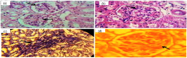 | |
| Fig. 4: | Kidney structures of STZ-induced diabetic rates after 60 days administration of L. reuteri (1x10⁸ CFU/rat/day), (a) Normal views of glomerulus of rats kidney that administration of L. reuteri (1x10⁸ CFU/rat/day) (H and E x100), (b) Hypercellularity and congestion in glomerular in diabetic rats no fed L. reuteri (H and E), (c) Interstitial fibrosis in diabetic rat’s kidney (Trichrome x400) and (d) Basement membrane thickness increasing in diabetic rats (PAS x100) |
In diabetic groups (D and D+I) the increase in glomerular epithelial cells and glomerular basement membrane thickening in glomerular capillary, to a severe extent was shown. Also some glomeruli showed glomerular atrophy with thickening of bowman capsule in tubular area, vacuolization of epithelium of proximal tubular and interstitial edema with increase in fibroblast and macrophage cells were observed. In Masson’s trichrome staining, often interspersed fibrosis seen in the tubules, comparable to normal groups. In STZ-induced diabetic rats that fed with L. reuteri (1x10⁸ CFU/rat/day) the kidney sections showed no pathological changes.
DISCUSSION
It is known that pathogenesis of DN is associated with abnormalities of renal NO generation (Kuno et al., 2011). The inhibition of mesangial cell growth is another effect of No. Nitric oxide also is a strong repressor of connective tissue growth factors that is important to development of interstitial glomerular fibrosis. It has been shown that hyperglycemia promoted oxidative stress in nephritic tissues, eventually leading to renal injury in diabetes (Chang et al., 2011). Present results suggested that when STZ-induced diabetic rats fed with L. reuteri (1x10⁸ CFU/rat/day) for 60 days, extension of hypercellularity, congestion in glomerulus and thickening of glomerular basement membrane did not occur, comparing to diabetic group. We also found the normal glomerulus and no pathological changes in the group that fed with insulin and normal groups. In diabetic rats in the absence of excessive NO mesangial matrix production will occur associated with type I collagen accumulation (Chiarelli et al., 2000). In this study, comparing with the control group the plasma level of NO was significantly decreased in diabetic group. Increased the Reactive Oxygen Molecules (ROMs) and decrease antioxidants may be responsible for decrease of NO production (Onozato et al., 2002). In the STZ-induced diabetic rats increase of renal oxidative stress caused by hyperglycemia have been shown (Cheng et al., 2010). Changes the production of NO associate with glomerular hyper filtration and vascular permeability (Dooley et al., 2012). A decrease in oxidative stress plays important role on the improvement of renal function by recovering renal BMP-7 expression in STZ-DN rats. BMP-7 plays an anti oxidative role in mesangial cells (Yeh et al., 2009). Due to the antioxidant role of BMP-7, it seems that Lactobacillus reuteri with ability to increase expression of BMP-7, prevented the progression of renal fibrosis in diabetic rats. TGF-β and the receptor together constitute a major biologic signal inducing a switch toward a profibrotic cellular phenotype (Wang et al., 2003). Our result shows the increase of TGF-β expression in diabetic group of rats are associated with decrease of BMP-7 gene expression. We also find that the expression of BMP-7 gene increased in diabetic rats fed with L. reuteri 1x10⁸ CFU/rat/day). In this case, it seems that BMP-7 could prevent oxidative stress hyperglycemia in the diabetic rats.
CONCLUSION
Present results showed that BMP-7 expression in kidney and serum levels of NO decreased in STZ-induced diabetic rats. But in diabetic groups that fed with L. reuteri (1x10⁸ CFU/rat/day)the kidney expression of BMP-7 and serum levels of NO increased. Perhaps the protective role of L. reuteri on rats kidney maybe due to their antioxidant properties. So it may be suggested that L. reuteri or other beneficial bacteria increase BMP-7 gene expression and improve DN. Further studies are required to evaluate the effect of different species of probiotic bacteria on the DN.
ACKNOWLEDGMENTS
This work was fully supported by a research grand from the Tehran University of Medical Sciences, Faculty of Medicine for which authors are thankful. We are also grateful to the members of the Department of Biochemistry, Faculty of Medicine, for their contributions.
REFERENCES
- Akbarzadeh, A., D. Norouzian, M.R. Mehrabi, S. Jamshidi and A. Farhangi et al., 2007. Induction of diabetes by streptozotcin in rats. Indian J. Clin. Biochem., 22: 60-64.
Direct Link - Andreasen, A.S., N. Larsen, T. Pedersen-Skovsgaard, R.M. Berg and K. Moller et al., 2010. Effects of Lactobacillus acidophilus NCFM on insulin sensitivity and the systemic inflammatory response in human subjects. Br. J. Nut., 104: 1831-1838.
PubMed - Dooley, A., K.R. Bruckdorfer and D.J. Abraham, 2012. Modulation of fibrosis in systemic sclerosis by nitric oxide and antioxidants. Cardiol. Res. Practice.
Direct Link - Bottinger, E.P. and M. Bitzer, 2002. TGF-beta signaling in renal disease. J. Am. Soc. Nephrol., 13: 2600-2610.
PubMed - Chang, C.C., C.Y. Chang, Y.T. Wu, J.P. Huang, T.H. Yen and L.M. Hung, 2011. Resveratrol retards progression of diabetic nephropathy through modulations of oxidative stress, proinflammatory cytokines and AMP-activated protein kinase. J. Biomed. Sci., Vol. 18.
CrossRef - Chiarelli, F., F. Cipollone, F. Romano, S. Tumini and F. Costantini et al., 2000. Increased circulating nitric oxide in young patients with type 1 diabetes and persistent microalbuminuria: Relation to glomerular hyperfiltration. Diabetes, 49: 1258-1263.
CrossRef - Yeh, C.H., C.K. Chang, M.F. Cheng, H.J. Lin and J.T. Cheng, 2009. The antioxidative effect of bone morphogenetic protein-7 against high glucose-induced oxidative stress in mesangial cells. Biochem. Biophys. Res. Commun., 382: 292-297.
CrossRefPubMedDirect Link - Helder, M.N., E. Ozkaynak, K.T. Sampath, F.P. Luyten, V. Latin, H. Oppermann and S. Vukicevic, 1995. Expression pattern of osteogenic protein-1 (bone morphogenetic protein-7) in human and mouse development. J. Histochem. Cytochem., 43: 1035-1044.
PubMed - Yuan, J.S., A. Reed, F. Chen and C.N. Stewart Jr., 2006. Statistical analysis of real-time PCR data. BMC Bioinform., Vol. 7.
CrossRefDirect Link - Cheng, M.F., L.J. Chen and J.T. Cheng, 2010. Decrease of klotho in the kidney of streptozotocin-induced diabetic rats. J. Biomed. Biotechnol.
CrossRef - Pickup, J.C., 2004. Inflammation and activated innate immunity in the pathogenesis of type 2 diabetes. Diabetes Care, 27: 813-823.
CrossRefDirect Link - Wang, S.N., J. Lapage and R. Hirschberg, 2001. Loss of tubular bone morphogenetic protein-7 in diabetic nephropathy. J. Am. Soc. Nephrol., 12: 2392-2399.
PubMed - Shoelson, S.E., J. Lee and A.B. Goldfine, 2006. Inflammation and insulin resistance. J. Clin. Invest., 116: 1793-1801.
CrossRefDirect Link - Simon, M., J.G. Maresh, S.E. Harris, J.D. Hernandez, M. Arar, M.S. Olson and H.E. Abbound, 1999. Expression of bone morphogenetic protein-7 mRNA in normal and ischemic adult rat kidney. Am. J. Physiol., 276: F382-F389.
PubMedDirect Link - Suzuki, D., T. Miyata, N. Saotome, K. Horie and R. Inagi et al., 1999. Immunohistochemical evidence for an increased oxidative stress and carbonyl modification of proteins in diabetic glomerular lesions. J. Am. Soc. Nephrol., 10: 822-832.
PubMedDirect Link - Vasavada, N. and R. Agarwal, 2005. Role of oxidative stress in diabetic nephropathy. Adv. Chronic Kidney Dis., 12: 146-154.
CrossRefDirect Link - Zahedi, F., H.M. Nasrabadi, M.T. Ebrahimi, M. Shabani and H. Aboutalebi, 2011. The effect of Lactobacillus brevis isolated from Iranian traditional cheese on cutaneous wound healing in rats. J. Cell Anim. Biol., 5: 265-270.
Direct Link - Kuno, Y., M. Iyoda, T. Shibata, Y. Hirai and T. Akizawa, 2011. Sildenafil, a phosphodiesterase type 5 inhibitor, attenuates diabetic nephropathy in non-insulin-dependent Otsuka Long-Evans Tokushima Fatty rats 0. Br. J. Pharmacol., 162: 1389-1400.
CrossRef - Yadav, H., S. Jain and P.R. Sinha, 2008. Oral administration of dahi containing probiotic Lactobacillus acidophilus and Lactobacillus casei delayed the progression of Streptozotocin-induced diabetes in rats. J. Dairy Res., 75: 189-195.
CrossRefPubMedDirect Link - Yadav, H., S. Jain and P. Sinha, 2007. Antidiabetic effect of probiotic dahi containing Lactobacillus acidophilus and Lactobacillus casei in high fructose fed rats. Nutrition, 23: 62-68.
CrossRefPubMedDirect Link