
S.G. Moharram
National Institute of Oceanography and Fisheries, Kaiet Bay, Alexandria, Egypt
O.M. Wahbi
National Institute of Oceanography and Fisheries, Kaiet Bay, Alexandria, Egypt
Z.A. El-Greisy
National Institute of Oceanography and Fisheries, Kaiet Bay, Alexandria, Egypt
Pakistan Journal of Biological Sciences
Year: 2011 | Volume: 14 | Issue: 12 | Page No.: 668-681


ABSTRACT
The impact of diluted levels of polluted seawater from the Egyptian Mediterranean coast on reproductive, toxicological and hematological characteristics of Siganus rivulatus were determined. Mature fish were exposed to diluted levels of 7.5, 10 and 15 ml L-1 polluted seawater. Hematological changes after 4, 15 and 30 days of exposure were measured. Erythrocytes decreased (p<0.05) as well as Packed Cell Volume (PCV) and Hemoglobin (Hb), indicating anemia developed to hypochromic macrocytic anemia at end of experiment. Leukocytes, increased (p<0.05), indicating susceptibility of fish to infection and stress. Granulocytic leukocytes, neutrophil and eosinophil increased. While lymphocytes decreased. Blood parameters of exposed fish revealed compensatory responses. The increase in developing hemocytoplast and myelocytes emphasize the compensatory and defensive reaction of fish to polluted water. Exposure to polluted water levels has a detrimental effect on gonads development, altered endocrine haemostasis, testosterone and progesterone levels decreased in females (p<0.05). While in male, progesterone level increased (p<0.01). Necrosis of spermatogenic cells and atresia of developing oocytes are pronounced at levels of 10 and 15 ml L-1 polluted seawater. Also, has necrotic effect on fish organs. Vacuolation and necrosis occurs in liver and kidney. Melanomacrophage aggregates can be seen. Gills showed epithetial lifting and vascular widening. Results showed that, polluted water has serious consequences on Siganus rivulatus blood characteristics as well as organs cellular structure. It rendered fish anemiatic, altered reproductive hormones level, leading to necrosis of males spermatogenic cells and atresia of developing oocytes.
PDF Abstract XML References Citation
Received: April 16, 2011;
Accepted: October 26, 2011;
Published: November 17, 2011
How to cite this article
S.G. Moharram, O.M. Wahbi and Z.A. El-Greisy, 2011. Effect of Polluted Water from the Egyptian Eastern Mediterranean Coast on Reproductive, Toxicological and Hematological Characteristics of Siganus rivulatus . Pakistan Journal of Biological Sciences, 14: 668-681.
DOI: 10.3923/pjbs.2011.668.681
URL: https://scialert.net/abstract/?doi=pjbs.2011.668.681
DOI: 10.3923/pjbs.2011.668.681
URL: https://scialert.net/abstract/?doi=pjbs.2011.668.681
P>
INTRODUCTION
It is well established that many chemicals, both natural and man-made, may adversely affect wildlife, including decrease in fertility, disrupted hormone secretion or reproductive histopathology. Damaged ovarian follicles were obser ved in female mussels exposed to tetrabromodiphenyl ether (Aarab et al., 2006). Reduced gonad size and delayed sexual maturity have been observed in perch exposed to leachate from Swedish refuse dumps (Noaksson et al., 2005). Inhibition of ovarian development, reduced egg weight and increased atresia, were found in white perch (Richard et al., 2004) in areas affected by domestic and industrial effluents. Inhibition of oogenesis and spermatogenesis were found in medaka exposed to isoflavones (Yiannis et al., 2003). Gonad differentiation and development of salmon were affected by sewage effluents (Luis et al., 2002). Fish living in Bizerta lagoon, downstream of anthropogenic pollution have been found to exhibit an array of altered features in their reproductive development, including sperm abnormality and ovarian atresia (Louiz et al., 2009).
Hormonal disruption in turn, affects reproductive function. Population decline has been demonstrated in wild freshwater fish as a result of exposure to organochlorine chemicals (Jobling and Tyler, 2003). A reduction in reproductive capacity and plasma steroid levels has been established in female zebrafish exposed to contaminated sediment and Atlantic salmon after exposure to sublethal doses of diazinon (Linderoth et al., 2006; Wall, 2000). Reduction in level of testosterone and 17 α-20 β progesterone was found in cod and turbot exposed to crude oil and alkyl phenols (Skilton et al., 2006). Depressions in sex steroids level was detected in cyprinid fish from three sampling sites along the Kor river (Ebrahimi and Taherianfard, 2011). In laboratory experiments, fathead minnows exposed to methyl mercury over their life cycle exhibited a parallel range of effects on circulating steroids (Paul et al., 2006).
Histological investigations may reveal toxic effects of pollutants on fish. The gill is the primary target organ for the toxic action of pollutants. Impairment of the respiratory and the ionoregulatory functions may occur due to the structural changes of gill epithelia. Gill lamellae lesions, including interstitial edema, rupture of pillar cells and necrosis of lamellar epithelium have been reported when fish were exposed to industrial effluent (Wynn and Waring, 2002) and copper (Mazon et al., 2002a). Fish liver not only perform an important role in pollutant metabolism, but also susceptible to damages by their action. The principal lesions observed in the hepatic tissue were focal vacuolation, necrosis, increased pool of macrophages aggregates and changes in hepatocytes structure and distribution (Noaksson et al., 2005; Grinwis et al., 2000). Pathological evidence in fish kidney attributed to pollutants, include breakdown of tubules, vacuolation, haemopoietic tissue reduction and melanomacrophage aggregation (Handy and Grosell, 2002).
Environmental pollution may change the structure of red and white cells (Katalay and Parlak, 2004). Haematological parameters as Red Blood Cells (RBC); Haemoglobin content (Hb); Packed Cell Volume (PCV) and White Blood Cells (WBC) are most commonly used to diagnose fish health (Davidson et al., 2002). Decreased levels of RBC, Hb, PCV and increased WBC count, neutrophilia and eosinophilia were previously recorded in some species of marine and freshwater fish (Barsiene et al., 2006; Jee et al., 2006; Ovuru and Ekweozor, 2004).
Pollution in Egyptian Eastern Coast had a detrimental effect on its fauna and flora. The main source of pollution there is El Tabia pump station in Abu Qir region that receives mixed waste products about 20.55x10⁴ m3/day. Untreated effluents, are pumped directly to seawater at the western part of Abu Qir bay that represent the most polluted part of the bay. Beside domestic waste the station receive industrial waste product from fertilizer, electrical, paper manufacture and petroleum factories as well as agriculture drainage (Nessim et al., 1994).
This study aimed to investigate the effect of diluted levels of seawater in front of the outfall of El-Tabia Pump Station effluents on gonadal activity, hematological parameters, as well as histopathological changes in some vital organs of Siganus rivulatus.
MATERIALS AND METHODS
Healthy Siganus rivulatus fish of average length of 15.4 cm and average weight of 44.2 g were acclimatized for 2 weeks in aerated aquarium (80 L). They were fed on artificial diet (40% protein and 8.5% fat) throughout the holding and experimental period.
Samples of seawater in front of the outfall was collected in plastic containers (during 2008-2009) and stored at 4°C. Analysis was clearly demonstrated in Table 1.
| Table 1: | Characteristics of effluent-water mixture |
 | |
The medium lethal concentration at which 50% of the fish died was determined (96 hr-LC50) (Franson, 1980).
For the chronic test, seventy two fish were randomly distributed among 8 glass tanks for concentrations of 7.5, 10 and 15 ml L-1 of polluted water and blank (seawater 36 ‰) for 30 days (2 replicates for each concentration). Blood samples were collected at intervals of 4, 15 and 30 days by caudal excision and stabilized with 50 IU sodium heparin. Erythrocyte count (RBC), Packed Cell Volume (PCV), Haemoglobin content (Hb) and White Blood Cell count (WBc) were determined Lewis et al. (2006). Blood sugar level was determined using diagnostic system (COBAS INTEGRA). Blood films and kidney prints were fixed and stained using Giemsa stain.
Unheparinized blood was collected from control and treated fish at end of 30 days of exposure and left at 4°C for 24 h to clot, then sera were collected and analyzed for steroids level using Radio-Immunoassay test (Pantex I125 kit).
Biopsy specimens were collected from gills, liver, kidney and gonads, fixed in formal saline, dehydrated, embedded in wax (melting point 56°C), sectioned at 4 μ, stained with Hematoxylin and Eosin and examined according to Culling (1974).
Statistical analysis: Results were analyzed for comparison among different polluted water treatments using one way Analysis of Variance (ANOVA) according to Snedecor and Cochran (1982). Duncan's Multiple Range-Test (Duncan, 1955) was applied to determine statistically significant differences, possibilities less than 0.05 were considered statistically significant (p<0.05). Program of MSTAT-C was used to compute the statistical analysis (MStat-C, 1988).
RESULTS
The physico-chemical characteristics of the effluent-water mixture are summarized in Table 1.
| Table 2: | Mean haematological values recorded at different periods in Siganus rivulatus exposed to different effluent-water levels (n = 5) |
 | |
| Values are mean±SE of 3 replicates. Mean values in a row with different superscript letters are significantly different (p<0.05) | |
| Table 3: | White blood cells count(mean±SE) in Siganus rivulatus exposed to different effluent-water levels for different periods (n = 3) |
 | |
| Values are mean±SE of 3 replicates. Mean values in a row with different superscript letters are significantly different (p<0.05). Agranulocytes: Agran., Large lymphocytes: L.L., Small lymphocytes: S.L., Granulocytes: Gran., Neutrophils: Nt., Eosinophils: Es., Basophils: Bs | |
It is clear that all the parameters values were higher than the Egyptian Agriculture Ministry standards limits (Law 48/1982). The LC50 (96 h) was found to be 40 ml L-1 of effluent.
Impact of pollutant on blood constituent: No mortality was observed in the control and treatments throughout the experiment. Table 2 shows the mean values of the haematological indices recorded from exposing Siganus rivulatus to effluent- seawater levels (7.5, 10 and 15 ml L-1) for 4, 15 and 30 days. After 4 days of exposure only in concentration 15 ml L-1, the number of Red Blood Cells (RBC) undergone a significant decrease 2.32±0.13 compared to 2.65±0.20 (106/mm3) in control. Moreover, fish exposed to the tested concentrations 7.5,10 and 15 ml L-1 for 15 and 30 days had significant lower number of RBC compared with their counterparts control. A maximum decrease in RBC number was at concentration of 15ml L-1 with values of 2.01±0.02 and 1.81±0.22 (106/mm3) after 15 and 30 days of exposure compared to 2.58±0.13 and 2.52±0.12 (106/mm3) in control ones. The Packed Cell Volume (PCV) data show a lower significant values for all treated groups, compared to control at all exposure periods. The greatest decrease was at concentration of 15 ml L-1 with values of 37.30±1.10, 36.91±0.20 and 36.88±0.05% after 4, 15 and 30 days of exposure compared to 43.73±0.03, 42.89±0.16 and 42.79±0.18% in control .On the other hand, a significant decrease was noted in the Haemoglobin concentration (Hb) after 4 days of exposure, became more pronounced after 15 and 30 days for levels 10 ml L-1 (5.89±0.27 and 5.74±0.13 mg/100 mL) and 15 ml L-1 with values of (5.60±0.10 and 5.50±0.22 mg/ 100 ml) compared to 6.64±0.08 and 6.48±0.02 mg/100 mL in control, respectively. The haematocrit and haemoglobin values in the treated fish showed a progressive decrease with time, leading to anaemic condition that progressed to a macrocytic hypochromic type after 30 days of exposure in higher levels (10 and 15 ml L-1).
After 4 days of exposure to effluent- seawater levels of 7.5, 10 and 15 ml L-1, blood sugar level showed a significant increase (74.80±0.47, 78.60±1.89 and 94.90±2.19 mg/100 mL), respectively, compared to (64.50±0.70 mg/100 mL) in control. In contrast, blood sugar level decreased after 15 and 30 days upon exposure to 10 and 15 ml L-1. It became more pronounced in fish exposed to 15 ml L-1 of effluent-water mixture (57.50±0.31, 51.37±1.25 mg/100 ml) in comparison to (64.75±0.14, 63.35±0.14 mg/100 mL) the control. A significant increase was observed in the White Blood Cells count (WBC) in all treated groups, compared to control at all exposure periods. A maximum increase in WBC number was at concentration of 15 ml L-1 with values of 16.43±0.71 103/mm3 after 30 days of exposure compared to 13.80±0.10 103/mm3 in control .
Leukocyte profile is given in Table 3. Exposure to effluent-water levels resulted in decrease in Agranulocytes percentage as a result of Small Lymphocytes (SL) decrease and an increase in granulocytes percentage due to increase in Neutrophils (Nt) and Eosinophils (Es) numbers. After 4 days small lymphocytes decreased significantly at 15 ml L-1 with value of 63.0±01.00 to 65.00±2.20% in control. In contrary, there was a significant increase in both neutrophilic and eosinophilic numbers at concentration 10 ml L-1 with value of 19.50±0.50 and 7.50±0.16% compared to 17.50±0.50 and 6.00±0.15% in control, respectively. While at concentration 15 ml L-1 they had values of 18.50±0.10 and 7.50±0.20% compared to 17.50±0.50 and 6.00±0.15% in control. Changes were more pronounced at higher levels (10 and 15 ml L-1) and persisted to the end of experiment where lymphocytes recorded lowest value of 56.50±3.20% compared to 64.50±2.00% in control after 30 days of exposure at concentration 15 ml L-1. While (Nt) and (Es) recorded the highest value 20.50±1.20 and 10.00±0.11% at the same concentration and the same period of exposure compared to values of 18.50±0.20 and 6.50±0.22% in control, respectively.
Blood smears of control fish are shown in Fig. 1. Red blood cells are oval shaped cells with homogenous cytoplasm and round nucleus with compact chromatin materials. Blood smears of fish exposed for 4 days at concentration 15 ml L-1 are shown in Fig. 2, revealed an increases in number of neutrophil (Nt) Fig. 2a. Higher percentage of deformed erythrocytes, pear and tear shaped erythrocytes can be seen Fig. 2b, also swelling erythrocytes with fading cytoplasm, indicating a decrease in its haemoglobin content were noticed Fig. 2c. After 15 days of exposure (Fig. 3), blood film of fish at the higher effluent-water levels 10 and 15 ml L-1 showed abnormality. An increase in neutrophils number (Fig. 3a), large lymphocytes (L.L) (Fig. 3b) also senile erythrocytes (Fig. 3c) were shown in fish exposed to concentrations of 10 ml L-1. Blood film of fish after 30 days of exposure are seen in Fig. 4. Fish at exposure concentrations of 10 and 15 ml L-1 showed neutrophilia. Neutrophils of different developmental stages could be seen in Fig. 4a. Erythrocytes with ragged outlines were seen in blood smears of fish at effluent-water concentrations of 10 ml L-1 (Fig. 4b) and 15 ml L-1 (Fig. 4c).
Kidney prints of fish exposed to concentrations of 15 ml L-1 of effluent-water mixture for 4 days are represented at (Fig. 5). It revealed great numbers of Haemocytoblast (Hcb) which is the mother cell of all blood cells (Fig. 5a) and great numbers of Myelocyte (M), which is the mother cell of white blood cells (Fig. 5b). After 15 days of exposure Fig. 6, Lymphocytes (L) showed high numbers at concentration of 10 ml L-1 of effluent-water mixture (Fig. 6a).
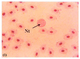 | |
| Fig. 1: | Blood film of control fish |
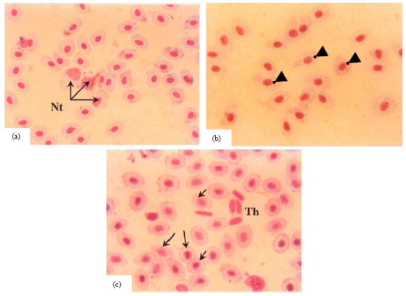 | |
| Fig. 2(a-c): | Blood film of fish exposed to 15 ml/l effluent for 4 days. (a) Increased neutrophils (Nt) thrombocytes (Th), (b) pear shaped erythrocytes [head arrows] and (c) Swelling erythrocytes (arrows) |
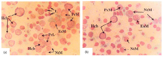
Also eosinophilic and neutrophilic myelocytes were higher in fish at 15 ml L-1 of effluent water mixture (Fig. 6b). At the end of the experiment, after 30 days of exposure (Fig. 7). Haemocytoblasts of variable sizes could be seen at concentrations of 10 ml L-1 of the effluent water mixture (Fig. 7a). The neutrophilic and eosinophilic myelocytes greatly increased at concentrations of 15 ml L-1 of the effluent water mixture (Fig. 7b).
Histopathological changes in fish organs
Gonads
Ovary: Histological changes in gonads of exposed fish occurred only at higher concentrations. Histological changes in ovaries are shown in Fig. 8.
 | |
| Fig. 3(a-c): | Blood film of fish exposed to 10 ml L-1 effluents for 15 days, notice (a) Neutrophils (Nt), (b) Eosinophils (Es), large lymphocytes (L) and (c) Senile erythrocytes (arrows) |
 | |
| Fig. 4(a-c): | Blood film of fish exposed to 15 ml L-1 effluents for 30 days, notice. (a) Neutrophilia and (b,c) Ragged vacuolated erythrocytes (arrows) |
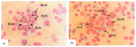 | |
| Fig. 5(a-b): | Kidney print of fish exposed to 15 ml L-1 effluent for 4 days Fig. 5a, b |
 | |
| Fig 6(a-b): | Kidney print of fish exposed to 10 and 15 ml L-1 effluent for 15 days, 10 ml L-1 for 15 days (a) 15 ml L-1 for 15 days |
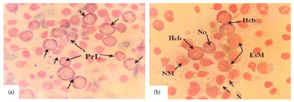 | |
| Fig. 7(a-b): | Kidney print of fish exposed to 10&15 ml/l effluent for 30 days, 10 ml/l for 30 days (a) 15 ml/l for 30 days, (b) Haemocytoblast (Hcb), Neutrophilic myelocyte (NtM), Basophilic myelocytes (BsM), Promyelocytes (PrM), prolymphocyte (PrL), Eosinophilic myelocyte (EsM), Lymphocyte (L), Neutrophil (N), Normoblast (No) |
In control fish, oocytes in the early perinucleolus and late perinucleolus stages are detected. Their cytoplasm is homogeneous and has purple color. The nuclei have distinct nucleoli and normal distribution of chromatin network (Fig. 8a). Ovary of fish exposed to different effluent-water levels showed abnormalities, including both nuclei and cytoplasm. The cytoplasm of early and late perinucleolus oocytes stained deep purple. The perinucleolus oocytes appeared as solid mass, while the late perinucleolus oocytes became atretic showing liquefied cytoplasm, deformed nucleoli and crumpled chromatin network at levels of 10 ml L-1 Fig. 8b and 15 ml L-1 of effluent-water mixture (Fig. 8c).
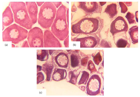 | |
| Fig. 8(a-b): | Cross section in ovary of fish after 30 days, (a) Control, (b) 10 ml L-1 and (c) 15 ml L-1of effluent-water mixture |
 | |
| Fig. 9(a-b): | Cross section in testis of fish after 30 days, (a) Control, (b) 10 ml L-1 and (c) 15 ml L-1 of effluent-water mixture |
Testis: Histological changes in testis are shown in (Fig. 9). In testis of control fish, seminiferous tubules have definite interlobular membrane. The spermatogenic cell layer posses primary and secondary spermatocytes (Fig. 9a). In males exposed to effluent-water levels of 10 ml L-1 the lobules have damaged membrane and spermatogenic cell layer (Fig. 9b). In case of 15 ml L-1, great necrosis of spermatogenic cell layer and complete disintegration of interlobular membrane were detected (Fig. 9c).
Sex hormones: No significant changes were found in levels of tested hormones in fish exposed to concentrations of 7.5 and 10 ml L-1 after exposure period of 30 days. Radio-immunoassay analysis of steroids level were shown in Table 4. Table 4 revealed that testosterone level in control females was higher than control males. Testosterone level has significantly decreased to 1.65±0.023 ng mL-1 in exposed females compared to 2.82±0.071 ng mL-1) in control females, while in exposed males it showed insignificantly decrease.
Progesterone level decreased significantly to 0.31±0.013 ng mL-1 in exposed females, it has value of 0.50±0.010 ng mL-1 in control females. In contrary, progesterone level in exposed males showed significantly increased value 0.50±0.018 ng mL-1 than control males 0.20±0.014 ng mL-1.
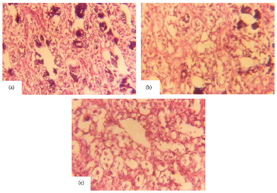
Liver: The changes existed in liver of fish due to exposure are seen in all effluent-seawater concentrations (Fig. 10). Liver of control fish showed the normal hepatic acini arrangement in cord of one layer thickening surrounding the bile duct. Hepatocytes are polygonal shaped cells with clear cellular border lines and rounded nuclei (Fig. 10a). In fish subjected to effluent- water concentration of 7. 5 ml L-1, the proper liver structure became obliterated, melanomacrophage aggregated, cytoplasmic vacuoles and pycnotic nuclei were observed (Fig. 10b). Hepatocytes necrosis occurred at concentrations of 10 and 15 ml L-1, cells were hypertrophied with vacuolated cytoplasm and pycnotic nuclei are seen at 15 ml L-1 effluent-seawater mixture (Fig. 10c).
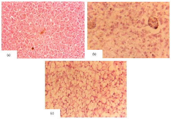
Kidney: The changes existed in kidneys of fish due to exposure are seen in all effluent-seawater concentrations (Fig. 11). Kidney of control fish showed uriniferous tubules with distinct epithelial cells and darkly stained nuclei. Haemopoietic tissue existed between the tubules Fig. 11a.
 | |
| Fig. 10(a-b): | Cross section in liver of fish after 30 days, (a) Control, (b): 7.5 ml L-1 and (c) 15 ml L-1 effluent-water mixture |
 | |
| Fig. 11(a-b): | Cross section in kidney of fish after 30 days, (a) Control, (b) 7.5 ml L-1 and (c) 15 ml L-1 effluent-water mixture |
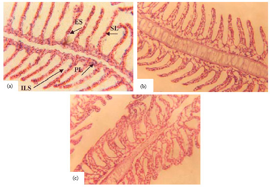 | |
| Fig. 12(a-c): | Frontal section in gills of fish after 30 days, (a) Control, (b) 10 ml L-1 effluent-water mixture and (c) 15 ml L-1 effluent-water mixture. Primary lamellae ( PL), Secondary lamellae (SL), Inter-lamellar space (ILS), Epithelial cells (ES) |
| Table 4: | Sex hormones levels in control and exposed Siganus rivulatus after 30 days of exposure to water-effluent level of 15 ml L-1 |
 | |
| Values are mean ±SE of 5 replicates. Mean values in a row with different superscript letters are significantly different (p<0.05) | |
At effluent-water concentration of 7.5 ml L-1, cells disintegration, vesicular highly staining cytoplasm, nuclear swelling, melanomacrophage aggregation and reduced haemapoietic tissue were detected in the kidney (Fig. 11b). At higher levels of 10 and 15 ml L-1, haemopoietic tissue was greatly reduced Severe destruction of tubular epithelium was observed at 15 ml L-1 effluent seawater mixture (Fig. 11c).
Gills: The changes existed in gills of fish due to exposure are seen in all effluent-seawater concentrations Fig. 12. Gills of control fish group exhibit normal architecture, consist of primary and secondary lamellae originated from either sides covered by epithelial cells. Underneath the epithelial covering of secondary lamellae is the supporting layer of pillar cells separated by lacunae where blood flow and gas exchange take place (Fig. 12a). Histological examination of fish gills at concentrations of 10 ml L-1 of effluent water mixture showed the most striking features being lamellar sinus widening and a pronounced lifting and desquamation of epithelium of secondary lamellae (Fig. 12b). At concentration of 15 ml L-1, complete denudation of cellular structure of lamellae occurred. Also, pillar supporting layer underneath was greatly reduced (Fig. 12c).
DISCUSSION
The present study revealed an interesting pattern of response of the haematological variables in exposed fish. In addition, different durations of exposure and different concentrations resulted in an anemic condition in fish, as shown from decreased RBC, PCV and Hb concentrations. From the present results, reduction in RBC count, PCV and Hb concentrations of tested fish compared to control may be due partly to the presence of heavy metals and other pollutants. These pollutants may cause RBC lysis . Previous studies had shown a decrease in RBC counts, relative PCV and Hb values when fish were exposed to Cassava effluent (Adekunle et al., 2007), diazinon (Svoboda et al., 2001) and textile dyes (Al-Sabti, 2000). These findings are in agreement with the present results. In contrary an increase in RBC, Hb and Ht value were recorded in African catfish on exposure to Gold crew (Alagoaa and Ekweozorb, 2009), to copper (Mazon et al., 2002b) and in Nile Tilapia and catfish exposed to lead (Al-Akela and Shamsia, 2000). Deformed RBCs were detected on exposure to cadmium (Witeska et al., 2006) and environmental pollution (Pacheco and Santos, 2002)
The distinct decrease in the level of Hb and PCV, clearly suggests haemodilution. In this instance, the change from normochromic anaemia after 5 days of exposure to hypochromic macrocytic anemia at higher levels of effluent- water mixture (10, 15 ml L-1), after 15 and 30 days of exposure is probably an adaptive response through the influx of immature erythrocytes from the haemopoietic tissue to the peripheral blood to make up to reduced RBC number and decreased haemoglobin concentration. These findings further support the hypothesis that haemodilution is a probable cause for decrease in Hb content in exposed fish. As a conclusion, the diluted effluent-water levels exert a profound influence on health condition of Siganus rivulatus after acute and chronic exposures. As a result, normochromic anemia has occurred, eventually leading to hypochromic, macrocytic anemic condition attributable to the swelling of RBC, haemodilution and impaired haemoglobin synthesis. Elahee and Bhagwant (2007) attributed this anemia to hypoxic conditions arising from gills degradation. Also, Kori-Siakpere and Ubogu (2008) reported normochronic anemia in Heteroclarias sp. due to zinc exposure.
In the present study, leukocytosis in Siganus rivulatus exposed fish may be attributed to increased leukocyte mobilization to protect the body against infection. This result is in agreement with previous studies of Prochilodus scrofa, due to copper exposure (Mazon et al., 2002b).
Neutrophils are matured leukocytes that control infections. The observed increase in percentage of neutrophils of the test fish may be due to the presence of pathogens in the polluted water as reflected by high BOD. This suggestion is supported by the increase in the WBC count in tested fish compared to control. WBC fights against infections, therefore the increase observed in the WBC of Siganus rivulatus in present work may be due to infection arising from pollution. Lymphopenia and both neutrophil and eosinophil ganulocytosis were reported in Oreochromis niloticus exposed to sublethal level of nickel (Alkahem, 1994). Also, lymphopenia was detected as a consequence of cypermethrin pesticide in Channa punctatus (Saxena and Seth, 2002). In contrary lymphocytosis was found on exposing Heteroclarias sp. to zinc by Kori-Siakpere and Ubogu (2008).
Exposure of Siganus rivulatus to the present effluent-water levels was found to have a detrimental effect on gonadal development. Histological changes in gonads were most pronounced at higher levels. Among the effects observed are inhibition of spermatogenesis, lobule boundary cells disintegration and atresia of early stages of oocytes. These morphological changes occurred in medaka exposed to sewage effluent (Yinnis et al., 2003.) and are qualitatively similar to those documented for Siganus rivulatus exposed to copper works effluent (Wahbi, 2004a). The present results predict atresia of developing oocytes. Similar effects have been demonstrated in white perch in areas affected by domestic and industrial effluents (Richard et al., 2004), in salmon chronically exposed to sewage effluent (Luis et al., 2002). Disintegration of lobule boundary cells and alteration of ovarian development were induced by lowering pH values in Diplodus sargus (Wahbi, 2004b)
In the present study, levels of testosterone and progesterone in both sexes have altered. This alteration is a direct result whether due to disruptions of the pituitary-gonadal axis or gonadal damage. As a result, it indirectly affects the development of oocytes and yolk formation, so fish can't reach maturity. Reduction in reproductive steroids were reported in cyprinid fish due to heavy metals (Ebrahimi and Taherianfard, 2011), zebra fish exposed to contaminated sediment (Linderoth et al., 2006) and white seabream exposed to lowered pH values (Wahbi, 2004b).
Histopathological alterations in exposed fish in this study were previously described by several authors (Olivaa et al., 2010; Wahbi et al., 2010; Van Dyka et al., 2009; Velmurugana et al., 2009; Fadel and Gaber, 2007), also following metals exposure (Giari et al., 2008; Figueiredo-Fernandes et al., 2007). Increased kidney necrosis was determined in Tilapia (Handy and Grosell, 2002). Macrophages aggregates in kidney of Prochilodus lineatus, subjected to a disturbed urban stream was described by Camargo and Martinez (2007). Vacuolation, necrosis and macrophages aggregates in liver of perch and flounder were detected (Noaksson et al., 2005; Grinwis et al., 2000) also liver necrosis were witnessed in Clarias gariepinus from polluted aquatic system (Marchand et al., 2009), due to zinc (Loganathan et al., 2006).
Gills alterations, as epithelial layer lifting, necrosis and frequent epithelial rupture were reported by Wynn and Waring (2002). In the present study, the changes recorded in gills of Siganus rivulatus, due to exposure, may represent a defense response, as it increase the distance across which pollutant must diffuse to reach the blood stream.
CONCLUSION
The effluent-water levels had negative direct or indirect effects on the haematological parameters, liver, kidney, gills and gonadal development in both males and females Siganus rivulatus. The blood parameters of Siganus rivulatus not only revealed cellular disturbances but also adaptive responses. Increase in leukocytes implies a mobilization of cell defense, although the reduction of the small lymphocytes percentage suggests a secondary effect of effluent. On the other hand, the decrease in red blood cell parameters (RBC, Hb and PCV) indicates an aneamic condition which gradually progresses upon prolonged exposure eventually cause hypochromic macrocytic anemia attributed to the swelling of the red blood cells, haemodilution and impaired haemoglobin synthesis. The increase in developing haemocytoplast and myelocytes emphasizes the compensatory and defensive reaction of fish to pollution. The pollutant altered reproductive hormones level, leading to necrotic changes in testis and atresia in oocytes. Also it has necrotic effect on fish organs.
REFERENCES
- Aarab, N., S. Lemaire-Gony, E. Unsuh, P.D. Hansen, O.K. Andersen and J.F. Narbonne, 2006. Preliminary study of responses in mussel (Mytilus edilus) exposed to tetrabromodiphenyl ether. Aquat. Toxicol., 78: S86-S92.
CrossRef - Adekunle, I.M., T.A. Arowolo, I.T. Omoniyi and O.T. Olubambi, 2007. Risk assessment of nile tilapia (Oreochromis niloticus) and African mud catfish (Clarias gariepinus) exposed to cassava effluent. Chem. Ecol., 23: 383-392.
CrossRefDirect Link - Alagoaa, K.J. and I.K.E. Ekweozorb, 2009. Sublethal effect of the dispersant Goldcrew on selected blood parameters of the African cat-fish Clarias gariepinus. Toxicol. Environ. Chem., 91: 339-343.
CrossRef - Al-Akela, A.S. and M.J.K. Shamsia, 2000. A comparative study of the toxicity of lead and its impact on the carbohydrate metabolism and some haematological parameters of cichlid fish, Oreochromis niloticus and catfish, Clarius gariepinus from Saudi Arabia. Toxicol. Environ. Chem., 74: 19-28.
CrossRef - Alkahem, H.F., 1994. The toxicity of nickel and the effects of sublethal levels on haematological parameters and behaviour of the fish, Oreochromis niloticus. J. Univ. Kuwait, 21: 243-251.
Direct Link - Barsiene, J., V. Dedonyte, A. Rybakovas, L. Andreikenaite and O.K. Anderson, 2006. Investigation of micronuclei and other nuclear abnormalities in peripheral blood and kidney of marine fish treated with crude oil. Aquat. Toxicol., 78: S99-S104.
CrossRef - Camargo, M.M.P. and C.B.R. Martinez, 2007. Histopathology of gills, kidney and liver of a Neotropical fish caged in an urban stream. Neotrop. Ichthyol., 5: 327-336.
CrossRefDirect Link - Ebrahimi, M. and M. Taherianfard, 2011. The effects of heavy metals exposure on reproductive systems of cyprinid fish from Kor River. Iran. J. Fish. Sci., 10: 13-26.
Direct Link - Elahee, K.B. and S. Bhagwant, 2007. Hematological and gill histopathological parameters of three tropical fish species from a polluted lagoon on the West Coast of Mauritius. Ecotoxicol. Environ. Saf., 68: 361-371.
CrossRef - Noaksson, E., M. Linderoth, U. Tjarnlund and L. Balk, 2005. Toxicological effects and reproductive impairments in female perch (Perca fluviatilis) exposed to leachate from Swedish refuse dumps. Aquat. Toxicol., 75: 162-177.
CrossRefDirect Link - Figueiredo-Fernandes, A., J.V. Ferreira-Cardoso, S. Garcia-Santos, S.M. Monteiro, J. Carrola, P. Matos and A. Fontainhas-Fernandes, 2007. Histopathological changes in liver and gill epithelium of Nile tilapia, Oreochromis niloticus, exposed to waterborne copper. Pesq. Vet. Bras., 27: 103-109.
CrossRefDirect Link - Giari, L., E. Simoni, M. Manera and B.S. Dezfuli, 2008. Histo-cytological responses of Dicentrarchus labrax (L.) following mercury exposure. Ecotoxicol. Environ. Saf., 70: 400-410.
CrossRefDirect Link - Jee, J.H., K.H. Park, Y.H. Keum and Y.C. Kang, 2006. Effect of 7, 12-dimethylbenz (a)anthracene on growth and haematological parameters in Korean rockfish, Sebastes schlegeli (Hilgendorf). Aquacult. Res., 37: 431-440.
CrossRef - Jobling, S. and C.R. Tyler, 2003. Endocrine disruption in wild freshwater fish. Pure Applied Chem., 75: 2219-2234.
CrossRef - Yiannis, K., C.B. Gordon, L.M. Tracy and D.M. Chris, 2003. Effect of isoflavones genestein in sewage on the gonadal development of japanese medaka, Oryzias latipes. Environ. Health Perspect., 111: 1158-1163.
PubMed - Linderoth, M., M. Ledesma and L. Balk, 2006. Sex steroids in the female zebrafish effects of cyproterone acetate and leachate-contaminated sediment extract. Aquat. Toxicol., 79: 192-200.
PubMed - Loganathan, K., B. Velmurugan, J.H. Howrelia, M. Selvanayagam and B.B. Patnaik, 2006. Zinc induced histological changes in brain and liver of Labeo rohita (Ham.). J. Environ. Biol., 27: 107-110.
PubMedDirect Link - Louiz, I., M. Ben-Attia and O.K. Ben-Hassine, 2009. Gonadosomatic index and gonad histopathology of Gobius niger (Gobiidea, Teleost) from Bizerta lagoon (Tunisia): Evidence of reproduction disturbance. Fish. Res., 100: 266-273.
CrossRefDirect Link - Luis, O.B.A., L.S. Jack, G.I. Michael and H.D. Robert, 2002. Y-chromosomal DNA markers for discrimination of chemical substances and effluent effects on sexual differentiation and gonadal development in salmon. Environ. Health Perspect., 110: 881-887.
PubMed - Marchand, M.J., J.C. van Dyk, G.M. Pieterse, I.E.J. Barnhoorn and M.S. Bornman, 2009. Histopathological alterations in the liver of the sharptooth catfish Clarias gariepinus from polluted aquatic systems in South Africa. Environ. Toxicol., 24: 133-147.
CrossRef - Mazon, A.F., C.C.C. Cerqueira and M.N. Fernandez, 2002. Gill cellular changes induced by copper exposure in the South American tropical freshwater fish Prochilodus scrofa. Environ. Res., 88: 52-63.
CrossRefDirect Link - Mazon, A.F., E.A.S. Monteiro, G.H.D. Pinheiro and M.N. Fernandes, 2002. Hematological and physiological changes induced by short-term exposure to copper in the freshwater fish, Prochilodus scrofa. Braz. J. Biol., 62: 621-631.
CrossRefDirect Link - Olivaa, M., M.L.G. de Canalesa, M.C. Garridob and S. Salesb, 2010. Lindane toxicity range-finding test in Senegal sole (Solea senegalensis) juvenile: Note on histopathological alterations. Toxicol. Environ. Chem., 92: 915-926.
Direct Link - Kori-Siakpere, O. and E.O. Ubogu, 2008. Sublethal haematological effects of zinc on the freshwater fish, Heteroclarias sp. (Osteichthyes: Clariidae). Afr. J. Biotechnol., 7: 2068-2073.
Direct Link - Ovuru, S.S. and I.K.E. Ekweozor, 2004. Haematological changes associated with crude oil ingestion in experimental rabbits. Afr. J. Biotechnol., 3: 346-348.
CrossRefDirect Link - Pacheco, M. and M.A. Santos, 2002. Biotransformation, genotoxic and histopathological effects of environmental contaminants in European eel (Anguilla anguilla L.). Ecotoxicol. Environ. Saf., 53: 331-347.
CrossRefPubMedDirect Link - Drevnick, P.E., M.B. Sandheinrich and J.T. Oris, 2006. Increased ovarian follicular apoptosis in fathead minnows (Pimephales promelas) exposed to dietary methylmercury. Aquat. Toxicol., 79: 49-54.
CrossRefDirect Link - Kavanagh, R.J., G.C. Balch, Y. Kiparissis, A.J. Niimi, J. Sherry, C. Tinson and C.D. Metcalfe, 2004. Endocrine disruption and altered gonadal development in white perch (Morone americana) from the lower Great Lakes region. Environ. Health Perspect., 112: 898-908.
PubMedDirect Link - Saxena, K.K. and N. Seth, 2002. Toxic effects of cypermethrin on certain hematological aspects of fresh water fish Channa punctatus. Bull. Environ. Contam. Toxicol., 69: 364-369.
CrossRefDirect Link - Martin-Skilton, R., R. Thibaut and C. Porte, 2006. Endocrine alteration in juvenile cod and turbot exposed to dispersed crude oil and alkylphenols. Aquat. Toxicol., 78: S57-S64.
CrossRefDirect Link - Van Dyka, J.C., M.J. Marchanda, G.M. Pietersea, I.E.J. Barnhoornb and M.S. Bornmanb, 2009. Histological changes in the gills of Clarias gariepinus (Teleostei: Clariidae) from a polluted South African urban aquatic system. Afr. J. Aquat. Sci., 34: 283-291.
CrossRef - Velmurugana, B., T. Mathewsa and E.I. Cengizb, 2009. Histopathological effects of cypermethrin on gill, liver and kidney of fresh water fish Clarias gariepinus (Burchell, 1822) and recovery after exposure. Envir. Technol., 30: 1453-1460.
CrossRefDirect Link - Witeska, M., B. Jezierska and J. Wolnicki, 2006. Respiratory and hematological response of tench, Tinca tinca (L.) to a short-term cadmium exposure. Aquacult. Int., 14: 141-152.
CrossRef - Svoboda, M., V. Luskova, J. Drastichova and V. Zlabek, 2001. The effect of diazinon on haematological indices of common carp (Cyprinus carpio L.). Acta Vet. Brno, 70: 457-465.
Direct Link