
Benjaporn Buranrat
Faculty of Medicine, Mahasarakham University, Talad, Mueang, Maha Sarakham 44000, Thailand
LiveDNA: 66.35857
ORCID: 0000-0002-1554-6327
Maleeruk Utsintong
Division of Pharmacy and Technology, Department of Pharmaceutical Care, School of Pharmaceutical Sciences, University of Phayao, Phayao 56000, Thailand
LiveDNA: 66.38303
ORCID: 0000-0001-6721-0887
Journal of Pharmacology and Toxicology
Year: 2023 | Volume: 18 | Issue: 1 | Page No.: 1-9
DOI: 10.3923/jpt.2023.1.9


ABSTRACT
Background and Objective: Zanthoxylum rhetsa (Roxb.) DC are spices for Thai food and it has been used for the treatment of several diseases. However, the activity of Z. rhetsa oil against cancer has not been fully investigated. In this study, the constituents of Z. rhetsa oil were characterized and explored the cytotoxic effects in cervical cancer cells. Materials and Methods: Zanthoxylum rhetsa oil was attained by hydro-distillation and was considered the active compound by Gas Chromatography-Mass Spectrometry (GCMS). The oil was evaluated for antioxidant, cytotoxicity, colony formation, apoptosis and migration in the human cervical cancer HeLa cell line. Results: Twenty-eight known compounds were identified in oil, including monoterpenes, sesquiterpenes and hydrocarbon. The oil inhibited the HeLa cell proliferation with a low IC50 value and decreased colony formation. Significantly, the induction of early apoptotic cell death was induced by the oil and a higher percentage was indicated at 250 μg mL–1. The cell number was reduced after incubating with the oil for 24 hrs and morphological change was observed at 250 μg mL–1. The mechanism of cell death and apoptosis was detected in the induction of ROS formation and were significant at the dose of 100 and 250 μg mL–1. Finally, the oil stimulated anti-migratory activities of the cervical HeLa. Conclusion: Zanthoxylum rhetsa oil was demonstrated to induce cancer cell death, activate early apoptosis and suppress the migration ability of cervical cancer cells through stimulating ROS formation.
PDF Abstract XML References Citation
How to cite this article
Benjaporn Buranrat and Maleeruk Utsintong, 2023. Zanthoxylum rhetsa (Roxb.) DC Oil Suppresses the Proliferation, Activates Apoptosis, Inhibits Migration of Human Cervical Cancer Cells. Journal of Pharmacology and Toxicology, 18: 1-9.
DOI: 10.3923/jpt.2023.1.9
URL: https://scialert.net/abstract/?doi=jpt.2023.1.9
DOI: 10.3923/jpt.2023.1.9
URL: https://scialert.net/abstract/?doi=jpt.2023.1.9
INTRODUCTION
Cervical cancer is the most common cancer that is reported in the fourth woman cancer worldwide1. In the early process of diagnosis, the surgical method is started with anticancer drugs which allow patients to receive highly effective treatment. However, the prognosis of early cervical cancer is still poor2. The surgical procedure is effective for the treatment of this cancer following the active anticancer drugs and leads the patient to a longer survival rate. Nevertheless, the standard anticancer drug treatment is still toxic in normal cells and death and serious complications take place. Cervical cancer death rates may be due to invasion, angiogenesis, metastasis and drug resistance3. Therefore, we immediately require to discover new natural compounds with less toxicity and high efficacy for treating or preventing cervical cancer.
Reactive oxygen species (ROS) lead to a variety of human cancers and antioxidants or scavenging agents are used to negate them4. Plants and other natural sources are significant origins of the encounter of anticancer remedies. Zanthoxylum rhetsa (Roxb.) DC belong to the Rutaceae family found in the Northern part of Thailand, locally called “Makhwaen”. Various parts and many extracts of Z. rhetsa were reported in numerous actions. The stem bark has been shown to possess anti-inflammatory activity5, sunscreen6, anticancer7, antinociceptive and antidiarrheal activity8. The part of the thorn that is rubbed with the water can be applied to the chest area to reduce pain and increase breast milk in lactating women9. The seed had photoprotective property10, antioxidant11, antibacterial12 and anticancer13. The fruit had anti-inflammatory14, antibacterial and anticancer15-17 and the volatile from the pericarp of fruit had antioxidant, antibacterial, antidiarrheal and non-selective spasmolytic activity18. For cytotoxicity, Z. rhetsa oil in Thailand was studied only against lung16 and breast cancer cell lines17.
From the data obtained, lignans and alkaloids from Z. rhetsa bark play a crucial role to activate melanoma B cell death with the highest activity from methanol extraction7. For breast cancer demonstrated that purified proteins from the pericarp of Z. rhetsa inhibited cell proliferation with a low IC50 value, decreased cell colonies and activated apoptosis19. The apoptotic induction was observed by detecting DNA fragmentation correlating with the arrested cell cycle19. Next, Z. rhetsa oil had a high ability to suppress human breast cancer cell proliferation and cell viability in both estrogen-positive MCF-7 cells and estrogen-negative MDA-MB-231 cells with equally IC50 values16.
Nonetheless, so far Z. rhetsa oil has been little tested for its anti-cancer effects. In present study aimed to evaluate the Z. rhetsa oil effects from fresh fruit on the growth, apoptosis and migration in human cervical cancer HeLa cells with the underlying mechanism of action through investigation of intracellular ROS formation.
MATERIALS AND METHODS
Study area: Fresh fruit was gathered from Nan Province on December, 2017 which was identified and deposited at Forest Herbarium, Thailand with a voucher specimen (BKF No. 194816).
Chemicals: The DPPH and L-ascorbic acid were obtained from Sigma Chemical Co., USA. All other chemicals used were obtained from local suppliers.
Plant extraction: Six hundred grams of fresh fruits were ground and distilled with water for 6 hrs. The Z. rhetsa oil was then added with anhydrous sodium sulfate to draw out the water and stored at 4°C before analyses.
Antioxidant assay: Antioxidant activity was tested using DPPH as previously reported20. Various concentrations of the oils were incubated with methanolic DPPH solution. After 30 min in dark at room temperature, the absorbance was measured at 515 nm. Blank and positive control (ascorbic acid) were also measured. All the determinations were performed in triplicate. The scavenging ability of DPPH was expressed as 50% of the inhibitory concentration (IC50).
GCMS assay: Zanthoxylum rhetsa oil was performed on a GC-MS using a GC 7890A Agilent joined to a mass spectrometer MSD5975C (EI) Agilent. Capillary column 5MS (30 m×0.25 mm×0.25 μm) was used. The conditions were as follows: Injection inlet was 250°C, injection volume was 0.5 μL in the split ratio (50:1), helium was the carrier gas, flow rate was 1.0 mL min–1. The column oven was programmed from 60-240 (4°C min–1). The total run time was 60 min. The temperature of the ion source and quadrupole were 230 and 150, respectively. The electron ionization was set at 70 eV with a mass scan of 30-500 amu. The mass of each component was done using the database of the National Institute Standard and Technology (NIST).
Cell lines and cytotoxicity assay: HeLa human cervical cancer cell was purchased from the ATCC and grown n DMEM (Gibco, Thermo Fisher Scientific, Inc with 10% fetal bovine serum (BSA) and 1% antibiotics. For the cytotoxic assay, cells were cultured onto 96-well culture plates with 1×10⁴ cells per well for 24 hrs and then treated with the Z. rhetsa oil with various concentrations (0-250 μg mL–1) for 24, 48 and 72 hrs. After incubation periods, the cells were fixed, stained with 0.4% sulforhodamine B (SRB), washed with PBS buffer and solubilized with 10 mM Tris base buffer. The optical density was read by spectrophotometry at 540 nm.
Colony formation assay: The 500 cells per well were cultured in 6-well culture plates for 24 hrs and then treated with the Z. rhetsa oil at indicated doses (0-250 μg mL–1) for 24 hrs. After 10 days, cells were fixed and stained with 0.25% crystal violet. The cells in culture plates were washed, captured and colonies.
Apoptosis by flow cytometry: The 2×10⁵ cells per well were cultured in 6-well culture plates for 24 hrs and incubated with various concentrations (0-250 μg mL–1) of Z. rhetsa oil for 24 hrs. Next, cells were collected and exposed to Annexin V-FITC and PI solution (BD Biosciences, San Diego, CA, USA) for 15 min. The cells were measured the apoptotic cells by flow cytometer (BD Biosciences, San Jose, CA, USA) using BD Accuri C6 Plus software.
Acridine Orange and Ethidium Bromide (AO/EB) staining assay: The 2×10⁵ cells per well were cultured in 6-well culture plates for 24 hrs and incubated with various concentrations (0-250 μg mL–1) of Z. rhetsa oil for 24 hrs. After that, cells were collected and exposed to AO/EB dye (1 μL AO/EB) for 15 min at room temperature. Then, the apoptotic cells were captured by an inverted fluorescence microscope (20× magnification, Olympus Corporation, USA).
Reactive oxygen species assay: The HeLa cells approximately (2×10⁵ cells per well) were plated in 6-well plates for 24 hrs. Next, the cell was exposed to various concentrations of Z. rhetsa oil (0-250 μg mL–1) for 24 hrs. Then cells were harvested and incubated with DCF-DA (25 μM) in DMEM medium for 30 min in dark at 37°C incubator. The fluorescent intensity was analyzed (488 nm excitation and 525 nm emission) by flow cytometry (BD Biosciences, San Jose, CA, USA) using BD Accuri C6 Plus software.
Wound healing assay: The 2×10⁵ cells per well were plated in 24-well culture plates for 24 hrs, made the wound by 0.2 mL sterile pipette tips and incubated with various concentrations (0-250 μg mL–1) of Z. rhetsa oil for 24 hrs. Wound size was measured at 0 and 48 hrs using a microscope imaging system (×4 magnification, Olympus Corporation, USA).
Statistical analysis: Results were expressed as Mean±SE and statistical analysis was conducted using GraphPrism version 5 software. The significance of the difference between control and treatment groups was examined with the student’s t-test, *p<0.05.
RESULTS
Active compounds in Z. rhetsa oil and antioxidant activity: The percentage yield of Z. rhetsa oil obtained by hydrodistillation displayed 9.5% v/w with pale-yellow colour. The GC-MS analysis was illustrated in Table 1. The oil contained twenty-nine known compounds and one unknown. The major components were limonene, beta-phellandrene and alpha-phellandrene with 26.61, 25.96 and 12.86%, respectively.
The DPPH assay of the Z. rhetsa oil was represented as IC50 values and compared to L-ascorbic acid. The results indicated that the IC50 values of L-ascorbic acid (10.70±0.78 μg mL–1) act as a standard for DPPH assay was significantly almost 1,745 times greater than the Z. rhetsa oil (76.45±6.93 mg mL–1).
| Table 1: | Chemical composition of Z. rhetsa oil | |
| Retention time (min) | Compounds | Relative area (%) |
| 5.294 | Alpha-thujenea,c | 0.17 |
| 5.512 | Alpha-pinenea,c | 5.37 |
| 6.547 | Sabinenea,c | 1.00 |
| 6.713 | Beta-pinenea,c | 0.13 |
| 7.022 | Beta-myrcenea,c | 8.17 |
| 7.491 | Pseudolimonena,c | 0.13 |
| 7.606 | Alpha-phellandrenea,c | 12.86 |
| 7.652 | 3-Carena,c | 0.45 |
| 7.909 | Alpha-terpinena,c | 0.21 |
| 8.201 | o-Cymenea,c | 5.84 |
| 8.447 | Beta-phellandrene | 25.96 |
| 8.510 | Limonenea,c | 26.61 |
| 8.910 | Trans-beta-ocimenea,c | 3.45 |
| 9.334 | Gamma-terpinena,c | 0.19 |
| 9.792 | 1-Cyclopropylpentanec | 0.21 |
| 10.335 | Alpha-terpinolena,c | 0.53 |
| 10.942 | Linaloola,d | 3.83 |
| 11.892 | p-Menth-2-en-1-ola,d | 0.18 |
| 14.186 | 1-Terpinen-4-ola,d | 0.45 |
| 14.444 | Cryptona,d | 0.52 |
| 14.815 | Alpha-terpineola,d | 0.76 |
| 15.279 | Decanalc,d | 0.29 |
| 15.456 | 1,2-Methyleneheptanec | 0.33 |
| 17.150 | Trans-geraniola,d | 0.22 |
| 18.272 | Phellandrala,d | 0.20 |
| 22.574 | Lavandulyl acetatea,d | 0.58 |
| 23.902 | Unknown | 0.18 |
| 24.148 | Beta-caryophyllenb,c | 0.37 |
| 26.654 | Germacrene Db,c | 0.82 |
| aMonoterpene, bSesquiterpene and cHydrocarbon | ||
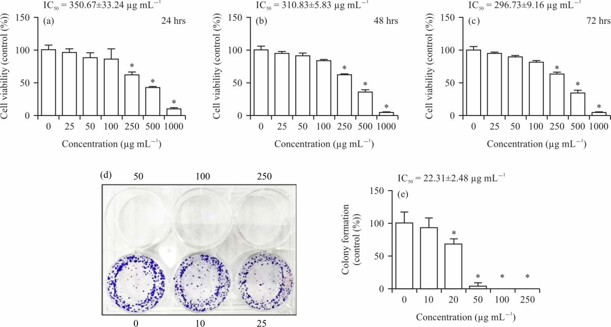 |
| Fig. 1: | Zanthoxylum rhetsa oil effects on HeLa cells proliferation and colony formation, (a-c) Cells were incubated with the extract for 24-72 hrs and measured the cell growth by SRB assay and (d-e) Cells were treated with the oil extract for 24 hrs, further cultured the cancer cells for 10 days and examined the cell colony formation *p<0.05 |
Effects of the Z. rhetsa oil on cell growth and colony formation: The antiproliferative activities of Z. rhetsa oil against human cervical HeLa cancer cell line SRB assay were shown in Fig. 1a-c. The extract exhibited antiproliferative activities against cancer cells in a concentration and time-dependent manner with the IC50 values of 350.67±33.24, 310.83±5.83, 296.73±9.16 μg mL–1 for 24, 48 and 72 hrs, respectively.
The colony formation assay of Z. rhetsa oil was shown in Fig. 1d-e. The extract concentration-dependently reduced the number of colonies in HeLa cells with a low IC50 value (22.31±2.48 μg mL–1) than the data of cell viability. These results demonstrated that the Z. rhetsa oil significantly suppressed the cell viability and colony formation of human cervical cancer cells.
Effects of the Z. rhetsa oil on cell number and cell apoptosis: To investigate whether the Z. rhetsa oil decreased cell growth by modulating apoptosis, cell apoptosis was conducted in HeLa cancer cells. The percentages of early and late phase apoptosis were significantly increased by the Z. rhetsa oil in a concentration-dependent manner (Fig. 2a-g). Especially late phase apoptosis induction, the oil stimulated late apoptosis in a dose-dependent manner with 3.0, 5.7, 24.2 and 47.8%, respectively. These results demonstrated that the Z. rhetsa oil significantly induced the cell apoptosis of human cervical cancer cells.
To confirm apoptosis induction by the oil, morphological changes were conducted with AO/EB staining. As shown in Fig. 3a-d, growth reduction was induced by the Z. rhetsa oil and viable cell number was greatly reduced in a dose-dependent manner and the number of apoptotic bodies was found at a high dose (250 μg mL–1) of the Z. rhetsa oil. Similarly, a 250 μg mL–1 dose of oil activated apoptosis on HeLa cells after detection with flow cytometry and AO/EB staining. These data indicated that the apoptosis of cells induced by the Z. rhetsa oil contributed to their antiproliferative effects.
Effects of the Z. rhetsa oil on ROS formation: Intracellular ROS formation was measured using a DCF-DA fluorescent probe in HeLa cervical cancer cells. After the cells were incubated with the Z. rhetsa oil for 24 hrs, the data from flow cytometry revealed that the oil significantly stimulated ROS production in a concentration-dependent manner (Fig. 3e-i). At the dose of 250 μg mL–1, the Z. rhetsa oil showed higher levels of ROS formation approximately 20.5% correlated with growth inhibition and apoptosis induction in HeLa cells. The results demonstrated that there is a reduction in cell growth and an increase in apoptosis when the level of ROS was increased with the Z. rhetsa oil.
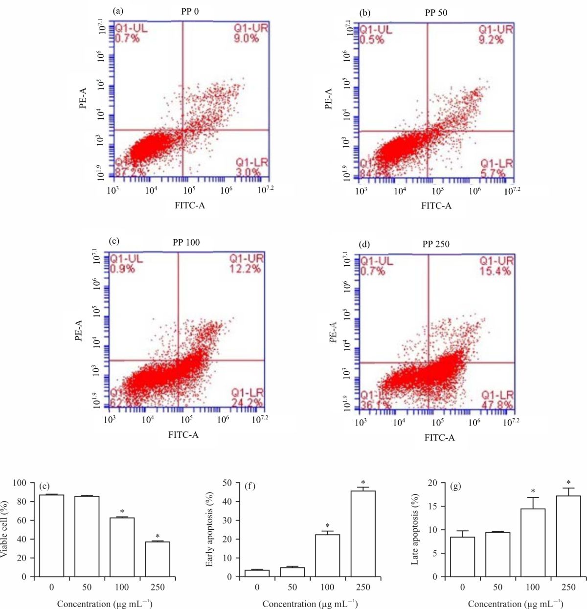 |
| Fig. 2(a-g): | Zanthoxylum rhetsa oil effects on HeLa cells apoptosis, (a-g) Cells were treated with the oil extract for 24 hrs and examined the apoptosis by flow cytometry *p<0.05 |
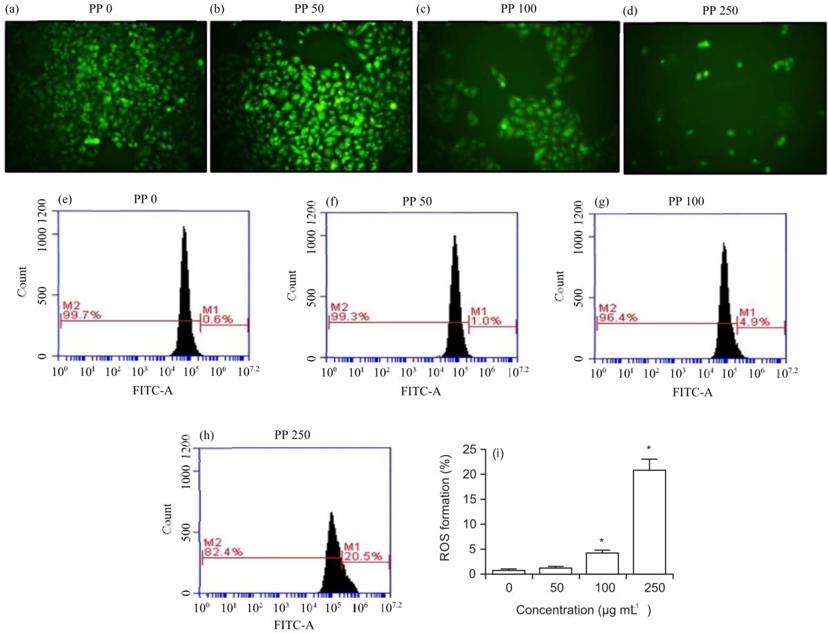 |
| Fig. 3(a-i): | Zanthoxylum rhetsa oil effects on HeLa cell morphology and ROS formation, (a-d) Cells were treated with the oil extract for 24 hrs, loaded with AO/EB solution and photographed the cell morphology by inverted fluorescence microscopy and (e-i) Cells were treated with the oil extract for 24 hrs, loaded with DCF-DA florescent-probe and examined the ROS formation by flow cytometry *p<0.05 |
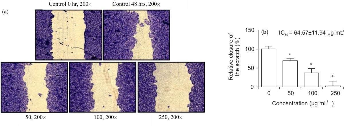 |
| Fig. 4(a-b): | Zanthoxylum rhetsa oil effects on HeLa cell migration, (a-b) Cells were made a wound, treated with the oil extract for 48 hrs and examined the cell migration *p<0.05 |
Effects of the Z. rhetsa oil on cell migration: To determine the effect of the Z. rhetsa oil on the HeLa cells migration by wound healing method. As shown in Fig. 4a-b, the percentage of cell migration rate was significantly reduced by the Z. rhetsa oil in HeLa cells in a concentration-dependent manner. Taken together, the results indicated that the Z. rhetsa oil had an inhibitory effect on cervical cancer cell migration.
DISCUSSION
This work demonstrated that Z. rhetsa oil significantly inhibited cervical cancer cells by attenuating proliferation, activating apoptosis and inhibiting migration in a dose-dependent manner. The standard anticancer drugs are the most frequent agents for treating cervical cancer, nevertheless, the resistance and toxic ability correlated with the agents are a matter of concern. Therefore, several compounds or natural products are discovered for exploring and evolving better than anticancer agents which exhibit high efficacy and low toxicity. Zanthoxylum rhetsa, a Thai vegetable with a spicy taste, is commonly found in the Northern part of Thailand and has been demonstrated with antioxidative activity and antiinflammation, however, the anticancer effects of Z. rhetsa oil is still less information.
Monoterpenes, hydrocarbon and sesquiterpenes were the main constituents found in Z. rhetsa oil. The leading compounds were limonene and beta-phellandrene (26.61 and 25.96%, respectively). Zanthoxylum rhetsa oil from northern Nan with high content of limonene exhibited strong cytotoxic properties against lung cancer H460 cells16. Recently, the leaf petiole extract of Z. rhetsa with a high percent of limonene showed strong inhibitory activity against MCF-7 (human breast adenocarcinoma cells)17. Zanthoxylum rhetsa oil grown in Vietnam with high content of sabinene showed an inhibitory effect against cervical cancer cells with the IC50 of more than 100 μg mL–1 17.
The effects of Z. rhetsa oil were investigated on cervical cancer HeLa cell growth by SRB and colony formation assay. Other Zanthoxylum species have also been demonstrated to be non-toxic to normal cell lines21. Zanthoxylum rhetsa oil inhibited cervical cancer cell growth with the IC50 value of 296.73 μg mL–1 and reduced the cancer cells in replication in a dose-dependent manner. As previous data found that several parts of Z. rhetsa had numerous actions on diseases and an anticancer effect was observed. Consistently, Z. rhetsa stem bark extract significantly suppressed melanoma B cells growth and the strongest growth inhibitory effects were demonstrated by chloroform, hexane, ethyl acetate, methanol and butanol fractions with IC50 values 156, 132.7, 174.3, 168 and 263.1 μg mL–1, respectively7. Additionally, the purified proteins from the pericarp caused inhibition of cell growth, reduction of cell colonies and activation of apoptosis19. The extract of Z. rhetsa showed the strongest anticancer effects in several cancer cells including cervical cancer cells.
For apoptosis in HeLa cells indicated that Z. rhetsa oil extract induced cancer cell early apoptosis in a dose-dependent manner from 50-250 μg mL–1. The observation of apoptosis was reported in human breast cancer MCF-7 cells by increasing DNA fragmentation and arresting the cell cycle at G2/M20. Several natural compounds from South-East Asian countries including Thailand, significantly induced cervical cancer cell apoptosis and suppressed the cell cycle distribution22,23. The Careya arborea leaf extract indicated that this extract induced cervical cells apoptosis at the high rate of late apoptosis and arrested the cell cycle at G0/G1 phase22. Confirmation with one of the most Thai vegetables, Oroxylum indicum, showed the anticancer effects in several cancer cell types including cervical cancer cells. At 250 μg mL–1 of seed, Oroxylum indicum demonstrated that it reduced viable cancer cells and stimulated late apoptosis by inducing ROS formation and mitochondrial dysfunction23. Finally, Thai vegetables had the strongest anticancer activities including Z. rhetsa oil extract.
Furthermore, this study indicated that Z. rhetsa oil extract significantly suppressed HeLa cell migration in a dose-dependent manner (50-250 μg mL–1) in in vitro study. Consistently, in vivo studies showed angiogenic properties suppression in the angiogenesis in the treated animal as compared to the control animal peritoneum, through reduction in the Vascular Endothelial Growth Factor (VEGF) level as compared to the control groups19. In conclusion, Z. rhetsa oil extract suppressed cervical cell migration.
The mechanism of Z. rhetsa oil extract-induced cell death and apoptosis was focused on ROS formation. The data demonstrated that these oil extracts stimulated the production of ROS formation in intracellular HeLa cells. Importantly, intracellular ROS act as messengers that play role in normal physiological functions. Nevertheless, ROS overproduction can cause damage to DNA, lipids, carbohydrates and proteins, leading to normal and cancer cell death24. Thai plant extract significantly activated cancer cells apoptosis which reduced the mitochondrial function and induced ROS formation22. The four Thai extracts of Careya sphaerica, Azadirachta indica, Piper nigrum and Oroxylum indicum stimulated ROS overgeneration with increasing caspase 3 activity and then lead to induced cancer cell apoptosis as Z. rhetsa oil-stimulated ROS25. Finally, Z. rhetsa oil may be useful for treating or preventing cervical cancer.
CONCLUSION
Monoterpenes were the predominant compound in Z. rhetsa oil from fresh fruits with a mild antioxidant effect. In addition, Z. rhetsa oil stimulates cervical cancer HeLa cell death, induces early apoptosis and inhibits migration. The mechanism of action indicated that Z. rhetsa oil activates ROS formation significantly at a high dose of the oil. The data showed that Z. rhetsa oil has potential effects for treating cervical cancer.
SIGNIFICANCE STATEMENT
This study explored the mechanism of Z. rhetsa oil in the cervical cancer HeLa cell line. The study reported that the oil-induced cell death via apoptosis and stimulated ROS system. This study will help the researcher to discover a natural compound for treating cervical cancer.
ACKNOWLEDGMENT
This research project was supported by the Thailand Science Research and Innovation Fund and the University of Phayao (Grant No. FF64-UoE017).
REFERENCES
- Torre, L.A., F. Bray, R.L. Siegel, J. Ferlay, J. Lortet-Tieulent and A. Jemal, 2015. Global cancer statistics, 2012. CA: Cancer J. Clinicians, 65: 87-108.
CrossRefDirect Link - Song, T.T., F. Xu and W. Wang, 2020. Inhibiting ubiquitin conjugating enzyme E2 N by microRNA-590-3p reduced cell growth of cervical carcinoma. Kaohsiung J. Med. Sci., 36: 501-507.
CrossRefDirect Link - Wang, T., J. Feng and A. Zhang, 2020. miR-584 inhibits cell proliferation, migration and invasion in vitro and enhances the sensitivity to cisplatin in human cervical cancer by negatively targeting GLI1. Exp. Ther. Med., 19: 2059-2066.
CrossRefDirect Link - Ziech, D., R. Franco, A.G. Georgakilas, S. Georgakila and V. Malamou-Mitsi et al., 2010. The role of reactive oxygen species and oxidative stress in environmental carcinogenesis and biomarker development. Chem. Biol. Interact., 188: 334-339.
CrossRefPubMedDirect Link - Thu, N.B., T.N. Trung, D.T. Ha, N.M. Khoi and N.V. Than et al., 2010. Zanthoxylum rhetsa stem bark extract inhibits LPS-induced COX-2 and iNOS expression in RAW 264.7 cells via the NF-κB inactivation. Nat. Prod. Sci., 16: 265-270.
Direct Link - Santhanam, R.K., S. Ahmad, F. Abas, I.S. Ismail, Y. Rukayadi and K. Shaari, 2013. Photoprotective properties of Zanthoxylum rhetsa: An in vitro analysis. J. Chem. Pharmaceut. Res., 5: 1512-1520.
Direct Link - Santhanam, R.K., S. Ahmad, F. Abas, I.S. Ismail, Y. Rukayadi, M.T. Akhtar and K. Shaari, 2016. Bioactive constituents of Zanthoxylum rhetsa bark and its cytotoxic potential against B16-F10 melanoma cancer and normal human dermal fibroblast (HDF) cell lines. Molecules, Vol. 21.
CrossRefDirect Link - Rahman, M.T., M. Alimuzzaman, S. Ahmad and A.A. Chowdhury, 2002. Antinociceptive and antidiarrhoeal activity of Zanthoxylum rhetsa. Fitoterapia, 73: 340-342.
CrossRefDirect Link - Lalitharani, S., V. Kalpanadevi and V.R. Mohan, 2013. Pharmacognostic studies on the spine of Zanthoxylum rhetsa (Roxb.) DC. Biosci. Discovery, 4: 5-11.
Direct Link - Kale, S.S., A.H. Rajmane, V.C. Urunkar, M.K. Gaikwad and S.B. Bhandare, 2011. Formulation and in-vitro evaluation of sun protection factor of methanolic extract of Zanthoxylum rhetsa DC. sunscreen lotion. Res. J. Pharmacog. Phytochem., 3: 206-210.
Direct Link - Palasuwan, A., S. Soogarun, T. Lertlum, P. Pradniwat and V. Wiwanitkit, 2005. Inhibition of heinz body induction in an in vitro model and total antioxidant activity of medicinal Thai plants. Asian Pac. J. Cancer Prev., 6: 458-463.
Direct Link - Reddy, L.J. and B. Jose, 2011. Statistical analysis of the antibacterial activity of Zanthoxylum rhetsa seed essential oil. J. Chem. Pharm. Res., 3: 440-444.
Direct Link - Nayaka S., B. Chakraborty, S.S. Pallavi, M.P. Bhat, K.N. Shashiraj and B. Ghasti, 2020. Synthesis of biogenic silver nanoparticles using Zanthoxylum rhetsa (Roxb.) DC seed coat extract as reducing agent and in-vitro assessment of anticancer effect on A549 lung cancer cell line. Int. J. Pharm. Res., 12: 306-314.
CrossRefDirect Link - van Hieu, N., N. van Hoa, K.T. Hoai, D.T. Hien and D.M. Tu et al., 2020. Inhibitory effect on nitric oxide production of essential oil from Zanthoxylum rhetsa (Roxb.) DC. fruit. J. Pharmacogn. Phytochem., 9: 67-70.
CrossRefDirect Link - Wongkatiya, N., C. Akekawatchai, P. Sanguansermsri, I.H. Fraser, C. Pratoomsoot and D. Sanguansermsri, 2018. Chemical compositions and biological properties of essential oils from Zanthoxylum rhetsa (Roxb.) DC and Zanthoxylum limonella alston. Afr. J. Traditional Complementary Altern. Med., 15: 12-18.
CrossRefDirect Link - Theeramunkong, S. and M. Utsintong, 2018. Comparison between volatile oil from fresh and dried fruits of Zanthoxylum rhetsa (Roxb.) DC. and cytotoxicity activity evaluation. Pharmacogn. J., 10: 827-832.
CrossRefDirect Link - Pham, C.B., T.I. Cam, T.T. Thi, P.M. Quan and T.Q. Toan et al., 2021. The chemical composition and biological activities of essential oils from Zanthoxylum rhetsa grown in Son La, Northwest Vietnam. J. Food Qual., Vol. 2021.
CrossRefDirect Link - Naik, R.R., A.K. Shakya, N.A. Khalaf, S. Abuhamdah, G.A. Oriquat and A. Maraqa, 2015. GC-MS analysis and biological evaluation of essential oil of Zanthoxylum rhesta (Roxb.) DC pericarp. Jordan J. Pharma. Sci., 8: 181-193.
Direct Link - Parrikar, P.D.N., B.K. Srinivas, D.K. Krishnappa and S. Jayarama, 2021. Apoptosis-inducing and antiangiogenic activity of partially purified protein from the pericarp of Zanthoxylum rhetsa in vitro and in vivo. Phcog. Mag., 17: 96-104.
CrossRefDirect Link - Brand-Williams, W., M.E. Cuvelier and C. Berset, 1995. Use of a free radical method to evaluate antioxidant activity. LWT-Food Sci. Technol., 28: 25-30.
CrossRefDirect Link - Lan, Y., H. Li, Y.Y. Chen, Y.W. Zhang, N. Liu, Q. Zhang and Q. Wu, 2014. Essential oil from Zanthoxylum bungeanum maxim. and its main components used as transdermal penetration enhancers: A comparative study. J. Zhejiang Univ. Sci. B, 15: 940-952.
CrossRefDirect Link - Buranrat, B., 2021. Growth inhibition, apoptosis induction and migratory suppression by Careya arborea leaf extract in HeLa cervical cells. Int. J. Pharmacol., 17: 339-349.
CrossRefDirect Link - Buranrat, B. and S. Boontha, 2022. The seed extract of Oroxylum indicum suppresses cell proliferation, migration and promotes apoptosis in cervical cancer cells. Pak. J. Pharm. Sci., 35: 553-559.
CrossRefDirect Link - Krishnan, S.N., J.L. Rosales and K.Y. Lee, 2019. Ros-mediated cancer cell killing through dietary phytochemicals. Oxid. Med. Cell. Longevity, Vol. 2019.
CrossRefDirect Link - Buranrat, B., A. Konsue and P. Wongsuwan, 2020. Extracts of edible, medicinal Thai plants inhibit the human breast cancer cells. Trop. J. Pharm. Res., 19: 595-601.
CrossRefDirect Link