

Vijay Pandi
Pharmacy group, Birla Institute of Technology and Science, Pilani-333031 Rajasthan, India
Anantha Naik Nagappa
Pharmacy group, Birla Institute of Technology and Science, Pilani-333031 Rajasthan, India
Prasad A. Thakurdesai
Department of Pharmacology, Poona College of Phannacy,
Bharati Vidyapeeth University, Erandwane, Pune-411 (138, India)
Journal of Pharmacology and Toxicology
Year: 2007 | Volume: 2 | Issue: 6 | Page No.: 551-558





ABSTRACT
The present study was designed to evaluate the effect of Losartan Potassium (LP) pretreatment at various time intervals against apomorphine (APM) induced stereotyped behavior and haloperidol (HP) induced catalepsy in mice. LP (100 mg kg-1, p.o.) reduced the intensity of the APM induced stereotyped behavior at when administrated 3 h and 6 h. prior to APM. However, such reversal was not observed when LP pretreatment time was 1, 12 or 24 h. LP (100 mg kg-1) was also found to potentiate HP-induced catalepsy both pretreated (2 h prior) and co-administrated with LP. However, onset of catalepsy in co-administrated LP group was 240 min which as drastically different when LP was administrated 2 h prior to HP (30 min). These results suggests that effects of LP induced modulation of dopaminergic functions are not because of LP per se (t1/2 = 2.12 h) but because of its active metabolite, EXP 3174 (t1/2 = 6-9 h).
PDF Abstract XML References Citation
How to cite this article
Vijay Pandi, Anantha Naik Nagappa and Prasad A. Thakurdesai, 2007. Effects of Losartan Potassium on Central Dopaminergic System in Mice. Journal of Pharmacology and Toxicology, 2: 551-558.
DOI: 10.3923/jpt.2007.551.558
URL: https://scialert.net/abstract/?doi=jpt.2007.551.558
DOI: 10.3923/jpt.2007.551.558
URL: https://scialert.net/abstract/?doi=jpt.2007.551.558
INTRODUCTION
The existence of the brain Renin Angiotensin System (RAS), which is independent of the circulating RAS, has been established. All enzymes and peptides necessary for the biosynthesis of these angiotensins have been recognized within the central nervous system (von Bohlen und Halbach, 2005; Sica, 1999). The brain RAS mediates several classic physiological effects including body water balance, maintenance of blood pressure, sexual behaviors and regulation of pituitary gland hormones (von Bohlen und Halbach, 2005) and also has more subtle functions involving complex mechanisms in central nervous system such as learning and memory (Sakai and Sigmund, 2005; Wright and Harding, 1997). Moreover, there is evidence to suggest that the RAS is involved in neurological disorders, such as Alzheimer's disease (Amouyel et al., 2000) and Parkinson's disease by mechanism of neuromodulation (von Bohlen und Halbach, 2005; Savaskan, 2005).
Angiotensin II (Ang II) acts on brain structures localized inside and outside the blood-brain barrier to induce drinking behavior and natriuresis, stimulate vasopressin release, modulate sympathetic outflow to the periphery and attenuate the baroreceptor reflex (Culman et al., 2002). The effector peptide of the RAS Ang II, binds at least to two G protein coupled receptor subtypes, referred as the AT1 and the AT2 receptors. Most of the classic actions of Ang II in the brain are mediated by AT1 receptors (Polidori et al., 1996) whereas AT2 receptors are involved in brain development and neuronal regeneration and protection (Wright and Harding, 1995). Animal studies have shown that AT1 receptor antagonists enable endogenous Ang II to stimulate neuronal regeneration via activation of AT2 receptors. There is substantial evidence that the AT2 receptor can offset or counteract the effects mediated by the AT1 receptor such as cell proliferation, water intake and blood pressure (Usberti et al., 1985).
Ang II is known to stimulate catecholamine release (Kimura et al., 1992; Corwin et al., 1985) including dopamine (Sawamura and Nakada, 1996; Jenkins et al., 1996) through AT1 subtype (Lucius et al., 1999). In addition, AT1 receptor antagonists (ARBs) modulate many functions through dopaminergic system (Bek et al., 2006; Maul et al., 2005; Grammatopoulos et al., 2005; Stragier et al., 2004). In the human brain prominent AR binding occurs in the substantia nigra pars compacta, especially overlying pigmented neurons and moderate binding occurs in the striatum (Allen et al., 1991) and past reports strongly suggest that angiotensin receptors are located on dopaminergic neurons in the substantia nigra and act presynaptically in the striatum (Grammatopoulos et al., 2005).
In vivo and in vitro studies on losartan (AT1 antagonist) have variable effects on striatal dopaminergic function (Culman et al., 2001, 2002). Acute peripheral administration of losartan reported to decrease striatal dopamine levels while chronic administration resulted in no alteration of brain tissue dopamine (DA) content, but causes a small rise in striatal dopamine metabolite DOPAC (Mendelsohn et al., 1993). Losartan (20 μmol kg-1, i.p) was reported to reduce Ang II-induced drinking behavior upto 24 h, i.e., beyond its half life of 2 h (Fitts et al., 2005; Stancheva et al., 2003; Bagi et al., 2003; Barbella et al., 1993). However, losartan have not been studied for its central effects (especially with respect to dopamine modulation) for over the period exceeding two hours. Therefore, the present study was designed to elucidate possible role of LP in the neuromodulation of dopamine using apomorphine (APM) induced stereotypy and haloperidol (HP) induced catalepsy in mice over a period exceeding 2 h.
MATERIALS AND METHODS
Materials
Losartan potassium (LP, Sun Pharma, Mumbai, India) and haloperidol (HP, RPG Life Sci Ltd., Halol, Gujarat, India) were obtained as gift samples and were dissolved in normal saline for the experiments. Apomorphine hydrochloride (APM) were purchased from Sigma-Aldrich, USA and solution was prepared in normal saline. All the drugs were administered intraperitoneally (except LP which was administered orally) in a maximum volume of 1 mL per 100 g of body weight of mice.
Animals
Albino mice (Swiss, 20-25 g, either sex) were used. They were allowed food and water ad libitum up to the time of experimentation. Prior to use, the mice were housed in polypropylene cages in groups of six to eight animals under natural light-dark cycle. Each animal was used only once under standard laboratory conditions. All the observations made at room temperature in a noiseless diffusely illuminated room. All observations were made between 9.00 to 17.00 h in a room with controlled temperature (23±1°C) and light intensity of 20 lux. All the experimental protocols were approved by Institutional Animal Ethics Committee (IAEC) as per provisions of Committee for the Purpose of Control and Supervision of Experimental Animals (CPCSEA), New Delhi, India
Effect of LP on APM Induced Stereotypy in Mice
Measurement of stereotyped behavior was done as per method described earlier (Battisti et al., 2000; Menge and Brand, 1971). In test groups, mice were pretreated with LP (100 mg kg-1, p.o.), either 1, 3, 6, 12 or 24 h prior to APM (2 mg kg-1, i.p.) and mice were observed for stereotypy behavior for next 50 min. Separate vehicle control group of mice was also maintained to which only APM was administrated. The intensity of stereotyped behavior was assessed at 5 min intervals throughout the duration of experiment. Behavior was scored as either 0 (no change than control), 1 (discontinuous sniffing, constant exploratory activity), 2 (continuous sniffing, periodic exploratory activity), 3 (continuous sniffing, discontinuous biting, gnawing or licking. Very brief periods of locomotor activity) or 4 (continuous biting, gnawing or licking; no exploratory activity). Mean stereotypy scores were calculated and presented as Fig. 1.
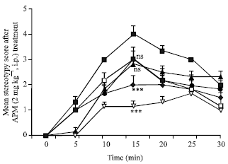 | |
| Fig. 1: | Effect of LP (100 mg kg-1, p.o.) on the APM (2 mg kg-1) induced stereotypy behavior ( ) on pretreatment with LP (100 mg kg-1) at 1 h ( ) on pretreatment with LP (100 mg kg-1) at 1 h ( ) , 3 h ( ) , 3 h ( ), 6 h ( ), 6 h ( ), 12 h ( ), 12 h ( ) and 24 h ( ) and 24 h ( ) prior to APM. Stereotypy behavior was assessed and scored. Data is represented as mean stereotypy score (6 mice per group) ±SEM and was analyzed by one-way ANOVA on ranks followed by Mann-Whitney U test on readings at 15 min after APM (where APM showed peak stereotypy score) ) prior to APM. Stereotypy behavior was assessed and scored. Data is represented as mean stereotypy score (6 mice per group) ±SEM and was analyzed by one-way ANOVA on ranks followed by Mann-Whitney U test on readings at 15 min after APM (where APM showed peak stereotypy score) |
Effect of LP on HP- Induced Catalepsy in Mice
In case of HP-induced catalepsy, to group I and II, HP (0.1 mg kg-1, i.p.) and LP (100 mg kg-1, p.o.) was administered respectively. In group III, mice were pretreated with LP (100 mg kg-1, p.o.) at 2 h prior to HP (0.1 mg kg-1, i.p.) administration. To group IV, concurrent administration of LP (100 mg kg-1, p.o.) and HP (0.1 mg kg-1, i.p.) was done.
Haloperidol-induced catalepsy was measured with the standard bar test (El Yacoubi et al., 2001) in a wooden chamber (length, 23 cm; width, 10.5 cm; height, 9 cm) with a horizontal metal bar (diameter, 0.4 cm; length, 10.5 cm) fixed at 9 cm above the floor and at 4 cm from the back of the box. Animals were used only once. After a 30-60 min habituation period to the testing room, each mouse was placed on bar. If the mouse maintained the imposed posture for at least 20 sec, it was said to be cataleptic and given the score of one point. For every 20 sec, one extra point was given for the continuation of the cataleptic posture. The animals were tested for catalepsy 30, 60, 120, 180, 240, 300 or 360 min after HP treatment. The test was considered complete when the front paw touched the ground or mouse climbed on the wooden block or after a lapse of 180 sec. The animals were awarded a score of zero if it failed to hold the wooden block for successive three attempts. Only those animals were selected for studies. The sub maximal cataleptic dose of HP (0.1 mg kg-1) was chosen so that potentiation/reversal effect of drug on catalepsy can be easily differentiated.
Statistical Analysis
The effect of LP on HP-induced catalepsy and APM-induced stereotypy was expressed as mean score ±SEM for stereotypy and catalepsy respectively. Data was analyzed by two-way repeated measure ANOVA on ranks followed by Dunn’s test. Statistical significance was set at p<0.05.
RESULTS
Effect of LP on APM- Induced Stereotypy
APM induced stereotypy behavior, which reached at peak at 15 min period (Fig. 1). LP (100 mg kg-1, p.o.) administration could not reverse stereotypy when LP was administrated 1 h prior to APM as shown in Fig. 1.
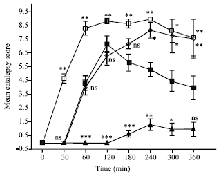 | |
| Fig. 2: | Effect of LP (100 mg kg-1, p.o.) () alone and after 2 h pre-treatment LP (100-Pr) () or concurrent administration, LP (100-Co) () on HP (0.1 mg kg-1) induced catalepsy (). Catalepsy was assessed and scored from 0 to 5 and mean stereotypy score (6 mice per group) ±SEM was presented. Data was analyzed by two-way ANOVA on ranks followed by Dunn’s test and was compared with HP treated group at respective time. * p<0.05, ** p<0.01, *** p<0.001 and ns-non-significant as compared with HP group at respective time |
On the other hand, stereotypy behavior was significantly reversed (p<0.001), when LP pretreatment was done 3 or 6 h. prior to APM but such reversal was not observed when LP pretreatment was done 12 or 24 h prior to APM (Fig. 1).
Effect of LP on Hp-Induced Catalepsy
Losartan Potassium (LP) at 100 mg kg-1, per se did not exhibit any cataleptic effects as HP (0.1 mg kg-1) exhibited with peak catalepsy score at 120 min (Fig. 2). However, pretreatment of LP significantly increased HP induced catalepsy score with onset of 30 min (p<0.01) and peak at 120 min (Fig. 2). However, co-administration of LP with HP, exhibited significant (p<0.05) increase in catalepsy only after 240 min (4 h), at which peak catalepsy score was seen (Fig. 2).
DISCUSSION
Anti-psychotic drugs like haloperidol and chlorpromazine (the so-called typical neuroleptics) induce abnormal motor behaviors in experimental animals and humans, including catalepsy in rats and mice (Sanberg et al., 1988). Neuroleptic- induced catalepsy in rodents is a robust behavioral method for the study of nigrostriatal dopaminergic function and its modulation by other transmitter systems (Sanberg et al., 1988; Pires et al., 1996). It is generally accepted that dopaminergic system in the brain is important for the mediation of drug induced stereotyped behavior. The nigrostriatal dopaminergic pathway has long been implicated in motor functioning (Sanberg et al., 1988). Dopamine is present in the region of nucleus accumbens and is responsible for locomotor activity, while stereotypy is mediated by striatal dopaminergic neurons (Sanberg et al., 1988). Stereotyped behavior may operate via a reciprocal balance between the dopaminergic and cholinergic systems, in favor of dopaminergic dominance.
The brain Renin-Angiotensin System (RAS) is reported to be important in cognition and anxiety and shown to reverse age-, scopolamine-, ethanol- and diabetes-induced deficits (Gard, 2004). Furthermore, AT1 receptor blockers appear to be able to enter the brain after peripheral administration and cause AT1 receptor blockade in the central nervous system (Wang et al., 2003). Role of the brain renin-angiotensin system in the development of hypertension is also reported earlier (Nakata et al., 2001).
There is a large body of in vitro evidence to support the concept of a relationship between brain Ang II and dopamine systems (Jenkins et al., 1996; Brown et al., 1996; Jenkins et al., 1995a). It also extends to the nigrostriatal dopaminergic system which bear AT1 receptors, both on their cell bodies in the substantia nigra presynaptically and on their terminals in the striatum, where Ang II can markedly potentiate DA release (Jenkins et al., 1996). This observation suggests that drugs which modulate central Ang II may be useful in regulating central dopaminergic activity.
Pires et al. (1996) investigated potentiation effect of losartan (10, 100 mg kg-1, i.p) on HP (1 mg kg-1, i.p) induced catalepsy in mice. Furthermore, Losartan (20 Fmol kg-1, i.p) was reported to reduced Ang II induced drinking behavior at 4, 12 and 24 h and suspected the sustained central effects of losartan due to its active metabolite (Polidori et al., 1996). But the central effects of losartan over the period of time exceeding 2 h on behavioral studies hitherto not had been reported in the literature.
In present study, we have investigated effects of LP against APM induced stereotypy and HP induced catalepsy behavior. Apomorphine directly activates dopamine receptors in the brain (Seeman, 1980; Stoof and Kebabian, 1984) and larger doses of the drug induced stereotyped behavior (sniffing, licking and gnawing) (Anden et al., 1967; Ernst, 1967). The stimulant effect of high doses of Apomorphine is attributed to activation of postsynaptic receptors in the central nervous system (Anden et al., 1967). The behavioral responses observed in animals after administration of the dopamine agonist, apomorphine are attributed to activation of D1 and D2 receptors (Seeman, 1980; Stoof and Kebabian, 1984). Mesolimbic and nigrostriatal dopaminergic pathways may be important in the mediation of locomotor activity and stereotyped behaviors. stereotyped behaviors is more closely associated with the caudate striatum area of brain (Kelly et al., 1975).
In this study, when LP was administrated 1 h prior, it failed to reverse the APM induced stereotypy but when it was administrated at 3 or 6 h prior to APM, it significantly reversed APM induced stereotypy. Similarly, at 12 or 24 pretreatment, LP could not affect APM induced stereotypy. These biphagic responses of LP against APM stereotypy could be explained on the basis of active metabolite of LP that has been reported earlier (Wong et al., 1990; Christen et al., 1991) and thought to be responsible for the longer action of LP beyond its half-life. The terminal half-life of losartan is 2.12 h and its active metabolite EXP 3174 (carboxylic acid derivative) exhibits half-life of about 6 to 9 h (Christen et al., 1991). These reports suggest that the reversal of APM induced stereotypy at 3 and 6 h in our study was due to active metabolite of LP (i.e., EXP 3174). This notion further confirmed by our results at 12 and 24 h when LP failed to show any effects on APM stereotypy. After 11 h of administration, plasma concentration of losartan becomes undetectable (Christen et al., 1991). This biphasic behavior of LP is supported by prior reports that Ang II (100 nM) caused a biphasic effect on electrophysiological response, which is delayed rectifier K+ current (Ik) of catecholaminergic transmission (Gelband et al., 1997).
LP, which influences the central angiotensinergic mechanisms have been found to affect stereotyped behavior induced by APM and suggested to be modulator of presynaptic dopaminergic neurons in nigro-striatal system. Since Ang II is also known to stimulate catecholamine release (Zimmerman, 1981) including dopamine (Jenkins et al., 1996; 1995b), angiotensin receptors mediates its effects in the brain through AT1 subtype (Mendelsohn et al., 1993) located on presynaptic nerve terminals (Raghavendra et al., 2001; Raghavendra et al., 1998).
Dopamine (D2) antagonist, haloperidol (HP) increases striatal dopamine release and induces catalepsy through its actions on striatal dopaminergic system (Jaskiw and Bongiovanni, 2004) and proved to be simple and reliable test for the investigation that involves D2 receptor (Fischer et al., 2002). In this study, LP alone did not show any catalepsy. However, difference between peak catalepsy effects after LP pretreatment (120 min) and co administration of LP+HP (i.e., 240 min) showed that these effects are of active metabolite and not of LP per se.
Losartan is a surmountable antagonist with relatively low affinity and rapid association at the AT1 receptor, whereas EXP 3174, the active metabolite of losartan, exhibits different degrees of insurmountable inhibition (Vauquelin et al., 2001). In present study, delayed onset and prolonged duration of responses (reduction of APM-induced stereotypy and increase in HP induced catalepsy) might be result of long lasting and insurmountable binding of active metabolite EXP 3174 of LP.
In conclusion, LP modulates dopaminergic (D2) neurotransmission in nigro-striatal neurons through its metabolite and provides a novel target for the development of better anti-psychotic as well as anti-parkinson’s agents.
ACKNOWLEDGMENTS
The authors wish to thank the Director, Birla Institute of Technology and Science, Pilani for his constant encouragement and support.
REFERENCES
- Amouyel, P., F. Richard, C. Berr, I. David-Fromentin and N. Helbecque, 2000. The renin angiotensin system and Alzheimer's disease. Ann. N Y Acad. Sci., 903: 437-441.
PubMedDirect Link - Bagi, E.E., E. Fekete and L. Lenard, 2003. Angiotensin II and III microinjections into the zona incerta influence drinking behavior. Brain Res., 977: 199-208.
Direct Link - Battisti, J.J., C.B. Shreffler, N.J. Uretsky and L.J. Wallace, 2000. NMDA antagonists block expression of sensitization of amphetamine- and apomorphine-induced stereotypy. Pharmacol. Biochem. Behav., 67: 241-246.
CrossRefDirect Link - Bek, M.J., X. Wang, L.D. Asico, J.E. Jones and S. Zheng et al., 2006. Angiotensin-II type 1 receptor-mediated hypertension in D4 dopamine receptor-deficient mice. Hypertension, 47: 288-295.
PubMedDirect Link - Brown, D.C., L.J. Steward, J. Ge and N.M. Barnes, 1996. Ability of angiotensin II to modulate striatal dopamine release via the AT1 receptor in vitro and in vivo. Br. J. Pharmacol., 118: 414-420.
PubMedDirect Link - Culman, J., J. Baulmann, A. Blume and T. Unger, 2001. The renin-angiotensin system in the brain: An update. J. Renin Angiotensin Aldosterone Syst., 2: 96-102.
PubMedDirect Link - Culman, J., A. Blume, P. Gohlke and T. Unger, 2002. The renin-angiotensin system in the brain: Possible therapeutic implications for AT(1)-receptor blockers. J. Hum. Hypertens, 16: S64-S70.
CrossRefDirect Link - El Yacoubi, M., C. Ledent, M. Parmentier, J. Costentin and J.M. Vaugeois, 2001. Adenosine A2A receptor knockout mice are partially protected against drug-induced catalepsy. Neuroreport, 12: 983-986.
PubMedDirect Link - Fischer, D.A., B. Ferger and K. Kuschinsky, 2002. Discrimination of morphine- and haloperidol-induced muscular rigidity and akinesia/catalepsy in simple tests in rats. Behav. Brain Res., 134: 317-321.
CrossRefDirect Link - Fitts, D.A., D.K. Zierath, E.E. Wilkins and J.E. Bassett, 2005. Losartan blocks drinking and cFos expression induced by central ornithine vasotocin in rats. Physiol. Behav., 86: 573-577.
CrossRefDirect Link - Gard, P.R., 2004. Angiotensin as a target for the treatment of Alzheimer's disease, anxiety and depression. Exp. Opin. Therap. Targets, 8: 7-14.
PubMedDirect Link - Gelband, C.H., M. Zhu, D. Lu, L.P. Reagan and S.J. Fluharty et al., 1997. Functional interactions between neuronal AT1 and AT2 receptors. Endocrinology, 138: 2195-2198.
PubMedDirect Link - Grammatopoulos, T.N., F. Ahmadi, S.M. Jones, M.W. Fariss, J.A. Weyhenmeyer and W.M. Zawada, 2005. Angiotensin II protects cultured midbrain dopaminergic neurons against rotenone-induced cell death. Brain Res., 1045: 64-71.
CrossRefDirect Link - Jaskiw, G.E. and R. Bongiovanni, 2004. Brain tyrosine depletion attenuates haloperidol-induced striatal dopamine release in vivo and augments haloperidol-induced catalepsy in the rat. Psychopharmacology, (Berl), 172: 100-107.
Direct Link - Jenkins, T.A., S.Y. Chai, D.W. Howells and F.A. Mendelsohn, 1995. Intrastriatal angiotensin II induces turning behaviour in 6-hydroxydopamine lesioned rats. Brain Res., 691: 213-216.
Direct Link - Jenkins, T.A., A.M. Allen, S.Y. Chai, D.P. MacGregor, G. Paxinos and F.A. Mendelsohn, 1996. Interactions of angiotensin II with central dopamine. Adv. Exp. Med. Biol., 396: 93-103.
PubMedDirect Link - Lucius, R., S. Gallinat, S. Busche, P. Rosenstiel and T. Unger, 1999. Beyond blood pressure: New roles for angiotensin II. Cell Mol. Life Sci., 56: 1008-1019.
CrossRefDirect Link - Maul, B., W. Krause, K. Pankow, M. Becker and F. Gembardt et al., 2005. Central angiotensin II controls alcohol consumption via its AT1 receptor. Faseb. J., 19: 1474-1481.
CrossRefDirect Link - Nakata, T., K. Takeda, S. Harada, A. Oguni and T. Hatta et al., 2001. Role of the central nervous system in the development of hypertension produced by chronic nitric oxide blockade in rats. Hypertens. Res., 24: 39-45.
Direct Link - Pires, J.G., S.R. Silva and H.A. Futuro-Neto, 1996. Effects of losartan on neuroleptic-induced catalepsy in mice. Brazilian J. Med. Biol. Res., 29: 1045-1047.
Direct Link - Polidori, C., R. Ciccocioppo, P. Pompei, R. Cirillo and M. Massi, 1996. Functional evidence for the ability of angiotensin AT1 receptor antagonists to cross the blood-brain barrier in rats. Eur. J. Pharmacol., 307: 259-267.
Direct Link - Raghavendra, V., K. Chopra and S.K. Kulkarni, 1998. Modulation of motor functions involving the dopaminergic system by AT1 receptor antagonist, losartan. Neuropeptides, 32: 275-280.
Direct Link - Raghavendra, V., K. Chopra and S.K. Kulkarni, 2001. Comparative studies on the memory-enhancing actions of captopril and losartan in mice using inhibitory shock avoidance paradigm. Neuropeptides, 35: 65-69.
CrossRefDirect Link - Sakai, K. and C.D. Sigmund, 2005. Molecular evidence of tissue renin-angiotensin systems: A focus on the brain. Curr. Hypertens. Rep., 7: 135-140.
CrossRefDirect Link - Savaskan, E., 2005. The role of the brain renin-angiotensin system in neurodegenerative disorders. Curr. Alzheimer Res., 2: 29-35.
PubMedDirect Link - Sawamura, T. and T. Nakada, 1996. Role of dopamine in the striatum, renin-angiotensin system and renal sympathetic nerve on the development of two-kidney, one-clip Goldblatt hypertension. J. Urol., 155: 1108-1111.
Direct Link - Stragier, B., S. Sarre, P. Vanderheyden, G. Vauquelin, M.C. Fournie-Zaluski, G. Ebinger and Y. Michotte, 2004. Metabolism of angiotensin II is required for its in vivo effect on dopamine release in the striatum of the rat. J. Neurochem., 90: 1251-1257.
PubMedDirect Link - Vauquelin, G., F. Fierens, I. Verheijen and P. Vanderheyden, 2001. Insurmountable AT(1) receptor antagonism: The need for different antagonist binding states of the receptor. Trends Pharmacol. Sci., 22: 343-344.
CrossRefDirect Link - Von Bohlen und Halbach, O., 2005. The renin-angiotensin system in the mammalian central nervous system. Curr. Protein Peptide Sci., 6: 355-371.
PubMed - Wright, J.W. and J.W. Harding, 1995. Brain angiotensin receptor subtypes AT1, AT2 and AT 4 and their functions. Regul. Pept., 59: 269-295.
CrossRefDirect Link - Wright, J.W. and J.W. Harding, 1997. Important role for angiotensin III and IV in the brain renin-angiotensin system. Brain. Res. Brain Res. Rev., 25: 96-124.
PubMedDirect Link - Zimmerman, B.G., 1981. Adrenergic facilitation by angiotensin: Does it serve a physiological function?. Clin. Sci., 60: 343-348.
PubMed