
L.E. Rodriguez-Tovar
Departamento de Inmunologia, Universidad Autonoma de Nuevo Leon, Mexico
Y. Valdez-Nava
Departamento de Inmunologia, Universidad Autonoma de Nuevo Leon, Mexico
A.M. Nevarez-Garza
Departamento de Inmunologia, Universidad Autonoma de Nuevo Leon, Mexico
D.K. Miller
Departamento de Inmunologia, Universidad Autonoma de Nuevo Leon, Mexico
G. Hernandez-Vidal
Departamento de Inmunologia, Universidad Autonoma de Nuevo Leon, Mexico
V. Riojas-Valdez
Departamento de Genetica, Facultad de Medicina Veterinaria y Zootecnia, Universidad Autonoma de Nuevo Leon, Mexico
R. Ramirez-Romero
Departamento de Inmunologia, Universidad Autonoma de Nuevo Leon, Mexico
Research Journal of Parasitology
Year: 2010 | Volume: 5 | Issue: 4 | Page No.: 231-235


ABSTRACT
In this study, attempts were made to identify the presence microsporidian spores in pigs faeces from technified farms in order to improve the diagnosis of intestinal microsporidiosis. Since the knowledge about swine intestinal microsporidiosis as a zoonotic agent is unknown, the aim of this study was to determine the presence of spores in pigs in farms in Mexico, by using the trichrome blue and calcofluor techniques. The stool collection was made between the months of May to August 2008. Thin smears were made from individual undiluted stool samples, stained and visualized at 1000x in an epifluorescente microscope. Microsporidian spores were detected in 96 out of 120 fecal samples of finishing stage healthy pigs (80%). Mature spores were observed as red or pink oval to round structures with trichrome blue and with calcofluor, spores showed an intense blue bright, while incomplete or germinated spores showed a faint glow and a transparent appearance. The definitive diagnosis was the identification of the polar tube, which appeared as a pink or reddish belt in the middle of the spore. Present study reports for first time the presence of intestinal microsporidiosis in finishing stage swine farms of Nuevo Leon, Mexico in pigs in Nuevo Leon, Mexico.
PDF Abstract XML References Citation
Received: April 13, 2010;
Accepted: June 03, 2010;
Published: August 07, 2010
How to cite this article
L.E. Rodriguez-Tovar, Y. Valdez-Nava, A.M. Nevarez-Garza, D.K. Miller, G. Hernandez-Vidal, V. Riojas-Valdez and R. Ramirez-Romero, 2010. Detection of Microsporidian Spores in Faeces of Pigs of Farms of Nuevo Leon, Mexico. Research Journal of Parasitology, 5: 231-235.
URL: https://scialert.net/abstract/?doi=jp.2010.231.235
URL: https://scialert.net/abstract/?doi=jp.2010.231.235
INTRODUCTION
Microsporidia are strictly intracellular spore-forming parasites belonging to the phylum Microspora that can infect both vertebrates and invertebrates. In the last years, research in microsporidians has been re-focused with great interest in veterinary medicine. Originally described as infections in mammals, fish and bees, microsporidians are now recognized in humans. Currently, the growing information about diarrhea caused by microsporidians in humans strongly suggests the risk of zoonosis. Microsporidia appears not only as an opportunistic causative agent of diarrhea in immunosuppressed individuals, but it has also been described in immunocompetent animals (Breitenmoser et al., 1999).
In Mexico there is no information about the possible genera and species of microsporidians that might be involved in animal infections. Most studies in animals have suggested the pig as one of the most important vectors and natural reservoir of the disease (Jeong et al., 2007). Microsporidian spores have been identified in swine by examining fecal material stained with chromotrope or fluorochrome. Despite the importance of microsporidians as causal agents of disease in animals, information in Mexico is scarce. Perhaps the small size of the spores and the absence of effective detection methods make the diagnosis difficult in clinical cases and subclinical infections (Didier et al., 1995).
The sources of infection and transmission of these organisms in animals are still far from been clarified and studies are still lacking to assess their involvement in human infections. Since the knowledge about swine intestinal microsporidiosis as a zoonotic agent is unknown, the aim of this study was to determined the presence of spores in pigs in farms in Nuevo Leon, Mexico, by using the Weber’s and Calcofluor white MR2 techniques.
MATERIALS AND METHODS
Animals and Samples
Stool samples were obtained from 120 finishing stage healthy pigs randomly selected from 6 farms (20 pigs per farm) located throughout Nuevo Leon. This stage was selected because of its proximity to human consumption. The stool collection was made between the months of May to August 2008. The faeces were directly collected from the rectum and fixed in 10% buffered formalin. The samples were stored for several weeks until use.
Staining Procedure and Observation
Microsporidia spores were microscopically identified in faeces using the modified trichrome blue stain (Volu-Sol, Inc.) and the calcofluor white MR2 (Sigma-Aldrich, St. Louis, MO, USA) procedures. Briefly, thin smears were made from individual undiluted stool samples, fixed in absolute methanol, stained and then spores were visualized at 1000x in an epifluorescente microscope (Axiostar Plus 50 HBO, Zeiss) and in the case of the fluorochrome, UV-light was used with a filter wavelength of 395 to 415 nm and observation light of 455 nm. According to a classification proposed previously (Clarridge et al., 1996), spore burdens (per slide) were counted as following: many (more than 10 spores), moderate (5 to 10 spores), rare (1 to 5 spores) and indeterminate (no spores). All samples were compared with control slides for Microsporidia (Scientific Device Laboratory).
RESULTS
Samples Stained with Modified Trichrome Blue
Most of the pigs examined, 96 out of 120 (80%), were positive for the presence of microsporidian spores with the modified trichrome blue technique. Spores were observed as red or pink oval to round structures. The spores showed the typical belt-like structure horizontally or diagonally oriented (Fig. 1). The quantity of spores estimated in stools was mainly between moderate (5 to 10 spores, n = 13) and rare (1 to 5 spores, n = 81). However, it was also possible observe more than 10 spores in some animals.
Samples Stained with Calcofluor White MR2
The staining of fecal samples with calcofluor White MR2 allowed the rapid observation of the microsporidian spores which showed different intensities of fluorescence (Fig. 2). The mature spores showed an intense blue bright, while incomplete or germinated spores showed a faint glow and a transparent appearance.
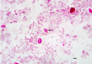 | |
| Fig. 1: | Smear of formalin-fixed stool sample. The microsporidian spore is pinkish-red and shows the distinct belt-like stripe (arrow). Bar = 1 μm (Modified Trichrome blue) |
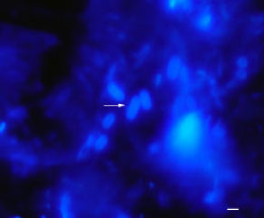 | |
| Fig. 2: | Smear of formalin-fixed stool sample. The microsporidian spore is ovoid and bluish (Arrow). Bar = 1 μm (Calcofluor white MR2) |
DISCUSSION
In this study the pig was chosen because it is the animal that harbors the pathogenic genera of microsporidia that have also been reported in humans (Sak et al., 2008). Unfortunately, in Mexico there is no information related to the presence of intestinal microsporidiosis in swine. The diagnosis of intestinal microsporidiosis has been difficult due to the small size of the spore and perhaps because it is easily unnoticed in routine diagnostic procedures. This is critical, since the definitive diagnosis of intestinal microsporidiosis depends, in most cases, on the direct observation of the spores in fecal samples (Didier et al., 1995). Microscopic examination of stained fecal smears for detection of microsporidia is a quick and noninvasive technique, which correlates with other improved techniques suggested for the determination of the parasite, such as ultrastructural (TEM), immunological (IFA) or molecular (PCR) methods (Ghosh and Weiss, 2009). However, these techniques are extremely costly, time consuming, require trained personnel and impractical when it comes to studying many samples.
For microscopic examination of microsporidian spores in stool samples, it is recommended to use special chitin stains, such as trichrome blue and calcofluor white MR2. In this study, we identified microsporidian spores in 96 fecal samples from a total of 120 adult pigs using those stains and represent the first finding of this parasite in Mexico in clinically healthy pigs. In this study, the definitive diagnosis was the identification of the polar tube, which appeared as a pink or reddish belt in the middle of the spore. Moreover, this morphological feature was a key to distinguish the microsporidian spores from yeasts and sporulated bacteria (Didier et al., 1995).
Both stains were effective in terms of detection of spores, but calcofluor white MR2 was more sensitive (Ghosh and Weiss, 2009). The identity of the parasite in this study remains uncertain; however, spores of E. bieneusi and E. intestinalis seem to be a common finding in pigs (Valencakova et al., 2006; Sak et al., 2008). Nonetheless, light-microscopy-based methods rarely make possible the complete identification of microsporidian species. Subsequent molecular studies will allow us to confirm the genotype of these parasites (Ghosh and Weiss, 2009).
Apparently, these parasites seem to be common in asymptomatic pigs in the studied farms and this coincides with the literature (Buckholt et al., 2002) which indicates that these parasites are most commonly identified in pigs, from clinically healthy farms. It was surprising that most of the pigs had intestinal microsporidiosis in a great proportion in all observed farms. These results are higher than that observed by others For example a study conducted by Breitenmoser et al. (1999), reported that 35% of pigs had Enterocytozoon bieneusi in faeces, whereas in another study only 18% had microsporidian spores (Buckholt et al., 2002). In present study, it is was not possible to determine whether the presence of these parasites in fecal samples are the result of an active infection or merely represent a traveling form of the parasite in the digestive tract of pigs. Nonetheless, this study demonstrates that intestinal microsporidiosis is widely diffused in the studied pig farms. This suggests that these animals could harbor infective spores and eliminating them into the environment. The possible source of infection at this stage remains undetermined, but it is likely that pigs are infected during an earlier stage of development by an unknown route. Colostrum or contaminated nipples with urine or faeces could be possible sources of transmission from the mother to the piglets (Sak et al., 2008). If this occurs, it is probable that pigs excrete spores throughout their lives and intestinal microsporidiosis remains undetectable in farms. Because the routine microscopic examination for microsporidia in faeces is not common in the country, we suggest using these stains as a practical first option for detection of infective spores.
Present study reports for first time the presence of intestinal microsporidiosis in pigs in Nuevo Leon, Mexico. However, this study covers only the stain identification of the spores and more studies are needed to establish the probable genotypes involved.
ACKNOWLEDGMENTS
We would like to thank to Martin Rivas Farias, DVM and Augusto Morales, DVM for their valuable assistance for help in collecting samples.
REFERENCES
- Breitenmoser, A.C., A. Mathis, E. Burgi, R. Weber and P. Deplazes, 1999. High prevalence of Enterocytozoon bieneusi in swine with four genotypes that differ from those identified in humans. Parasitology, 118: 447-453.
CrossRef - Buckholt, M.A., J.H. Leem and S. Tzipori, 2002. Prevalence of Enterocytozoon bieneusi in swine: An 18-month survey at a slaughterhouse in Massachusetts. Applied Environ. Microbiol., 68: 2595-2599.
CrossRef - Clarridge, J.E., S. Karkhanis, L. Rabeneck, B. Marino and L.W. Foote, 1996. Quantitative light microscopic detection of Enterocytozoon bieneusi in stool specimens: A longitudinal study of human immunodeficiency virus-infected microsporidiosis patients. J. Clin. Microbiol., 34: 520-523.
Direct Link - Didier, E.S., J.M. Orenstein, A. Aldras, D. Bertucci, L.B. Rogers and F.A. Janney, 1995. Comparison of three staining methods for detecting microsporidia in fluids. J. Clin. Microbiol., 33: 3138-3145.
Direct Link - Ghosh, K. and L.M. Weiss, 2009. Molecular diagnostic tests for microsporidia. Interdisciplinary Perspective Infect. Dis., 2009: 13-13.
CrossRef - Jeong, D.K., G.Y. Won, B.K. Park, J. Hur and J.Y. You et al., 2007. Occurrence and genotypic characteristics of Enterocytozoon bieneusi in pigs with diarrhea. Parasitol. Res., 102: 123-128.
PubMed - Sak, B., M. Kvac, D. Hanzlikova and V. Cama, 2008. First report of Enterocytozoon bieneusi infection on a pig farm in the Czech Republic. Vet. Parasitol., 153: 220-224.
PubMed - Valencakova, A., P. Balent, M. Huska, F. Novotny and L. Luptakova, 2006. First report on Encephalitozoon intestinalis infection of swine in Europe. Acta Vet. Hung., 54: 407-411.
PubMedDirect Link