
B. Venkatrajah
Department of Microbiology and Bioinformatics, CMS College of Science and Commerce, Coimbatore-642 006, Tamil Nadu, India
V. Vanitha Malathy
Department of Zoology, Kunthavai Naachiyar Government Arts College for Women, Tanjore, Tamil Nadu, India
B. Elayarajah
Department of Microbiology and Bioinformatics, CMS College of Science and Commerce, Coimbatore-642 006, Tamil Nadu, India
Mohan
Department of Science and Humanities, Sri Shanmuga College of Engineering and Technology, Sangagiri, Erode, Tamil Nadu, India
R. Rajendran
Department of Microbiology, PSG College of Arts and Science, Coimbatore, Tamil Nadu, India
Ram Rammohan
Department of Microbiology and Bioinformatics, CMS College of Science and Commerce, Coimbatore-642 006, Tamil Nadu, India
Journal of Medical Sciences
Year: 2012 | Volume: 12 | Issue: 6 | Page No.: 148-160


ABSTRACT
Wound healing is a complicated process with haemostasis, neovascularisation and re-epithelisation. To accelerate the process, moisture, sterile condition without microbial intervention and plant metabolites to influence the wound healing related metabolic functions are needed. The 40s Ne yarn was used to prepare the wound dressing cotton gauze. The physical properties such as areal density, air permeability and stiffness of gauze were analysed. Mixed polymer formulations like chitosan-sodium alginate, sodium alginate-calcium alginate and chitosan-sodium alginate-calcium alginate were prepared and coated separately on cotton gauze surface using pad-dry-cure method. The herbal extract of Bletilla striata was immobilized on the polymer coated fabrics to increase the rate of wound healing. The qualitative and quantitative antibacterial assays of coated cotton gauze was evaluated against the bacteria S. aureus ATCC 6538 and E. coli ATCC 10229 using the standard EN ISO 20654:2004 and AATCC testing methods. Animal model was used to reveal the wound closure effect of the prepared specimens. A novel design of various polymer coated cotton gauze fabric for wound healing with antimicrobial activity was developed using 40s Ne yarn. The cotton gauze with all the three polymers and herbal extract showed rapid wound healing effect due to excellent antibacterial activity by chitosan, moisture retaining by alginate and cell proliferation and tissue regeneration effect from B. striata extract in albino rat animal models.
PDF Abstract XML References Citation
Received: December 27, 2012;
Accepted: February 10, 2013;
Published: April 24, 2013
How to cite this article
B. Venkatrajah, V. Vanitha Malathy, B. Elayarajah, Mohan, R. Rajendran and Ram Rammohan, 2012. Biopolymer and Bletilla striata Herbal Extract Coated Cotton Gauze
Preparation for Wound Healing. Journal of Medical Sciences, 12: 148-160.
DOI: 10.3923/jms.2012.148.160
URL: https://scialert.net/abstract/?doi=jms.2012.148.160
DOI: 10.3923/jms.2012.148.160
URL: https://scialert.net/abstract/?doi=jms.2012.148.160
INTRODUCTION
Wound management is based on physiological support and the hygienic condition of the bandage material. Bandages are designed to perform specific functions depending upon the final medical requirements (Mi et al., 2002). As infections delay healing and increase scar formation, achieving closure as soon as possible is important (Yang and Lin, 2004). An effective wound dressing typically has the following properties: the dressing material must absorb any bodily fluids exuded from the wounded area and should permit evaporation of moisture to certain rate. It should not excessively adhere to the surrounding tissue to facilitate easy removal of used guaze. If not, additional damage to the wound may prolong recovery (Liu et al., 2008). An easily stripped wound dressing material must be developed to reduce the pain suffered by patients when wound dressings are frequently changed (Borkow and Gabbay, 2010).
Chitosan is a natural biopolymer that is derived from chitin is known in the wound management field for its haemostatic properties (Kumar, 2000). Its bioactive property of affecting macrophage prevalence in the wound site and helps in faster wound healing was noticeable (Mi et al., 2001). The other properties like bacteriostatic and fungistatic are useful in wound treatment. The important action of chitosan against microbial growth is the interaction of positively charged chitosan with the negatively charged residues at the cell surface of many fungi and bacteria which causes extensive cell surface alterations like cell wall permeability change. This results in the inhibition of normal microbial metabolism and cell death (Wang et al., 2006). Nanoparticles of chitosan have efficient bacterial growth inhibiting property when analysed in vitro against Staphylococcus aureus and Escherichia coli. Because of the smaller size (±400 nm), large surface area is available to have proper contact with bacterial pathogens and alter their cell membrane integrity to affect their growth. This study of Katas et al. (2011) revealed the antimicrobial function of chitosan in its various size forms. Similarly chitosan with varied molecular weight can exhibit difference in bacterial killing action. The observations of Batista et al. (2011) revealed the main inhibitory action of medium molecular weight chitosan is more effective by damaging the cell wall and membranes of bacteria.
Chitosan solubilized in weak acids can form intermolecular bonds to form a network which exhibits good tensile strength and elasticity to avoid breaking of the polymeric film structures made (Niamsa and Baimark, 2009). This shows the physical property enhancing nature of chitosan when bound with any natural fibers like cotton. Chitosan has enormous free amino group in its structure providing reactive nature to interact with other chemical compounds. The interlinking ability of chitosan to various chemicals is not limited. As shown in the work of Rana et al. (2009) chitosan can interact with heavy metals like arsenic for its removal in environmental clean-up process. The edible nature of chitosan extends its usage in organic substance preservation. Coating of chitosan on papaya fruit surface delays ripening, colour change and other quality loss. This is an evidence for the biocompatibility of chitosan and its protective action from external environment (Aleryani-Raqeeb et al., 2008). Hence the chitosan can act as protective covering polymeric materials of organic substances.
The synergistic role of chitosan with organic and inorganic substances was observed by many researchers. The edible film of chitosan immobilized with thyme essential oil showed enhanced antimicrobial effects by establishing synergistic effect (Hosseini et al., 2008). This reveals the ability of chitosan to remain unaltered when mixed with other antimicrobial agents.
Alginate derived from brown seaweeds is used as a reinforcing element for cell walls. It is used in various industries like food, textile and chemical as a thickening agent because of its gel forming ability (Skjak-Braek and Espevik, 1996). This bio-fibre have unique gel forming characteristics; on contact with wound exudates. The sodium ions in the wound exudates can exchange ions with the calcium in the fibres and by absorbing more water can form gel. The formed gel helps to keep a moist interface between the dressing and wound surface which can positively assist the healing process (Knill et al., 2004). According to the work of Gupta et al. (2007), the sodium alginate was used as an edible gelling agent with mango pulp. Alginate texturization of mango pulp improves its shelf life without nutrient loss and also microbiologically safe for up to 12 months. Synthetic seeds prepared for plant tissue culture using sodium alginate are advantageous according to Maqsood et al. (2012). The explants encapsulated by the polymer, grow embryos by nourishing and protecting from damages. Germination of embryos will also get advent. By the research work of Devendra et al. (2011) revealed the sodium alginate encapsulated embryos of rare hybrid plants can be propagated safely.
Based on the work of Francis Borgio (2011), Calcium alginate is an excellent encapsulating compound, the cell bound amylase entrapped by calcium alginate showed higher activity than the coconut coir fiber immobilized enzyme. This work shows the compatible nature of alginate polymer to enzymes.
Medical practice over a long period of time has demonstrated that the traditional Chinese medicine is highly biocompatible when it is in contact and interacts with human body tissues, body fluid or blood. The traditional Chinese medicine Bletilla striata has been used extensively to treat alimentary canal mucosal damage, ulcers, bleeding, bruises, wounds and burns. Chinese Pharmacopoeia states that B. striata supports functions of hemostasia and detumescence and promotes recovery (Diao et al., 2008). According to Lin et al. (2010) an efficient wound dressing is highly needed for the wounds, burns, surgical wounds or acute wounds with excessive bleeding. B. striata has high wound healing, haemostatis, detumescence and tissue granulating ability. It is used to recover patients from cuts, abrasion, toxins of wound tumescence, pulmonary haemorrhage and gastric ulcer. The presence of glysan, volatile oil and starch in B. striata can activate platelets, rapid thrombin production and stops protease activity. Hence, it shortens thrombin time, restricts fibrinolysis and forms artificial thrombus to yield haemostasia.
This study deals with the development and characterization of surgical bandage coated with polymer and herbal extract. It is expected that the coating of sodium alginate, calcium alginate, chitosan and their blends will impart moisture necessary for wound healing, easy removal of bandage while changing without tissue damage and antibacterial properties to the bandage for accelerated wound healing process without microbial intervention. Also the immobilization of Bletilla striata aqueous extract will improve the rate of wound healing property.
MATERIALS AND METHODS
Materials: Chitosan (deacetylation = 75%) was received from Sigma-Aldrich, USA. Calcium alginate and sodium alginate were procured from Hi-Media, India. Bletilla striata was obtained from Sing Long Pharmaceutical Co., Ltd. Taichung, Taiwan. The cotton yarns of 40s Ne was procured from Lakshmi Mills Ltd., Coimbatore, Tamil Nadu, India.
Gauze fabric production: The gauze bandage loom machine with 300 picks/min capacity was used to spin cotton gauze from the yarns procured from spinning mills. The cotton gauze was prepared at Department of Fashion Technology, Kumaraguru College of Technology, Coimbatore.
Properties of gauze fabric: Using the counting glass, physical properties of the cotton gauze like ends per inch and picks per inch were measured. As per ASTM D3775, areal density was measured using gsm cutter method. The other physical properties of cotton gauze such as thickness and stiffness were measured as per ASTM D 1777-96 and ASTM D 6828 standard methods, respectively.
Scouring and bleaching of fabrics: The cotton fabrics were scoured according to the method of (Mirjalili et al., 2011), using sodium hydroxide 3.0% (owm), sodium carbonate 2.0% (owm) and other auxiliaries as per the standard procedure using distilled water. These fabrics were then bleached with hydrogen peroxide of 4.0% volume and with other auxiliaries as per the standard procedure. The distilled water was used for the above processes. The processed cotton was designated as C1 (without any polymer coating).
Coating of cotton with chitosan-sodium alginate (C2): A modified method of Shanmugasundaram (2012) was used for polymer coating on cotton gauze surface. In brief, chitosan (8.0 g) was dissolved in 2.0% (v/v) aqueous acetic acid solution by stirring for 1 h at 60°C. Sodium alginate (2.0 g) was added to the chitosan solution and stirred for 10 min. The chitosan-sodium alginate coated cotton gauze was padded twice with the solution of same concentration to a wet pick of 80%. The padded fabric was dried at 80°C for 5 min and cured at 140°C for 3 min.
Coating of cotton with sodium alginate-calcium alginate (C3): Calcium alginate (8.0 g) was dissolved in 4.0% (w/v) sodium carbonate solution (100 mL) for 30 min at 40°C. Sodium alginate (2.0 g) was added to the calcium alginate solution and stirred for 10 min. The sodium alginate-calcium alginate coated cotton gauze (C3) was padded twice with the prepared polymer solution to a wet pick of 80%. The padded fabric was dried at 70°C for 3 min and cured at 120°C for 5 min.
Coating of cotton with chitosan, sodium alginate and calcium alginate (C4): Chitosan (4.0 g) was dissolved in 2.0% (v/v) aqueous acetic acid solution for 1 h at 60°C. Sodium alginate (2.0 g) was added to the chitosan solution and stirred for 10 min. The cotton gauze was padded twice with the solution of same concentration to a wet pick of 80%. The padded fabric was dried at 80°C for 5 min and cured at 140°C for 3 min. Calcium alginate (8.0 g) in was dissolved by stirring in 4.0% (w/v) sodium carbonate solution (100 mL) for 30 min at 40°C. The chitosan-sodium alginate coated gauze was now padded twice with the calcium alginate solution of same concentration to a wet pick of 80% to prepare chitosan-sodium alginate-calcium alginate (C4) cotton gauze. The padded fabric was dried at 70°C for 3 min and cured at 120°C for 5 min.
Bletilla striata extract preparation: The shade dried Bletilla striata whole plant was grounded and 40 g of ground specimen was homogenized and dispersed in 80°C double-distilled water for 4 h. The aqueous extracts were filtered to remove impurities and concentrated at 40°C using vacuum evaporation. The condensates were then freeze-dried to obtain the final powder used in this experiment (Liu et al., 2009).
Immobilization of Bletilla striata extract in polymer coated cotton: The herbal extract of Bletilla striata (6 mL) at a concentration of 20% was used for immobilization. The various polymer coated cotton fabric (C2, C3 and C4) was immersed separately in the herbal solution and incubated at room temperature for 24 h. After incubation, the cotton samples were washed with distilled water and dried at room temperature for 2 days.
Calculating the add-on percentage: Based on the method proposed by Shanmugasundaram (2012), the polymer and the herbal solution add-on percentage on the samples were calculated using the following relationship:
| (1) |
where, W1 is the weight of coated sample and W2, the weight of un-coated sample.
The polymer add-on percentage was calculated individually for all the polymer coated cotton samples. Similarly the herbal add-on percentage was also calculated after immobilizing the herbal solution in various polymer coated cotton samples.
SEM analysis: To analyze the presence of coated materials on the cotton fiber surface, scanning electron microscopic images were used. The samples were prepared by mounting them on aluminium stubs and using Sputter Cater they were coated with gold and magnified in SEM.
Assay for antibacterial properties: The antibacterial effect of the coated and uncoated cotton gauze was determined qualitatively by agar diffusion plate test using EN ISO 20645:2004 method against Staphylococcus aureus ATCC 6538 and Escherichia coli ATCC 10229. For quantitative analysis of bacterial reduction, AATCC 100-2004 test method was employed.
EN ISO 20645:2004 method employs a two layered agar plate for qualitative antimicrobial determination. The lower layer and upper layer have sterile culture medium (10±1 mL of Trypticase Soy Agar-TSA) and agar layer (TSA precooled to 45°C) with individual test bacteria (1x10⁸ cells) respectively (Erdem and Yurudu, 2008). On the surface of the solidified TSA plates the test gauze specimens (25±5 mm in diameter) were imprinted using sterile forceps. The agar plates with cotton gauze specimens were incubated for 18-24 h at 37°C. The antibacterial activity was determined by the extent of bacterial growth in the contact zone between agar and gauze specimen.
Bacterial growth inhibition zone was calculated using the formula:
| (2) |
where, H is the zone of inhibition in mm, D is the total diameter of specimen and inhibition zone in mm, d is the diameter of specimen in mm.
The standard bacterial strain which gets effectively inhibited by EN ISO 20654:2004 method was further analysed for its quantitative measurement by AATCC 100-2004 test method proposed by Pinho et al. (2011). The cotton samples both coated and uncoated in a size of 4.25±0.1 cm in diameter were placed in separate 250 mL glass jar with screw cap and inoculated with 1.0±0.1 mL of bacterial inoculum (1x10⁵ cells mL-1). The inoculated bacterial cells have contact with cotton specimen for 24 h. Sterilized distilled water (100 mL) was added into the jar and stirred vigorously for 1 min. After serial dilution, 1mL of the solution, from every dilution was plated on nutrient agar and incubated for 24 h at 37±2°C.
Wash fastness test (AATCC Test Method 124; Version-1996): Wash fastness test was performed using AATCC test method 124. The persistence of bound polymers and herbal extract on the surface of cotton gauze after certain number of washes can be determined by this test. By washing the treated cotton gauzes with AATCC Standard Reference Detergent WOB (without bleaching agent) the durability of antibacterial effect and the persistence of coated polymers after washing the gauzes were evaluated. After consecutive 1st and 3rd laundering cycles, using AATCC 100-2004 test method the antibacterial activity of the specimens were analyzed for bacterial reduction percentage.
Bacterial reduction percentage: The recovered bacterial colonies from the tested cotton gauze surface on the agar plates were counted and used in the following equation to calculate bacterial reduction percentage. The bacteria remain survive on the surface of before and after washed cotton gauze can be calculated.
| (3) |
where, A is the number of bacterial colonies from coated cotton gauze after inoculation over a contact time of 18 h and B is the number of bacterial colonies from uncoated cotton gauze after inoculation at 0 contact time.
Wound healing analysis: The prepared specimens (C1, C2, C3 and C4), for control the drug Teramycin ointment (Pfizer, USA) (Tarun and Gobi, 2012) and cotton gauze coated only with B. striata extract were used as wound healing dress material in female albino rats. The study was permitted by Institutional Animal Ethics Committee and was performed according to the international rules relating to animal experiments and biodiversity right. A group of 3 rats with more or less equal weight (150±10 g) were selected for every specimen. After acclimatization for a minimum of 2 days with standard pellet feed and water, the animals were anesthetized by intramuscular injection with 0.15 cc of Ketalar. To make the skin evident for wound creation, the hairs were removed and 70% ethanol was used to sterilize the skin surface. Using a 10 mm biopsy punch, the skin outer layer was excised in all the animal models and care was taken to make the wound with similar size and depth at dorsal interscapular region of each animal. The prepared cotton gauze specimens and standard ointment were placed on the wound and the progressive changes in the wounded areas during healing were monitored every other day for 15 days using a digital camera (14MP, Nikon, Japan).
Statistical methods: The results of every in vitro analysis of coated and uncoated cotton gauze formulations were compared statistically to understand the level of significance using SBSS (ver. 11.0) statistical software. The results were presented as Mean±standard deviation (SD). The mean values of different polymer mix and herbal solution coated cotton gauze were compared to analyze their antibacterial activity using Chi Square test method. Statistical significance was set at p<0.05. Using one-way analysis of variance (ANOVA) the analysis of wound closure was done and the values of p = 0.001 were considered statistically significant.
RESULTS
The properties of cotton gauze fabric prepared were listed in Table 1. The results revealed that the prepared cotton gauze has similar physical properties like the cotton gauze normally used in medical practices.
| Table 1: | Properties of cotton gauze fabric |
 | |
| The physical properties of cotton gauze was analysed according to ASTM standards methods | |
Polymer add-on in cotton gauze fabric: The polymer add-on percentage in cotton gauze fabric and given in Table 2. Using Eq. 1, the percentage of polymer added on to the surface of cotton gauze C2, C3 and C4 can be calculated. The results of polymer add-on percentage showed 42.4, 36.8 and 33.4%, respectively. Figure 1 reveals the presence of added polymer formulations on the surface of prepared cotton gauze.
Herbal extract add-on in cotton gauze fabric: The add-on percentage of Bletilla striata extract immobilized on various polymer coated cotton using Eq. 1 and listed in Table 3. The cotton gauze C2, C3 and C4 showed the herbal solution add-on percentage of 93.25, 92.15 and 95.76, respectively. From the results it is evident that the presence of different polymer mix in cotton gauze was not affecting the herbal extract uptake. To check whether the herbal solution uptake efficiency can be influenced or retarded by the coated polymers, uncoated cotton gauze was immobilized with herbal extract and the add-on percentage was found to be 97.83. When compared there was negligible difference in herbal solution add-on percentage of control gauze with herbal solution and the polymer mix cotton gauzes. The extract of B. striata influences tissue regeneration in wound areas and can promote cell proliferation.
SEM results: At different magnification from 100X to 5000X, the SEM image of specimens were recorded. Only at higher magnification, the coated molecules were clearly observed as seen in Fig. 2. The presence of coated material reveals the efficiency of the method used to add them on cotton gauze surface.
Antibacterial property assay: The antibacterial activity of the polymer mix and herbal extract coated cotton gauze was expressed as zone of inhibition diameter as an average (±Standard Deviation) of duplicate determination using the Eq. 2.
| Table 2: | Polymer Add-on in cotton gauze |
 | |
| Polymer add-on percentage calculation analyze the coating efficiency of the cotton gauze used, An average (±SD) of duplicate specimen add-on % were expressed | |
| Table 3: | Herbal solution Add-on in polymer coated cotton gauze |
 | |
| The herbal extract uptake efficiency of the cotton gauze decides the improved wound healing function. An average (± SD) of duplicate specimen add-on % were expressed | |
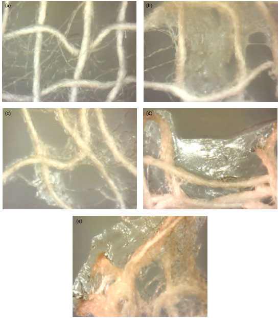 | |
| Fig. 1(a-e): | Stereo-zoom microscopic images of mixed polymer coated cotton gauze, (a) Normal cotton gauze (C1), (b) Chitosan-sodium alginate coated gauze (C2), (c) Sodium alginate and calcium alginate coated gauze (C3), (d) Chitosan, sodium and calcium alginate coated gauze (C4) and (e) Cotton gauze coated with polymer and B. striata extract |
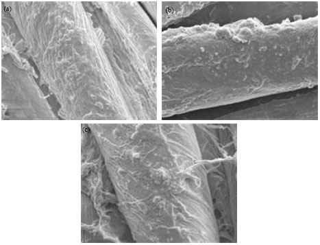 | |
| Fig. 2(a-c): | SEM analysis of polymer and B. striata herbal extract coated cotton gauze, (a) Chitosan-sodium alginate coated gauze (C2), (b) Sodium alginate and calcium alginate coated gauze (C3) and (c) Chitosan, sodium and calcium alginate coated gauze (C4) |
| Table 4: | EN ISO 20645:2004 antibacterial assay method |
 | |
| An average (±SD) of duplicate agar plate values were expressed, aGood effect, b Limited effect | |
The results tabulated in Table 4 indicate the bioefficacy of the used polymer mix along with herbal extract. The test exhibits good antibacterial activity against S. aureus ATCC 6538 and limited effect on E. coli ATCC 10229 (Fig. 3, 4).
Since the coated cotton gauze was effective against S. aureus ATCC 6538 than E. coli ATCC 10229, the bacterial reduction percentage was analysed for S. aureus ATCC 6538 by AATCC 100-2004. Using equation (3) the reduction percentage of S. aureus ATCC 6538 in C1 (uncoated cotton gauze) was found to be 0, whereas the cotton gauze C2, C3 and C4 showed 78, 45 and 79% bacterial reduction respectively before wash. Similarly, the first and the third washes exhibits admitted bacterial reduction percentage (Table 5).
Wash fastness test (AATCC Test Method 124; Version-1996): The colonizing ability of the bacterial cells which resist and survive the antibacterial attack of the coated polymer was analysed for the laundered specimens.
| Table 5: | Bacterial reduction percentage of S. aureus ATCC 6538 |
 | |
| An average (± std. deviation) of duplicate agar plate values were expressed | |
The coated and uncoated cotton gauze were washed for 1st and 3rd launderings to determine whether the polymers and herbal extract bound to the fabric would be durable through normal washings. After washing, the cotton gauze were sterilized and then assayed for antibacterial properties. Following Eq. 3 the reduction percentage of S. aureus (ATCC 6538) and E. coli (ATCC 10229) after the first and third wash of fabric was calculated (Table 5) and showed more than 70% bacterial reduction for chitosan-sodium alginate coated and chitosan-sodium alginate-calcium alginate coated cotton gauze. This provided the evidence for the antimicrobial activity persistence in the treated fabric after washes. Whereas, the cotton coated with calcium alginate-sodium alginate showed less than 50% bacterial reduction. This may be due to the absence of chitosan in the mixture coated on cotton gauze which was the bacterial killing agent. According to the AATCC standards, less than 50% of bacterial reduction is considered to be insignificant and more than 70% of bacterial reduction is acceptable.
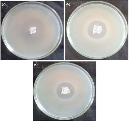 | |
| Fig. 3(a-c): | EN ISO 20645:2004 method of antibacterial assay against S. aureus ATCC 6538, (a) Growth inhibition zone of C2, (b) Growth inhibition zone of C3 and (c) Growth inhibition zone of C4 |
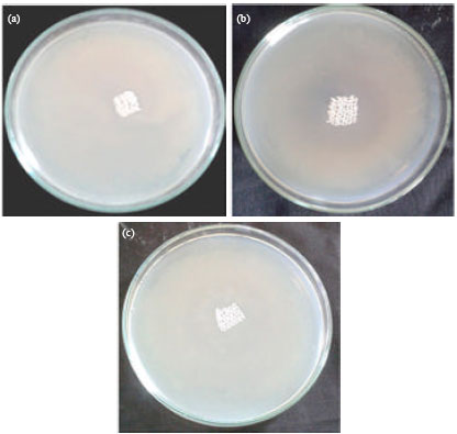 | |
| Fig. 4(a-c): | EN ISO 20645:2004 method of antibacterial assay against E. coli ATCC 10229, (a) Growth inhibition zone of C2, (b) Growth inhibition zone of C3 and (c) Growth inhibition zone of C4 |
Wound healing analysis: Wound healing analysis was done by measuring the area of wound closure. The albino rats in each group (n = 3) were analysed for wound closure and the mean values were used to calculate the efficiency of wound closure (Fig. 5).
| Table 6: | Wound healing test in animal model |
 | |
| The average mean values of triplicate sample result were used for statistical analysis | |
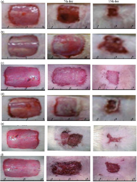 | |
| Fig. 5(a-f): | Wound healing test in animal model, (a) Wound healing by Teramycin ointment, (b) Wound healing ability of C1 (uncoated), (c) Wound healing ability of C2, (d) Wound healing ability of C3, (e) Wound healing ability of C4 and (f) Wound healing ability of B. striata extract |
From Table 6 it was evident that rapid wound closure occurs in animals treated with cotton gauze coated with chitosan-sodium alignate-calcium alginate and herbal extract solution. The efficiency of the remaining test specimens and standard ointment has healing effects with difference in wound closure among them.
DISCUSSION
The looming process adopted has 300 picks/min, prepared the cotton gauze with needed areal density, thickness and stiffness. These physical properties are analysed as per the standard ASTM methods to compare with normal cotton gauze used in hospitals. This ensures that the prepared cotton gauze has the prescribed physical properties as like normal wound dressing gauze. The physical properties of cotton gauze have direct influence with the add-on percentage of the coating drugs, their analysis are needed for further study.
The add-on percentage of the polymer mix with cotton gauze showed not much difference in their uptake values. As the dissolved substances are polymers, the thickness of the polymer solution is slightly denser than the water consistency. From the obtained results it is evident that the presence of sodium and calcium alginates can form thick gel than chitosan. Hence the uptake value of the polymer mixes with both sodium and calcium alginates showed lesser add-on percentage. The gel and sticky nature of the polymer mix prepared can show good adsorbent property with the gauze surface. Moisture is an important factor for wound healing and the alginate ions presence will maintain moisture due to their gel forming ability by ion exchange mechanism. The wound with dryness will cause damage to regenerated tissues while change of wound dressing and cause severe pain and fear of wound dressing to patients (Gupta and Saxena, 2011). The sodium ions of wound secretions will exchange ions with calcium in coated cotton gauze surface and form gel which can retain moisture for long duration. The prevalence of moisture enables change of wound dressing without damage the healing wound. The presence of alginate provides a moist environment for rapid granulation and reepithelialisation of wounded tissues (Kim et al., 1999).
The add-on percentage of herbal extract by prepared cotton gauze was efficient in chitosan present polymer mix and exhibits the uptake efficiency of chitosan. The coated herbal extract consists only of minute herbal particles and hence the B. striata extract add-on percentage is less comparatively with polymer add-on percentage. As the cotton fibers and coated polymers can absorb herbal extract, the add-on percentage of herbal solution is influenced. When the result of the uncoated cotton gauze herbal solution uptake efficiency was compared with polymer coated gauzes there was negligible difference in herbal solution add-on percentage. This reveals that polymer presence was not affecting herbal solution uptake efficiency of cotton gauze rather influenced. The immobilization of B. striata aqueous extract on polymer coated gauzes will influence tissue regeneration in wounded areas and can promote cell proliferation. The aqueous extract has been used instead of solvent extract, as the wound healing polysaccharides in B. striata will be extracted effectively using aqueous solution (Liu et al., 2009).
The magnification of the prepared cotton gauze with polymer mix and herbal solution revealed their presence and the efficiency of coating method used in this analysis. Under optimal conditions, the magnification of the specimen will be appropriate and a high resolution image can be produced (Gomes et al., 2010). The SEM images created with the used specimen at higher magnification can be a good evidence for the coated substance presence. Hence, the cotton gauze becomes functionalised by retarding bacterial colonization with rapid wound closure function.
EN ISO 20645:2004 standard test method evaluates the sensitivity pattern of the bacterial pathogens used in this study. Based on this method, = 1-0 mm sized inhibition zone and no growth under specimen are accepted as effective, whereas 0 mm sized inhibition zone and slight growth are evaluated as limited effect. According to the standards prescribed by early works (Erdem and Yurudu, 2008), among the two bacterial pathogens used for analysis, the sensitivity pattern of E. coli ATCC 10229 is high against the coated polymers.
Agar diffusion test is a preliminary test to detect the diffusive antimicrobial finish. Antibacterial activity of tested gauze against Gram positive cocci is effective than Gram negative rods. Due to presence of pores in peptidoglycan layer of Gram positive cocci cell wall, they are sensitive to chitosan and Gram negative rods have strong cell wall arrangement to resist chitosan attack (Je and Kim, 2006). The membrane-active microbiostatics action of chitosan is responsible for bacterial inhibitory effect by altering the cell membrane permeability leading to bacterial killing action.
The bacterial reduction percentage of uncoated and polymer coated cotton showed significant differences in their antibacterial ability. This indicated that even when mixed with other polymers and herbal extract, the chitosan bound to the cotton fabric retained its antibacterial effect. According to Chun and Gamble (2007), the reactive nature of the antibacterial compounds is the main concern when coated in mixed form on fabric surfaces. The bacterial morphology has a known importance in their analysis. The bacterial cell morphology get alters in its size and shape when exposed to antibiotics indicating its antibiotic resisting capability. Based on their resisting capability, the cell size will get decrease or enlarge. When exposed to bacteriostatic doses of antibiotics the bacterial structures alters drastically rather exposed to natural antimicrobial agents like chitosan (Gomes et al., 2010). A permanent coating of natural agents is actually essential for long term beneficial action against microbes. Based on the work of El-tahlawy et al. (2005) for a permanent antibacterial finish in cotton, cross linking agents are essential. As the cotton retains the coated drugs after launderings, cross linker addition is suitable for the fabrics that are reused. In this work cross linkers are not used on cotton gauze to have a permanent antibacterial finish, as they are not clinically reused.
In animal models the wound closure analysis is done using the prepared cotton gauze and compared with the standard ointment every other day (Tarun and Gobi, 2012). The wound healing effect by C4 test specimen is observed rapid by checking the closure of wounded area. C4 test specimen coated with both calcium and sodium alginate along with chitosan and herbal extract of Bletilla striata helps to maintain moisture than the C1 test specimen (uncoated). The presence of polymer leads to the formation of gel through ion exchange process that influences moisture essential for wound healing. The bioactive property of chitosan affects macrophage prevalence in the wound site for improved wound healing. The other properties like bacteriostatic and fungistatic are particularly useful for wound treatment. The presence of chitosan will provide antibacterial effect which helps the wounded cells to regenerate without any microbial intervention. Hence, the presence of both sodium-calcium ions along with alginate and chitosan accelerates wound healing effect. The herbal extract of B. striata enhances the wound healing action. The presence of polysaccharide in its extract is responsible for enhancing vascular endothelial cell proliferation and vascular endothelial growth factor expression (Diao et al., 2008). This leads to the healing of wound in accelerated fashion along with the antibacterial action of chitosan and moisture retaining property of alginate.
One of the complex processes with series of metabolic functions and changes in human body is the wound healing mechanism. Blood clotting is the initial change that takes place once a wound was made. This was followed by slight inflammation leading to haemostasis. In the present study, during initial hours after wounds were made, they were associated with the same reaction changes. The development of fibrous tissues and blood vessels are the wound healing process that take place in the first week. At the start of second week, soft tissues consisting mainly of tiny blood tissues and fibres were formed over the wound. In the end of second week, new epithelial cells were formed to form a skin cover to the wound. In the third week, new extracellular matrix and remodelling of tissues takes place to make wound closure.
CONCLUSION
The wound dressing material used in this study was cotton gauze prepared from cotton yarns of 40s Ne. Their physical properties were analysed to ensure proper polymer add-on percentage with cotton fibers. Three different polymer mix were prepared containing chitosan-sodium alginate, chitosan-calcium alginate and chitosan-sodium-calcium alginate and the aqueous extract of B. striata was immobilized on their surface. The add-on percentage of both polymer and herbal extract was calculated and revealed appropriate. SEM analysis showed the presence of added drugs along with cotton fibers in the images. The antibacterial activity of the coated cotton gauze was both qualitatively and quantitatively analysed against S. aureus (ATCC 6538) and E. coli (ATCC 10229). The ability of coated polymers to resist the abrasive force by laundering procedure was analysed using wash fastness test. The coated herbal solution enhances cell proliferation and growth factor expression. This results in rapid wound healing action as there was no microbial intervention due to chitosan presence and moisture prevalence by alginate presence. The efficiency of the functionalised cotton gauze was analysed using albino rats as animal models. The test results reveals better wound healing efficiency for the cotton gauze coated with chitosan, alginates of sodium and calcium and B. striata herbal extract.
ACKNOWLEDGMENT
We are grateful to acknowledge Hi-Wave Pharmaceuticals, Puducherry for providing the needed facilities. We also thank CMS Educational and Charitable Trust for helping us to conduct this research work and the Principal, Kunthavai Nachiar Govt. Arts College for Women to finish the work.
REFERENCES
- Aleryani-Raqeeb, A., T.M.M. Mahmud, S.R.S. Omar and A.R.M. Zaki, 2008. Effects of calcium infiltration and chitosan coating on storage life and quality characteristics during storage of papaya (Carica papaya L.). Int. J. Agric. Res., 3: 296-306.
CrossRefDirect Link - Gomes, A.P., J.F. Mano, J.A. Queiroz and I.C. Gouveia, 2010. Assessment of Bacteria-Textile Interactions using Scanning Electron Microscopy: A Study on LbL Chitosan/Alginate Coated Cotton. In: Microscopy: Science, Technology, Applications and Education, Mendez-Vilas, A. and J. Diaz (Eds.). Formatex Research Center, Badajoz, Spain, ISBN-13: 9788461461899, pp: 286-292.
Direct Link - Batista, A.C.L., G.C. Dantas, J. Santos and R.V.S. Amorim, 2011. Antimicrobial effects of native chitosan against opportunistic gram-negative bacteria. Microbiol. J., 1: 105-112.
CrossRef - Gupta, B. and S. Saxena, 2011. Chitosan-polyethylene glycol coated cotton membranes for wound dressings. Indian J. Fibre Text. Res., 36: 272-280.
Direct Link - Chun, D.T.W. and G.R. Gamble, 2007. Textile technology using the reactive dye method to covalently attach antibacterial compounds to cotton. J. Cotton Sci., 11: 154-158.
Direct Link - Gupta, D.K.D., N. Roopa and R.K. Leela, 2007. Development of stable restructured mango gel. Am. J. Food Technol., 2: 176-182.
CrossRefDirect Link - Devendra, B.N., N. Srinivas and G.R. Naik, 2011. Direct somatic embryogenesis and synthetic seed production from Tylophora indica (Burm.f.) Merrill an endangered, medicinally important plant. Int. J. Bot., 7: 216-222.
CrossRefDirect Link - Diao, H., X. Li, J. Chen, Y. Luo and X. Chen et al., 2008. Bletilla striata polysaccharide stimulates inducible nitric oxide synthase and proinflammatory cytokine expression in macrophages. J. Biosci Bioeng., 105: 85-89.
CrossRefDirect Link - El-Rahlawy, K.F., M.A. El-Bendary, A.G. El-Hendawy and S.M. Hudson, 2005. The antimicrobial activity of cotton fabrics treated with different crosslinking agents and chitosan. Carbohydr. Polym., 60: 421-430.
CrossRef - Pinho, E., L. Magalhaes, M. Henriques and R. Oliveira, 2011. Antimicrobial activity assessment of textiles: Standard methods comparison. Ann. Microbiol., 61: 493-498.
CrossRef - Borgio, J.F., 2011. Immobilization of microbial (wild and mutant strains) amylase on coconut fiber and alginate matrix for enhanced activity. Am. J. Biochem. Mol. Biol., 1: 255-264.
CrossRefDirect Link - Borkow, G. and J. Gabbay, 2010. Preventing pathogens proliferation and reducing potential sources of nosocomial infections with biocidal textiles in developing countries. Open Biol. J., 3: 81-86.
Direct Link - Je, J.Y. and S.K. Kim, 2006. Antimicrobial action of novel chitin derivative. BBA-Gen. Subjects, 1760: 104-109.
PubMed - Lin, J.H., C.T. Lu, J.J. Hu, Y.S. Chen, C.H. Huang and C.W. Lou, 2010. Property evaluation of Bletilla Striata/ Polyvinly alcohol nano fibers and composite dressings. J. Nanomater., Vol. 2010.
CrossRefDirect Link - Kim, H.J., H.C. Lee, J.S. Oh, B.A. Shin and C.S. Oh et al., 1999. Polyelectroylte complex composed of chitosan and sodium alginate for wound dressing application. J. Biomater. Sci. Polym., 10: 543-556.
PubMedDirect Link - Erdem, A.K. and N.O.S. Yurudu, 2008. The evaluation of antibacterial activity of fabrics impregnated with dimethyltetradecyl (3-(trimethoxysilyl) propyl) ammonium chloride. IUFS J. Biol., 67: 115-122.
Direct Link - Knill, C.J., J.F. Kennedy, J. Mistry, M. Mi-raftab, G. Smart, M.R. Groocock and H.J. Williams, 2004. Alginate fibres modified with unhydrolysed and hydrolysed chitosans for wound dressings. Carbohydr. Polym., 55: 65-76.
CrossRef - Liu, B.S., C.H. Yao and S.S. Fang, 2008. Evaluation of a non-woven fabric coated with a chitosan bi-layer composite for wound dressing. Macromol. Biosci., 8: 432-440.
CrossRef - Kumar, M.N.V.R., 2000. A review of chitin and chitosan applications. React. Funct. Polym., 46: 1-27.
CrossRefDirect Link - Maqsood, M., A. Mujib and Z.H. Siddiqui, 2012. Synthetic seed development and conversion to plantlet in Catharanthus roseus (L.) G. Don. Biotechnology, 11: 37-43.
CrossRefDirect Link - Mi, F.L., S.S. Shyu, Y.B. Wu, S.T. Lee, J.Y. Shyong and R.N. Huang, 2001. Fabrication and characterization of a sponge-like asymmetric chitosan membrane as a wound dressing. Biomaterials, 22: 165-173.
CrossRef - Mi, F.L., Y.B. Wu, S.S. Shyu, J.Y. Schoung, Y.B. Huang, Y.H. Tsai and J.Y. Hao, 2002. Control of wound infections using a bilayer chitosan wound dressing with sustainable antibiotic delivery. J. Biomed. Mater. Res., 59: 438-449.
CrossRef - Hosseini, M.H., S.H. Razavi, S.M.A. Mousavi, S.A.S. Yasaghi and A.G. Hasansaraei, 2008. Improving antibacterial activity of edible films based on chitosan by incorporating thyme and clove essential oils and EDTA. J. Applied Sci., 8: 2895-2900.
CrossRefDirect Link - Niamsa, N. and Y. Baimark, 2009. Preparation and characterization of highly flexible chitosan films for use as food packaging. Am. J. Food Technol., 4: 162-169.
CrossRefDirect Link - Rana, M.S., M.A. Halim, S.A.M. Waliul Hoque, K. Hasan and M.K. Hossain, 2009. Bioadsorbtion of arsenic by prepared and commercial crab shell chitosan. Biotechnology, 8: 160-165.
CrossRefDirect Link - Shanmugasundaram, O.L., 2012. Development and characterization of cotton and organic cotton gauze fabric coated with biopolymers and antibiotic drugs for wound healing. Indian J. Fibre Text. Res., 37: 146-150.
Direct Link - Tarun, K. and N. Gobi, 2012. Calcium alginate/PVA blended nano fibre matrix for wound dressing. Indian J. Fibre Text. Res., 37: 127-132.
Direct Link - Yang, J.M. and H.T. Lin, 2004. Properties of chitosan containing PP-g-AA-g-NIPAAm bigraft nonwoven fabric for wound dressing. J. Membr. Sci., 243: 1-7.
CrossRef - Mirjalili, M., S.S. Nasirian and L. Karimi, 2011. Effects of corona discharge treatment on some properties of wool fabrics. Afr. J. Biotechnol., 10: 19436-19443.
CrossRefDirect Link