
A.S.A. Haffor
Department of Zoology, College of Science, P.O. Box 2455, King Saud University, Asian Network for Scientific InformationRiyadh-11451, Saudi Arabia
Journal of Medical Sciences
Year: 2004 | Volume: 4 | Issue: 2 | Page No.: 164-169


ABSTRACT
The purpose of this study was to examine the effects of oxygen breathing on cardiac mitochondrial injury, GOT and free radical (FR) production. Twelve rats (SRW) were exposed to oxygen (100% O2) breathing, for 4-6 h daily for 4.5 weeks. Ultrastructure examination was conducted by transmission electron microscope, GOT and FR determinations were conducted using the kinetic method. Myocardial ultrastructure examination showed mitochondrial hyperplasia, cristae dislocation, matrical lamina formation and secondary lysosomal structure. Baseline FR and GOT were 179.5 Carr U and 35.43 U L-1, respectively. Following exposure to oxygen breathing FR increased to 348.50 Carr U and GOT increased to 75.38 U L-1. Both the changes in FR and in GOT were significant (P<0.05). The regression of GOT on FR after oxygen breathing was significant (P<0.05). Mitochondrial and cellular pathological abnormalities were attributed to oxidative damage, induced by oxygen breathing that resulted in increased rate of free radicals production and the subsequent elevation in GOT.
PDF Abstract XML References Citation
How to cite this article
A.S.A. Haffor, 2004. Effects of O2 Breathing on Cardiac Mitochondrial, GOT and Free Radical Production. Journal of Medical Sciences, 4: 164-169.
DOI: 10.3923/jms.2004.164.169
URL: https://scialert.net/abstract/?doi=jms.2004.164.169
DOI: 10.3923/jms.2004.164.169
URL: https://scialert.net/abstract/?doi=jms.2004.164.169
INTRODUCTION
Oxygen breathing has variety of medical applications such as treatment for intoxication, soft tissue infections, traumatic ischemia, intensive care and post surgery. However, 100% oxygen breathing causes oxidative stress which is related to free radical production and peroxidation of biomolecules. Damages from cells, especially myocardium, leak into circulation and contribute to Glutamate-Oxaloacetate transaminase (GOT/ASAT) concentration in the blood.
The chemistry of oxygen and its derivatives has been extensively reviewed[1,2]. The diatomic oxygen (O2) is reactive itself due to the fact that its two unpaired electrons are located in different molecular orbital and possess a parallel spins. As a result, O2 accepts electrons, one at a time, from other biological radicals such as hydroxyl radical (*OH) and the superoxide radical (*O2) which together with the hydrogen peroxide (H2O2) and the singlet oxygen (1* O2), are the reactive oxygen species (ROS).
Most authorities are in agreement that mitochondria are the main source for free radicals (FR) production[3-9]. In addition, mitochondria DNA (mtDNA) damage occurs much faster than does nuclear DNA because mtDNA is not protected by proteins, rather it is attached to the inner mitochondrial membrane[10-13]. Further, it has been shown that mitochondria are easily affected by oxygen toxicity in birds[14] and in mammals[15-16].
Information regarding the side effects of 100% oxygen breathing on cardiac cellular and mitochondria has not been reported. The purpose of the present study was to examine mitochondria pathological changes on the myocardium induced by 100% oxygen in relation to GOT and FR.
MATERIALS AND METHODS
Experimental design: Eight rats (SRW) male with mean age and weight of 14 weeks and 267 g, respectively, underwent 100% oxygen exposure for 4.5 h daily for 4 weeks period. All measurements were taken before and after 100% oxygen treatment.
Statistical analysis: Mean group differences for FR and GOT were evaluated using paired t-test. A multiple regression model for GOT versus FR relationship was generated using curve linear regression. Probability plot was used to estimate the population hypothesized predicted values. Durbin-Watson statistic was used to evaluate the validity of the regression model.
Oxygen exposure: Animals were placed in a closed box that has an inlet and outlet. The inlet was connected to 100% O2 medical grade tank in which the flow was maintained at 5 L per min (LPM). The regulator of the tank was provided with a humidifier in order to saturate the inspired air with H2O. The outlet of the box was connected to vacuum line that was adjusted at 3 LPM to ensure that the concentration of oxygen in the box would be approximately equal to the concentration in the tank.
Tissue samples collection: Eight rats were sacrificed and cardiac tissue samples were collected to serve as control. Another eight rats were exposed to 100% oxygen for 4.5 h daily for six week, then sacrificed. Tissues samples were selected by random from each group and prepared as described below.
Ultrastructure procedures: Tissue samples were immediately fixed in 3% buffered glutaraldhyde (0.1 M cacoddylate buffer at pH 7.4 for 4 h at 2 to 4°C. The tissue samples were washed then were post fixed in 1% osmium peroxide in the buffer for 2 h at 2-4°C, then washed and kept overnight. Fixed tissue samples were dehydrated in graded concentrations of ethyl alcohol, (30, 50, 70 and 90%) for 30 min each and finally in absolute ethanol (100%) for 40 min. Tissues were infiltrated gradually in resin and embedded in plastic capsules in fresh full strength agar 100 epoxy resins before being cured at 60-70°C for 2 days. Dehydrated tissue samples were embedded in epon and araldite mixture. Polymerized resin blocks containing tissue samples were prepared for sectioning, first semi-thin sections (1 μm) which were stained with toluidine blue for purpose of orientation. Accordingly ultra sections (70 nm) were made and double stained with uranyl acetate and lead citrate. Ultra sections were mounted on carbon-coated grids, then examined and photographed by Transmission Electron microscope (JEOL-100 CX) at 80KV.
Blood samples collection: Blood samples were collected into heparin chilled glass vile. Twenty μl of whole blood was analyzed for FR immediately. The remaining of sample was centrifuged immediately at 3,000 rpm for 10 min and then analyzed for GOT concentration.
Kinetic test for free radical determination: Free radical was measured according to d-ROMs test procedure (FERAS-II, Italy). The test measures the levels of hydrogen peroxide (R-OOH) which are generated by peroxidation of biological compounds; lipid, amino acids and nucleic acids.
Assay principle: The test is a colorimetric test and based on the principle of the ability of hydrogen peroxides to generate free radicals after reacting with some transitional metals (Fe2+/Fe3+), according to Fenton’s Reaction as follows:
Thus, the hydrogen peroxides of biological sample (whole blood) generate free radicals (alcoxy and peroxyl radicals) after exposure to a transitional metal (Fe++/Fe+++). When a correctly buffered chromogen substance (N, N-diethyl-phenylendiamine) lead to the reduction of hydrogen peroxides which in turns colored as radical cation. Color intensity was read using IRAM photometer, at 505 nm. Alcoxyl and peroxyl radicals derived from the hydrogen peroxides, in the d-ROMs test were expressed in “CARR UNITS” (CARR U), where one CARR U correspond to 0.08 mg 100-1 ml H2O2.
Kinetic determination of GOT: Kinetic rate method providing quantitative determination of aspartate aminotransferase in plasma. Aspartate amiontransferase catalyzes the reversible transamination of L-aspartate and a-ketoglutarate to oxaloacetate and L-glutamate. The oxaloacetate is then reduced to malate (via malate dehydrogenase) with concurrent oxidation of reduced NADH to b-nicotinamide adenine dinucleotide. Sample and reagent were mixed and changes in absorbance were read using UV/Visible spectrophotometer (Pharmacia-UltraSpec-2000).
RESULTS
Cardiac muscle before oxygen breathing: Arrangement of the thin and thick filaments, mitochondria and the sarcoplasmic reticulum (T-system) were clearly observed (Fig. 1). The regularity of the Z-lines that were separating the boundaries at the beginning and end of the sarcomeres were also observed. Mitochondria distributions were homogenous and their locations and size were normal.
Mitochondrial swelling and secondary lysosomal structure after oxygen breathing: The cloudy swelling (Fig. 2) reflected water accumulation and hydrogen peroxide, H2O2 accumulation. Although, unable to measure water accumulation but able to measure FR production based on H2O2. Thus the observed cloudiness reflected malfunction of osmotic control secondary to a failure of various hydro and lipid peroxidase movement across the inner mitochondria membrane leading to flooding of inner and outer membrane.
| Table 1: | GOT and FR before and after 100% oxygen breathing |
 | |
| *One Carr U is equivalent to 0.08 mg H2O2; values are average of triplicate measurements | |
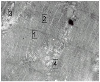 | |
| Fig. 1: | Heart muscle; 1) regular Z-line, 2) normal I band, 3) normal mitochondria size, 4) normal SR orientation (X = 10,000) |
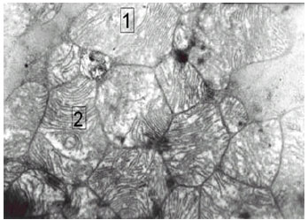 | |
| Fig. 2: | Swelling of the mitochondria (MS) accompanied by an increase in mitochondria mass and an increase in the proliferation of the cristae. Flooding of inner and outer membranes. Note, mitochondria swelling (MS) with increase in size and dilution of the inner membrane (X=20, 000) |
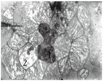 | |
| Fig. 3: | Lysosomal secondary structure indicating a detoxication mechanism of survival; 1) Remainants of digested organelles, 2) deterioration of the cristae and formation of lamellar bodies |
 | |
| Fig. 4: | The regression of GOT on FR after oxygen breathing |
 | |
| Fig. 5: | Normal probability plot which shows 50% probability are likely to be oxidatively stressed at 300 Carr U |
 | |
| Fig. 6: | Residual plot showing that the unexplained variance departed symmetrically from zero |
In addition, mitochondria swelling which was accompanied by an increase in mitochondria mass and hence reflect an increase in the concentration proliferation of the cristae as well as lamina formation. Evidently these mitochondrial pathological changes represent the cellular basis accounted for changes in the permeability of the outer and inner mitochondria membrane as evident by the rise in free radicals production rate.
Furthermore, secondary lysosomal structure was obvious indicating a detoxication mechanism for survival. The remainants of digested organelles, as well as the deteriorated cristae and formation of lamellar bodies were clearly shown (Fig. 3).
Effects of oxygen breathing on GOT and FR: Baseline means±SE GOT were 35.43±4.25 U L-1. Results of correlated t-test showed that, following exposure to 100% oxygen breathing GOT increased significantly (P<0.05) to 75.38±10.96 U L-1 (Table 1). Baseline mean free radical was 179.5±15.17 Carr U (Table 1), which corresponds to 14.40 mg 100 ml-1 H2O2. Following exposure to oxygen breathing, FR increased significantly (P<0.05) to 348.50±38.22 Carr U (Table 1).
GOT and FR regression model after oxygen breathing: The relationship between GOT and FR production after exposure to 100% oxygen breathing was significant (P<0.05). The regression model was nonlinear and can be described as follows:
Coefficient of determination (R2) was 0.55, which means there were 55% of the variability in GOT explained by the variability in FR after oxygen breathing (Fig. 4). The probability plot of the predicted population value, based on sample proportion (Fig. 5) which indicated that the success rate for oxidative stress is more likely to occur at or above 50%, when FR production at or above 300 Carr U. This value of FR is equivalent to 24 mg H2O2 100 ml-1 of blood.
The regression model was evaluated using residual plot. Figure 6 illustrates that the residual variance was symmetrically depart from zero at all levels. In other words the residual variance of GOT stayed about constant al all levels thus the errors associated with the model was random. As the residual is negative at low and high values thus the model was better described by nonlinear rather than linear regression model.
DISCUSSION
The major findings of the present study showed that oxygen breathing resulted in variety of pathological mitochondrial changes in the myocardial that was associated with repeated hyperplasia which were interpreted as a compensatory replacement mechanism. These cellular damages induced by 100% oxygen breathing had been reported in liver tissue[14] and in other tissues[17] which were related to elevation in GOT concentration. In this regard GOT measurement has been used in the diagnosis and treatment of certain liver diseases and heart diseases. It has been shown that the growth of houseflies in an atmosphere of 100% O2 markedly reduced their mean and maximum life span and increases the rate of accumulation of protein carbonyls in whole body extracts[18] and in isolated mitochondria[8]. Similarly, elevated atmospheric O2 decreased the mean and maximum life spans of nematodes[19].
The second major finding of this study was the increased rate of free radicals production following exposure to 100% oxygen breathing. Most authorities are in agreement that mitochondria are the major oxygen radicals producing sites. Mitochondria release hydrogen peroxide to the cytosol, leading to imbalance between its generation and elimination by cellular antioxidants[4,20-23] and DNA[13]. Results of the present study showed a significant (P<0.05) regression model between FR and GOT which confirmed the relationship between cellular damages, GOT activity and FR production.
It has been reported[15-16] when tissue antioxidant were directly studied as a function of mean life span (MLSP) had proved that FR production is lower in long-lived than in short-lived species. Pigeon MLSP is nine folds higher (35 years) than rats (4 years), whereas BMR and body size are similar. It was found that pigeons had less mitochondrial reactive oxygen species (ROS) generation than rats in all the organs studied that include brain, liver, lungs, heart and kidney[6,18]. It was also found that pigeons had less ROS in respiratory chain. When pigeon were exposed to hyperoxia[14], oxidative stress was more profound as evident by variety of mitochondrial pathology, giant, swelling, proliferation, fusion, golf-court shape and ring shape. Thus it is the rate of ROS production rather than removal which correlates negatively with MLSP. It can believe that the continued rise in free radical production along with failure of endogenous defense mechanisms to effectively neutralize H2O2 toxic intermediates and to prevent significant free radical injury have become an important issue as it apply to environmental medicine studies.
In conclusion, exogenous 100% oxygen breathing resulted in mitochondrial pathological changes, an elevation in GOT and an increased rate of FR production. Oxygen therapy should be titrated carefully to minimize its toxic effects.
ACKNOWLEDGMENTS
This project was supported by a Grant Number ZOO/1423/03 for Scientific Research in Physiology from the Faculty of Science, Office of Research, King Saud University, Riyadh, Saudi Arabia. Special appreciation is extended to the Director of the Office of Research and the Dean of the Faculty of Science at King Saud University, Riyadh.
REFERENCES
- Halliwell, B. and M. Dizdaroglu, 1992. Commentary the measurement of oxidative damage to DNA by HPLC and GC/MS techniques. Free Radic. Res. Commun., 16: 75-87.
CrossRefDirect Link - Sohal, R.S. and A. Dubey, 1994. Mitochondrial oxidative damage, hydrogen peroxide release and aging. Free Radic. Biol. Med., 16: 621-626.
PubMed - Barja, G., 1999. Mitochondrial free radical generation sites of production in state 4 and 3, organ specificity and relationship with aging rate. J. Bioenergy Biomembr., 31: 347-366.
CrossRefDirect Link - Epe, B., 1996. DNA damage profiles induced by oxidatizing agents. Rev. Physiol. Biochem. Pharmcol., 127: 223-249.
PubMedDirect Link - Berlett, B.S. and E.R. Stadtman, 1997. Protein oxidation in aging, disease and oxidative stress. J. Biol. Chem., 272: 20313-20316.
Direct Link - Beuchat, C.A. and C.R. Chong, 1998. Hyperglycemia and its consequences for hemoglobin glycation. Comp. Biochem. Physiol., 120: 409-416.
PubMedDirect Link