
S. Chandra Mohan
Research and Development Centre, Bharathiar University, Coimbatore, 641 046, Tamil Nadu, India
K. Sasikala
Department of Chemistry, Saranathan College of Engineering, Tiruchirappalli, 620 012, Tamil Nadu, India
T. Anand
Department of Biochemistry, PRIST University, Thanjavur, 613 403, Tamil Nadu, India
P.C. Vengaiah
Horticultural Research Station, Y S R Horticultural University, Pandirimamidi, 533288, Rampachodavram (PO), East Godavari Dist, Andhra Pradesh, India
S. Krishnaraj
Department of Physics, PRIST University, Thanjavur, 613 403, Tamil Nadu, India
Research Journal of Microbiology
Year: 2014 | Volume: 9 | Issue: 3 | Page No.: 142-150


ABSTRACT
This study aims to synthesis of silver nanoparticles using aqueous extract of Canthium comandelicum leaves and evaluates its antibacterial activity, as well as antioxidant activity. Characterizations of silver nano particles (AgNPs) were determined by using ultraviolet-visible (UV-Vis) spectrophotometry, Scanning Electron Microscopy (SEM) and X-ray diffraction. The ultraviolet and visible absorption spectroscopy results show a strong resonance centered on the surface of silver nanoparticles at 430 nm. SEM showed the formation of silver nanoparticles were polycrystalline with nano sized grains. X-ray diffraction analysis showed that the particles were crystalline in nature with face centered cubic structure of the bulk silver with the broad peaks at 38.16, 44.44 and 64.20. The bactericidal properties of the synthesized AgNPs were investigated using the agar cup plate method. The results conclude that silver nanoparticles have good antibacterial activity against different microorganisms such as Bacillus subtilis, E. coli and S. aureus. The synthezied AgNPs shows higher antioxidant activity found by DPPH assay. The synthesized silver nanoparticcles and the leaf extract the plants were found to exhibit antibacterial as well as antioxidant property.
PDF Abstract XML References Citation
Received: January 10, 2014;
Accepted: February 15, 2014;
Published: May 06, 2014
How to cite this article
S. Chandra Mohan, K. Sasikala, T. Anand, P.C. Vengaiah and S. Krishnaraj, 2014. Green Synthesis, Antimicrobial and Antioxidant Effects of Silver Nanoparticles using Canthium coromandelicum Leaves Extract. Research Journal of Microbiology, 9: 142-150.
URL: https://scialert.net/abstract/?doi=jm.2014.142.150
URL: https://scialert.net/abstract/?doi=jm.2014.142.150
INTRODUCTION
Many research studies reported the synthesis of silver nanoparticles using plant extracts such as Zea mays (Leela and Vivekanandan, 2008), pine, persimmon, ginkgo, magnolia and platanus leaves (Song and Kim, 2009), Jatropha curcas seeds (Bar et al., 2009), banana peel (Bankar et al., 2010), Acalypha indica leaf (Krishnaraj et al., 2010), Chenopodium album leaf (Dwivedi and Gopal, 2010), Rosa rugosa (Dubey et al., 2010), Trianthema decandra roots (Geethala kshmi and Sarada, 2010), Ocimum sanctum stems and roots (Ahmad et al., 2010), Sesuvium portulacastrum leaves (Nabikhan et al., 2010), Murraya koenigii (curry) leaf (Christensen et al., 2011), Macrotyloma uniflorum seeds (Vidhu et al., 2011), Ocimum sanctum (Tulsi) leaf (Singhal et al., 2011), Garcinia mangostana (mangosteen) leaf (Veerasamy et al., 2011), Stevia rebaudiana leaves (Yilmaz et al., 2011), Nicotiana tobaccum leaf (Prasad et al., 2011), Soap nuts (Ramgopal et al., 2011), Ocimum tenuiflorum, Solanum trilobatum, Syzygium cumini, Centella asiatica and Citrus sinensis leaves (Logeswari et al., 2012), Arbutus unedo leaf (Kouvaris et al., 2012), Ficus benghalensis leaf (Saxena et al., 2012), mulberry leaves (Awwad and Salem, 2012), Olea europaea leaves (Awwad et al., 2012), Eucalyptus chapmaniana (Sulaiman et al., 2013), Saraca indiaca bark (Garg et al., 2014), Calophyllum inophyllum (Malarvizhi and Ramakrishnan, 2014) leaves. Various phytochemical, antimicrobial and wound healing studies have already been carried out with canthium coromandelicum leaf extract (Mohan et al., 2014). In this study, we have synthesized silver nanoparticles using Canthium coromandelicum leaf extract for reduction of Ag+ ions to Ag nanoparticles from silver nitrate solution. It was also shown that the size of silver nano particles in between 10-40 nm and the morphological characterizations are performed using Scanning Electron Microscope (SEM) and X-Ray Diffractometer (XRD). The optical absorption properties are measured using UV-Visible spectrophotometer and observed the absorption peaks in 430 nm region which are close to the characteristics Surface Plasmon Resonance (SPR) wavelength of metallic silver.
MATERIALS AND METHODS
Chemicals: AR grade silver nitrate (AgNO3) and DPPH purchased from Merck, India. Standard solution as Chloromphenical (25 mg mL-1 distilled water) and ascorbic acid was purchased from SISCO Research Laboratories Pvt. Ltd., India. All other chemicals and solvents used were of analytical grade available commercially.
Plant materials: The plant Canthium coromandelicum leaves were dried and extracted with ethanol using soxhlet apparatus for 24 h. The ethanol extract is used for the determination of antimicrobial and antioxidant activity.
Preparation of plant extract, 1 mM AgNO3 and AgNPs: The dried leaves extract of Canthium coromandileum were weighed and dissolved in sterile distilled water (10 mg 100 mL-1). For the preparation of 1 mM AgNO3, 0.016 g of AgNO3 weighed accurately and made upto 100 mL-1 using sterile distilled water. For the preparation of AgNPs, 90 mL of 1 mM silver nitrate was added to10 mL of plant extract to make up a final solution 200 mL and centrifuged at 18,000 rpm for 25 min.
UV-Vis spectra analysis: The reduction of pure Ag+ ions was monitored by measuring the UV-Vis spectrum of the reaction medium at 5 h after diluting a small aliquot of the sample into distilled water. UV-Vis spectral analysis was done by using UV-Vis spectrophotometer UV-1800.
SEM analysis of silver nano particles: Scanning Electron Microscopic (SEM) analysis was done using VEGA3 TESCAN machine, Japan. Thin films of the sample were prepared on a carbon coated copper grid by just dropping a very small amount of the sample on the grid. Extra solution was removed using a blotting study and then the films on the SEM grid were allowed to dry by putting it under a mercury lamp for 5 min.
Microorganisms: Staphylococcus aureus (Gram positive), Escherichia coli (Gram negative) and Bacillus subtilis (Gram positive) were the microorganisms used and they were obtained from the Microbiology Laboratory of the Thanjavur Medical College Hospital,Thanjavur.
| Table 1: | Antimicrobial activity of AgNPs |
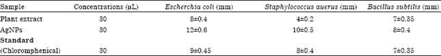 | |
| Values were expressed as Mean±SD | |
These microorganisms were identified and confirmed by Microbiologists, Department of Microbiology, Thanjavur Medical College, Thanjavur.
Determination of antimicrobial activity: The antimicrobial activity was performed by agar cup plate method.
Antimicrobial assay: Antibiogram was done by disc diffusion method (NCCLS, 1993; Awoyinka et al., 2007) using plant extracts. Petri plates were prepared by pouring 30 mL of NA medium for bacteria/fungi. The test organism was inoculated on solidified agar plate with the help of micropipette and spread and allowed to dry for 10 min. The surfaces of media were inoculated with bacteria/fungi from a broth culture. A sterile cotton swab is dipped into a standardized bacterial/ fungi test suspension and used to evenly inoculate the entire surface of the nutrient agar/PDA plate. Briefly, inoculums containing Staphylococcus aureus, Escherichia coli and Bacillus subtilis on nutrient agar plates for bacteria. Using sterile forceps, the sterile filter studies (6 mm diameter) containing each 30 μL of plant extract, AgNO3 solutions. AgNPs and standard solution as chloromphenical were laid down on the surface of inoculated agar plate. The plates were incubated at 37°C for 24 h for the bacteria and at room temperature (30±1)°C for 24-48 h for yeasts strains. Each sample was tested in triplicate.
Measurement of zone of inhibition: The antimicrobial potential of test compounds was determined on the basis of mean diameter of zone of inhibition around the disc in millimeters. The zones of inhibition of the tested microorganisms by the extracts were measured using a millimeter scale. The diameter sizes in mm of the zone of inhibition are shown in the Table 1.
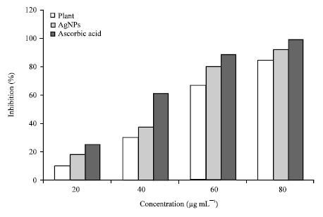
Free radical scavenging activity of Canthium coromandelicum: DPPH (1,1-diphenyl-2-picrylhydrazyl) radical-scavenging activity was determined by the method of Portillo et al. (2001). Briefly, a 2 mL aliquot of DPPH methanol solution (25 mg mL-1) was added to 0.5 mL sample solution at different concentrations (20, 40, 60 and 80 μg mL-1, respectively). The mixture was shaken vigorously and allowed to stand at room temperature in the dark for 30 min. Then the absorbance was measured at 517 nm in a spectrophotometer. Lower absorbance of the reaction mixture indicated higher free-radical scavenging activity. The scavenging activity of sample was expressed as percentage.
RESULTS
Synthesis and characterization of AgNPs: Addition of plant extract resulted in the gradual change of the color of AgNO3 solution from colorless to brown (Fig. 1), indicating the synthesis of AgNPs.
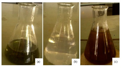 | |
| Fig. 1: | Photographs showing, (a) Plant extract, (b) AgNO3 before the addition of plant extract and (c) Color changes after adding AgNO3 after reaction time of 5 h |
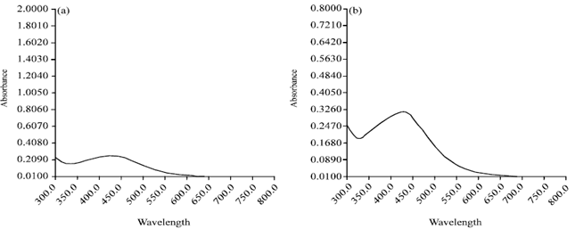 | |
| Fig. 2(a-b): | UV-Vis absorption spectra of aqueous silver nitrate with leaf extract, (a) After 3 h and (b) After 5 h |
The color intensity also increased with the duration of incubation. The plant extracts without AgNO3 show pale yellow in color and AgNO3 solution as colorless. The plant-broths, yield of AgNPs as revealed by spectral analysis was best by broth of Canthium coromandelicum that exhibited the highest peak. The absorbance was monitored at a wave length in the range of 517 nm and the resulting absorption spectrum showed the peak at 430 nm (Fig. 2) that was characteristic of AgNPs.
Scanning electron microscope: Scanning electron microscope analysis shows that the synthesized AgNPs are polydispersed (Fig. 3), with particle size range from 10-40 nm.
XRD studies: Figure 4 showed the XRD confirming the existence of silver colloids in the sample. The Braggs reflections were observed in the XRD pattern at 2 Ø = 38.16, 44.44 and 64.20. These Braggs reflections clearly indicated the presence of (111), (200) and (220) sets of lattice planes and further on the basis that they can be indexed as Face-Centered-Cubic (FCC) structure of silver.
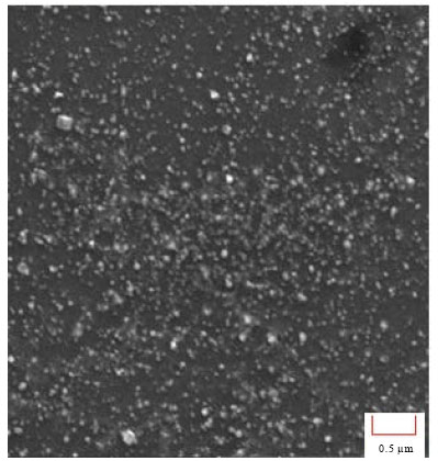 | |
| Fig. 3: | SEM image of silver nano particles |
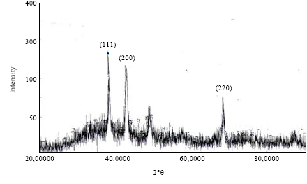 | |
| Fig. 4: | XRD pattern of silver nano particles |
Hence, XRD pattern thus clearly illustrated that the silver nan oparticles formed in this present synthesis are crystalline in nature.
Antimicrobial activity: The AgNPs of Canthium coromandileum shows highest antibacterial activity was observed against E. coli, Bacillus subtilis and S. aureus.
 | |
| Fig. 5: | DPPH antioxidant activity |
The inhibitory activities in culture media of the Ag nanoparticles reported in Table 1 were comparable with standard antimicrobiotic viz. chloromphenical.
In vitro antioxidant activity: The yield of the ethanol extract of the plant extract and AgNPs and its DPPH capacity are given in Fig. 5. The antioxidant capacity of Canthium coromandelicum is expressed as the number of equivalents of ascorbic acid. The study reveals that the antioxidant activity of the extract is in the increasing trend with the increasing concentration of the plant extract and AgNPs. The observed scavenging effect of plant extract, AgNPs and standard decreases in the following order: Ascorbic acid>AgNPs>plant extract. Thus, we found AgNPs possess potential antioxidant activity as compared with ascorbic acid.
DISCUSSION
Synthesis of silver nanoparticles: The green synthesis of silver nanoparticles through Canthium coromandelicum plant extract was carried out. The phytochemicals present in the plant extract were considered responsible for the reduction of silver ions. After the addition of plant extract, the silver nitrate solution turned to brown color within 30 min. The appearances of brown color suggest the formation of silver nanoparticles (SNPs). The UV-Visible spectra spectra showed strong Surface Plasmon Resonance (SPR) band at 430 nm, indicating the formation of silver nano particles and the broadening of peak indicated that the particles are polydispersed. SEM and XRD analysis were carried out to understand the size and structure morphology of the AgNPs, the SEM analysis showed the particle size between 10-40 nm and XRD showed the face-centered cubic (fcc) structure of the AgNPs.
Antimicrobial activity: In this study, to evaluate the antimicrobial effects AgNPs against various microorganisms, we used three representative microorganisms, Bacillus subtilis, E. coli and S. aureus. There were distinct differences among them. When AgNPs were tested they effectively inhibited bacterial growth. In our results, AgNPs showed antimicrobial activity against E. coli that was similar to that found by Sondi and Salopek-Sondi (2004). In contrast, the inhibitory effect of AgNPs was mild in S. aureus and Bacillus subtilis as compared with other microorganisms; these results suggest that the antimicrobial effects of AgNPs may be associated with characteristics of certain bacterial species. The growth of microorganisms was inhibited by the green synthesized SNPs showed variation in the inhibition of growth of microorganisms may be due to the presence of peptidoglycan which is a complex structure and after contains teichoic acids or lipoteichoic acids which have a strong negative charge. This charge may contribute to the sequestration of free silver ions. Thus gram positive bacteria may allow less silver to reach the cytoplasmic membrane than the gram negative bacteria (Ahmad et al., 2011). We think that the lower efficacy of the AgNPs against S. aureus and Bacillus subtilis may derive from the difference as a point of membrane structure. To confirm this hypothesis, further comparative study between various gram-negative and gram-positive bacterial species is needed. The peptidoglycan layer is a specific membrane feature of bacterial species and not mammalian cells. Therefore, if the antibacterial effect of AgNPs is associated with the peptidoglycan layer, it will be easier and more specific to use AgNPs as an antibacterial agent. The SNPs synthesized from plant species are toxic to multi-drug resistant microorganisms. It shows that they have great potential in biomedical applications.
Antioxidant activity
DPPH radical scavenging activity: Free radicals are harmful by-products generated during normal cellular metabolism which could initiate oxidative damage to body. Antioxidants are believed to play a significant role in the body’s defense system against free radicals. The phenolic and flavanoid compounds derived from plants were proved to be potent anti oxidant and free radicals scavengers . Some of these plants have shown potent antioxidant activity (Mohan et al., 2012). The DPPH radical was widely used to evaluate the free-radical scavenging capacity of antioxidants. The DPPH antioxidant assay is based on the ability of DPPH a stable free radical, to decolorize in the presence of antioxidants. The DPPH radical contains an odd electron which is responsible for the absorbance at 517 nm and also for visible deep purple color. When DPPH accepts an electron donated by an antioxidant compound, the DPPH is decolorized which can be quantitatively measured from the changes in absorbance. The antioxidant activity of Canthium coromandelicum and AgNPs were shown in Fig. 5. The Canthium coromandelicum and AgNPs exhibited a significant dose dependent inhibition of DPPH activity. AgNPs possess probable antioxidant activity as compared with plant extract.
CONCLUSION
The present study included the bio-reduction of silver ions through plant extract and testing for their antimicrobial activity and antioxidant activity. The aqueous silver ions exposed to the extract, the synthesis of silver nano particles were confirmed by the change of colour of plant extract, silver nano particles were further confirmed by using UV-Vis spectroscopy and SEM analysis. The results indicated that silver nanoparticles have good antibacterial activity against different microorganisms such as Bacillus subtilis, E. coli and S. aureus. It is confirmed that silver nano particles are capable of rendering high antibacterial efficacy and hence has a great potential in the preparation of drugs used against bacterial diseases. Based on the DPPH assay, silver nano particles found to be good antioxidant, as comparable with ascorbic acid. Applications of Ag nanoparticles based on these findings may lead to valuable discoveries in various fields such as medical devices and antimicrobial systems.
ACKNOWLEDGMENTS
The authors wish to acknowledge Dr. S. Velavan, Harman Research Centre, Thanjavur and also thankful to the National Institute of Technology, Tiruchirappalli for XRD.
REFERENCES
- Ahmad, N., S. Sharma, M.K. Alam, V.N. Singh, S.F. Shamsi, B.R. Mehta and A. Fatma, 2010. Rapid synthesis of silver nanoparticles using dried medicinal plant of basil. Colloids Surf. B: Biointerfaces, 81: 81-86.
CrossRefDirect Link - Ahmad, N., S. Sharma, V.N. Singh, S.F. Shamsi, A. Fatma and B.R. Mehta, 2011. Biosynthesis of silver nanoparticles from Desmodium triflorum: A novel approach towards weed utilization. Biotechnol. Res. Int.
CrossRefDirect Link - Awwad, A.M. and N.M. Salem, 2012. Green synthesis of silver nanoparticles by mulberry leaves extract. Nanosci. Nanotechnol., 2: 125-128.
CrossRefDirect Link - Awwad, A.M., N.M. Salem and A.O. Abdeen, 2012. Biosynthesis of silver nanoparticles using Olea europaea leaves extract and its antibacterial activity. Nanosci. Nanotechnol., 2: 164-170.
CrossRefDirect Link - Bankar, A., B. Joshi, A.R. Kumar and S. Zinjarde, 2010. Banana peel extract mediated novel route for the synthesis of silver nanoparticles. Colloids Surf. A: Physicochem. Eng. Aspects, 368: 58-63.
CrossRefDirect Link - Bar, H., D.K. Bhui, G.P. Sahoo, P. Sarkar, S. Pyne and A. Misra, 2009. Green synthesis of silver nanoparticles using seed extract of Jatropha curcas. Colloids Surf. A: Physicochem. Eng. Aspects, 348: 212-216.
CrossRefDirect Link - Mohan, C.S., V. Balamurugan, R. Elayaraja and A.S. Prabakaran, 2012. Antioxidant and phytochemical potential of medicinal plant Kalanchoe pinnata. Int. J. Pharm. Sci. Res., 3: 881-885.
Direct Link - Mohan, S.C., K. Sasikala and T. Anand, 2014. Antimicrobial and wound healing potential of Canthium coromandelicum leaf extract-a preliminary study. Res. J. Phytochem., 8: 35-41.
CrossRefDirect Link - Christensen, L., S. Vivekanandhan, M. Misra and A.K. Mohanty, 2011. Biosynthesis of silver nanoparticles using Murraya koenigii (curry leaf): An investigation on the effect of broth concentration in reduction mechanism and particle size. Adv. Mater. Lett., 2: 429-434.
Direct Link - Dwivedi, A.D. and K. Gopal, 2010. Biosynthesis of silver and gold nanoparticles using Chenopodium album leaf extract. Colloids Surf. A: Physicochem. Eng. Aspects, 369: 27-33.
CrossRefDirect Link - Dubey, S.P., M. Lahtinen and M. Sillanpaa, 2010. Green synthesis and characterizations of silver and gold nanoparticles using leaf extract of Rosa rugosa. Colloid Surf. A: Physicochem. Eng. Aspects, 364: 34-41.
CrossRefDirect Link - Geethalakshmi, R. and D.V.L. Sarada, 2010. Synthesis of plant-mediated silver nanoparticles using Trianthema decandra extract and evaluation of their anti microbial activities. Int. J. Eng. Sci. Technol., 2: 970-975.
Direct Link - Sulaiman, G.M., W.H. Mohammed, T.R. Marzoog, A.A.A. Al-Amiery, A.A.H. Kadhum and A.B. Mohamad, 2013. Green synthesis, antimicrobial and cytotoxic effects of silver nanoparticles using Eucalyptus chapmaniana leaves extract. Asian Pac. J. Trop. Biomed., 3: 58-63.
CrossRefDirect Link - Kouvaris, P., A. Delimitis, V. Zaspalis, D. Papadopoulos, S.A. Tsipas and N. Michailidis, 2012. Green synthesis and characterization of silver nanoparticles produced using Arbutus Unedo leaf extract. Mater. Lett., 76: 18-20.
CrossRefDirect Link - Krishnaraj, C., E.G. Jagan, S. Rajasekar, P. Selvakumar, P.T. Kalaichelvan and N. Mohan, 2010. Synthesis of silver nanoparticles using Acalypha indica leaf extracts and its antibacterial activity against water borne pathogens. Colloids Surf. B: Biointerfaces, 76: 50-56.
CrossRefDirect Link - Leela, A. and M. Vivekanandan, 2008. Tapping the unexploited plant resources for the synthesis of silver nanoparticles. Afr. J. Biotechnol., 7: 3162-3165.
Direct Link - Malarvizhi, P. and N. Ramakrishnan, 2014. Biogenic silver nano particles using Calophyllum inophyllum leaf extract: Synthesis, spectral analysis and antimicrobial studies. World J. Pharm. Res., 3: 2258-2269.
Direct Link - Nabikhan, A., K. Kandasamy, A. Raj and N.M. Alikunhi, 2010. Synthesis of antimicrobial silver nanoparticles by callus and leaf extracts from saltmarsh plant, Sesuvium portulacastrum L. Colloids Surf. B: Biointerf., 79: 488-493.
CrossRefPubMedDirect Link - Prasad, K.S., D. Pathak, A. Patel, P. Dalwadi, R. Prasad, P. Patel and K. Selvaraj, 2011. Biogenic synthesis of silver nanoparticles using Nicotiana tobaccum leaf extract and study of their antibacterial effect. Afr. J. Biotechnol., 10: 8122-8130.
Direct Link - Portillo, A., R. Vila, B. Freixa, T. Adzet and S. Canigueral, 2001. Antifungal activity of Paraguayan plants used in traditional medicine. J. Ethnopharmacol., 76: 93-98.
CrossRefPubMedDirect Link - Ramgopal, M., C. Saisushma, I.H. Attitalla and A.M. Alhasin, 2011. A facile green synthesis of silver nanoparticles using soap nuts. Res. J. Microbiol., 6: 432-438.
CrossRefDirect Link - Saxena, A., R.M. Tripathi, F. Zafar and P. Singh, 2012. Green synthesis of silver nanoparticles using aqueous solution of Ficus benghalensis leaf extract and characterization of their antibacterial activity. Mater. Lett., 67: 91-94.
CrossRefDirect Link - Garg, S., A. Chandra, A. Mazumder and R. Mazumder, 2014. Analgesic potential of hydrogels of silver nanoparticles using aqueous extract of Saraca indica bark. Int. J. Pharm. Sci. Res., 5: 240-245.
Direct Link - Singhal, G., R. Bhavesh, K. Kasariya, A.R. Sharma and R.P. Singh, 2011. Biosynthesis of silver nanoparticles using Ocimum sanctum (Tulsi) leaf extract and screening its antimicrobial activity. J. Nanoparticle Res., 13: 2981-2988.
Direct Link - Sondi, I. and B. Salopek-Sondi, 2004. Silver nanoparticles as antimicrobial agent: A case study on E. coli as a model for Gram-negative bacteria. J. Colloid Interface Sci., 275: 177-182.
CrossRefDirect Link - Song, J.Y. and B.S. Kim, 2009. Rapid biological synthesis of silver nanoparticles using plant leaf extracts. Bioprocess Biosyst. Eng., 32: 79-84.
CrossRefDirect Link - Veerasamy, R., T.Z. Xin, S. Gunasagaran, T.F.W. Xiang, E.F.C. Yang, N. Jeyakumar and S.A. Dhanaraj, 2011. Biosynthesis of silver nanoparticles using mangosteen leaf extract and evaluation of their antimicrobial activities. J. Saudi Chem. Soci., 15: 113-120.
CrossRefDirect Link - Vidhu, V.K., A. Aromal and D. Philip, 2011. Green synthesis of silver nanoparticles using Macrotyloma uniflorum. Spectrochim. Acta A Mol. Biomol. Spectros., 83: 392-397.
CrossRefDirect Link - Yilmaz, M., H. Turkdemir, M.A. Kilic, E. Bayram, A. Cicek, A. Mete and B. Ulug, 2011. Biosynthesis of silver nanoparticles using leaves of Stevia rebaudiana. Mater. Chem. Phys., 130: 1195-1202.
CrossRefDirect Link