
E. Jeroh
Department of Physiology, Faculty of Postgraduate Studies, Delta State University, Abraka, Nigeria
LiveDNA: 234.2474
C.P. Aloamaka
Department of Physiology, College of Basic Medical Sciences, Delta State University, Abraka, Nigeria
E.K. Nwangwa
Department of Physiology, College of Basic Medical Sciences, Delta State University, Abraka, Nigeria
Journal of Biological Sciences
Year: 2022 | Volume: 22 | Issue: 2 | Page No.: 65-69


ABSTRACT
Background and Objective: Stress has been observed to alter male reproductive function through the effect of HPA on the adult male Wistar rat. Humans are consistently being exposed to stressful conditions on daily basis. The objective of this study was to determine the role of rotatory cage induced stress on the alteration of liver function in male Wistar rats. Materials and Methods: The effect of exposure to a stressor on liver function test of male Wistar rats was carried out in this study using 72 adult male Wistar rats. The rats used were weighed between 180 and 240 g. The rats were obtained from the Animal House of Delta State University, Abraka. The animals were allowed to acclimatize to the laboratory condition for 2 weeks. They were divided into group 1 (1 week exposure) group 2 (2 weeks exposure) and group 3 (3 weeks exposure) to the stressor. Each of the groups was further divided into control, with stress duration of 1, 3 and 5 hrs containing six (6) rats each. Data generated were expressed as Mean±SEM. Data generated were statistically analyzed using One-way Analysis of Variance (ANOVA) and a student t-test to compare the means, p<0.05 was considered significant. Results: The results showed a significant (p<0.05) stepwise decrease in cholestasis markers as evidenced by the decrease in Alanine Phosphatase (ALP) due to exposure of the rats to stressors for 1, 3 and 5 hrs relative to control rats. The results for ALT showed upon exposure to stressors for 1, 3 and 5 hrs, there was a significant (p<0.05) stepwise decrease in ALT when compared to the control irrespective of the duration of treatment with the 5 hrs exposure group showing a greater decrease in liver ALT relative to the rat's control group. The result for liver Aspartate Transaminase (AST) showed a significant (p<0.05) decrease in serum AST relative to 1 hr and control treatment group. Conclusion: However, the result of the rats following exposure to stressors for 5 hrs shows a greater significant (p<0.05) decrease relative to 1, 3 hrs and the control exposure group irrespective of the duration of exposure.
PDF Abstract XML References Citation
Copyright: © 2022. This is an open access article distributed under the terms of the creative commons attribution License, which permits unrestricted use, distribution and reproduction in any medium, provided the original author and source are credited.
How to cite this article
E. Jeroh, C.P. Aloamaka and E.K. Nwangwa, 2022. Role of Rotatory Cage Induced Stress on Alteration of Liver Function in Male Wistar Rats. Journal of Biological Sciences, 22: 65-69.
DOI: 10.3923/jbs.2022.65.69
URL: https://scialert.net/abstract/?doi=jbs.2022.65.69
DOI: 10.3923/jbs.2022.65.69
URL: https://scialert.net/abstract/?doi=jbs.2022.65.69
INTRODUCTION
Health they say is wealth therefore, any stress that would have a serious impact on our health status must be avoided. Stress is a strong risk factor for the poor health of women1. They stated that the mechanism of how stress affects the key neurohormones like cortisol and oxytocin that influence stress-related risk and resilience is yet to be properly understood. Stress from different sources can lead to serious diseases due to wear and tear on the body as a result of stressful events2.
Stress is a double edge phenomenon that can be productive or counterproductive. Quick response to stress is a promotion of survival3. They also observed an increased level of corticosterone in rats exposed to physical stress as well as psychological stress. They further stated that physical and psychological stress can induce cognitive disorders which may result in a mood swing. Physical stress triggers psychiatric disorders which are responsible for amygdala dysfunction4. They further stated that the male and female sexes respond differently to stress.
Stress is a collective occurrence that attracts a cascade of events. This has been implicated in the development of numerous diseases5. They observed that stress increases the production of free radicals which culminates in the increased level of MDA and reduced level of SOD and CAT in all the rats used for their study. They further observed that kidney, liver and brain SOD decreased more in the 3rd week when compared to the control.
Acute stress can lead to more third party helping behaviour but not more punishing behaviour6. They stated that acute psychological stress promotes altruistic behaviour towards an irrelevant third party. Chronic stress at work has been associated with cardiovascular diseases, especially for people older than 50 years7,8.
Serum Aspartate Transaminase (AST), Alanine Transaminase (ALT) and Alkaline Phosphatase (ALP) are known as the biomarkers for liver injuries9-11. An increased level of liver enzymes is associated with liver injury and damage which leads to a decrease in ALT, AST and ALP12. Aspartate aminotransferase which used to be called Serum Glutamic-Oxaloacetic Transaminase (SGOT) is an enzyme found in the cells of the heart and liver. Healthy individuals have a low level of AST hence it is used to determine the health status of animals. Alanine aminotransferase (ALT) used to be called Serum Glutamic Pyruvate Transaminase (SGPT) is found mainly in the liver and kidney with a small amount found in the heart. Upon liver damage, the elevation of the enzyme is seen in the serum or plasma hence, it is a major marker of liver injury while Alkaline Phosphatase (ALP) is an enzyme in the blood which helps to break down protein. It plays a role in liver function hence, used for Liver Function Tests (LFT). An abnormal level is an indication of a medical condition. Unexplained weight loss can be determined by the level of ALP. The amount of ALP is increased if bones are developing or the liver is not functioning adequately. A low level of ALP is an indication that the Zn level is low and there is weight loss, tiredness as well as weakness hence, it is used as a biostatistics marker for liver injury and weakness in animals. ALP, AST and ALT are basic biological markers of Liver Function Test (LFT). Their levels in the serum are indications of liver injury. The main aim of this study was to investigate the role of rotatory cage induced stress on the alteration of liver function in Wistar rats.
MATERIALS AND METHODS
Study area: This research was carried out at the Delta State University, Abraka, Nigeria from May 1st and ended on 15th October, 2021.
Animal care ethics: Seventy two adult male Wistar rats which weighed between 180-240 g were obtained from the Animal House of Delta State University, Abraka. The animals were allowed to acclimatize to the laboratory condition (temperature 24-28°C and 12 hrs light-dark cycle) for 2 weeks. After 2 weeks the research commenced. The experimental rats had free access to rat chow (Top Feeds Nigeria) and clean drinking water throughout the study. All the animals were treated according to the Helsinki declaration of guiding principles in the care and use of animals. Approval was sought and obtained from the Research Ethical Committee of Delta State University, Abraka, before the commencement of the study. The study was carried out within 3 weeks.
Research design/experimental procedure: The experimental research approach was used for this study. The study was carried out on 72 male rats, divided randomly into 3 groups, with each group containing 4 sub-groups of 6 rats each. The groups are shown below:
| • | Group 1: Rats were exposed to stressors for the duration of 1, 3 and 5 hrs, respectively for 1 week | ||
| • | Group 1A: Control | ||
| • | Group 1B: 1 hr per day exposure to stress | ||
| • | Group 1C: 3 hrs per day exposure to stress | ||
| • | Group 1D: 5 hrs per day exposure to stress | ||
| • | Group 2: Rats were exposed to stressors for the duration of 1, 3 and 5 hrs for 2 weeks | ||
| • | Group 2A: Control | ||
| • | Group 2B: 1 hr per day exposure to stress | ||
| • | Group 2C: 3 hrs per day exposure to stress | ||
| • | Group 2D: 5 hrs per day exposure to stress | ||
| • | Group 3: Rats were exposed to stressors for the duration of 1, 3 and 5 hrs for 3 weeks | ||
| • | Group 3A: Control | ||
| • | Group 3B: 1 hr per day exposure to stress | ||
| • | Group 3C: 3 hrs per day exposure to stress | ||
| • | Group 3D: 5 hrs per day exposure to stress | ||
Stressor model: Rotatory cage was used as the stressor.
Sample collection: At the end of the experimental period, the animals were fasted overnight and euthanized by cervical dislocation. Blood was collected through ventricular punctures for biochemical assays. The liver was carefully dissected and weighed on an electronic weighing balance. The blood sample collected was centrifuged and the serum was collected for biochemical assays.
Biochemical analysis: The biochemical assay was carried out using commercial kits for enzyme assay.
Statistical analysis: Data generated were expressed as Mean±SEM. Data generated were statistically analyzed using One-way Analysis of Variance (ANOVA), followed by Tukey and Bonferroni’s post hoc test. The p<0.05 was considered significant while p>0.05 was considered not significant. Values not sharing a common alphabet as superscripts are significantly different from each other at the level of p<0.05. The *p<0.05 relative to control, ap<0.05 relative to 1 hr stress responses and bp<0.05 relative to 3 hrs stress responses.
RESULTS
Effect of exposure to stress on Alanine Phosphatase (ALP) of male wistar rats: Figure 1 shows Alanine Phosphatase (ALP) also called the cholestasis marker of the rats used for the experiment. The results show a significant (p<0.05) stepwise decrease in cholestasis markers as evidenced by a decrease in Alanine Phosphatase (ALP) due to exposure of the rats to stressors for 1, 3 and 5 hrs relative to control rats. However, there was a greater significant (p<0.05) decrease in ALP for the 3 and 5 hrs treatment group in weeks 2 and 3 relative to 1 and control with the 5 hrs treatment group showed a greater decrease in ALP at week 3 relative to control group.
Effect of exposure to stressor on serum ALT of male wistar rats: The results in Fig. 2 show the serum alanine aminotransferase (ALT) of rats used for the experiment. Upon exposure to stressors for 1, 3 and 5 hrs, the result shows that there was a significant (p<0.05) stepwise decrease in ALT when compared to the control irrespective of the duration of treatment. However, upon exposure of rats to stressors for 3 and 5 hrs, there was a significant (p<0.05) decrease while the rats in the 5 hrs treatment group showed a greater decrease in liver ALT relative to the rat control group.
Effect of exposure to stressor on serum AST of male wistar rats: Figure 3 shows the liver Aspartate Transaminase (AST) of rats used for the experiment.
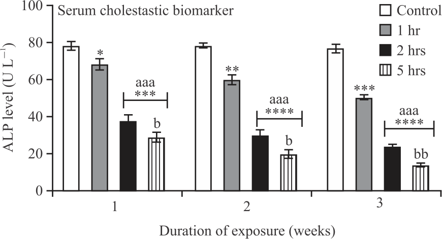 |
| Fig. 1: | Effect of exposure to stressor on ALP of male wistar rats *Bars represent Mean±SEM (n = 6), p<0.05, *p<0.05, ***p<0.001, ****p<0.0001 relative to control, aaap<0.001, relative to 1 hr stress responses and bp<0.05, bbp<0.01relative to 3 hrs stress responses |
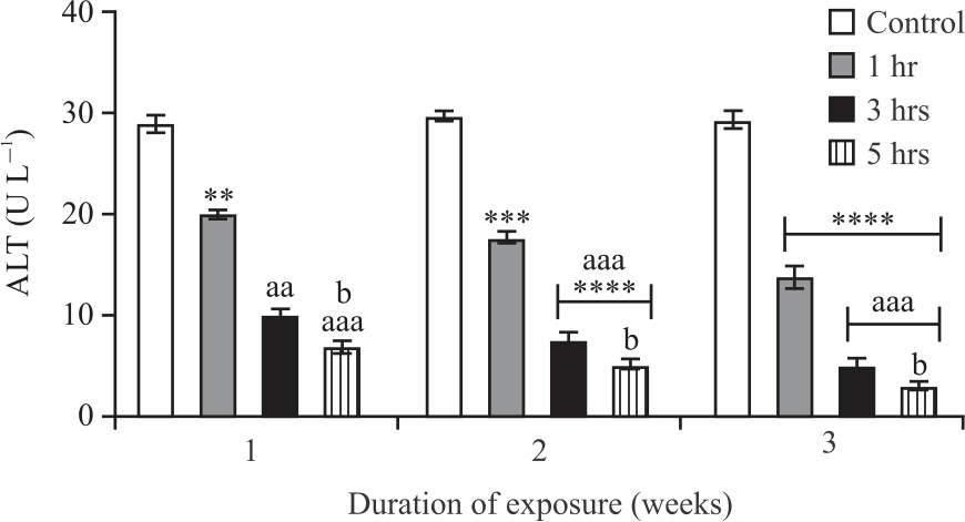 |
| Fig. 2: | Effect of exposure to stressor on serum ALT in male wistar rats *Bars represent Mean±SEM (n = 6), p<0.05, **p<0.01, ***p<0.001, ****p<0.0001 relative to control, aap<0.01, aaap<0.001 relative to 1 hr stress responses and bp<0.05 relative to 3 hrs stress responses |
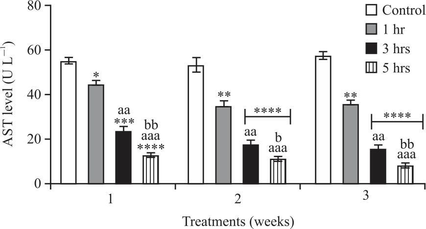 |
| Fig. 3: | Effect of exposure to stressor on serum AST in male wistar rats *Bars represent Mean±SEM (n = 6), p<0.05, *p<0.05, ***p<0.001, ****p<0.0001 relative to control, aap<0.01, aaap<0.001, aaaap<0.0001 relative to 1 hr stress responses and bp<0.05, bbp<0.001 relative to 3 hrs stress responses |
The results in Fig. 3 which represents liver aspartate amino transaminase (AST) upon exposure of rats to stressor for 1, 3 and 5 hrs, shows a significant (p<0.05) decrease in serum AST relative to 1 hr and control treatment group. However, the result of the rats following exposure to stressors for 5 hrs shows a greater significant (p<0.05) decrease relative to 1 and 3 hrs and the control treatment group irrespective of the duration of treatment.
DISCUSSION
Stress can alter liver function which may cause liver disease3,5. Previous studies have reported a decrease in ALP11. The results of this research showed a significant (p<0.05) stepwise decrease in cholestasis markers as evidenced by the decrease in Alanine Phosphatase (ALP) due to exposure of the rats to stressors for 1, 3 and 5 hrs relative to control rats. The results for ALT showed upon exposure to stressor for 1, 3 and 5 hrs, there was a significant (p<0.05) stepwise decrease in ALT when compared to the control irrespective of the duration of treatment with the 5 hrs exposure group showing a greater decrease in liver ALT relative to the rat’s control group. The result for liver Aspartate Transaminase (AST) showed a significant (p<0.05) decrease in serum AST relative to 1hr and control treatment group. However, the result of the rats following exposure to stressors for 5 hrs shows a greater significant (p<0.05) decrease relative to 1 hr, 3 hrs and the control treatment group irrespective of the duration of treatment. This study further reaffirms the report from previous studies10,11.
In this study, the rats exposed to stress in weeks 1, 2 and 3 at a stress duration of 5 hrs produced a highly significant (p<0.05) decrease in ALP, ALT and AST. These findings imply that stress can alter liver function which may culminate in liver disease. This research is in agreement with the previous studies2,10-12 which stated that stress from different sources can lead to serious diseases due to wear and tear of the body as a result of stressful events.
CONCLUSION
This study investigated the effect of exposure to a stressor on liver function test of male Wistar rats. The results from the study showed that the rats exposed to stress in weeks 1, 2 and 3 at a stress duration of 5 hrs produced a highly significant (p<0.05) decrease in ALP, ALT and AST. These findings imply that stress can alter liver function which may culminate in liver disease thus affecting the health of the rats.
SIGNIFICANCE STATEMENT
This study discovered that rotatory cage induced stress can cause alteration of liver function which can lead to liver disease. A low level of liver enzymes most times is an indication of poor health status. Health disorders are measured by the functionality of the liver, health challenges lead to liver injury. Serum Alanine Transaminase (ALT), Aspartate Transaminase (AST) and Alkaline Phosphatase (ALP) are known biomarkers for liver injuries.
REFERENCES
- Kuchenbecker, S.Y., S.D. Pressman, J. Celniker, K.M. Grewen and K.D. Sumida et al., 2021. Oxytocin, cortisol, and cognitive control during acute and naturalistic stress. Int. J. Biol. Stress, 24: 370-383.
CrossRefDirect Link - Mauss, D. and M.N. Jarczok, 2021. The streamlined allostatic load index is associated with perceived stress in life-findings from the MIDUS study. Int. J. Biol. Stress, 24: 404-412.
CrossRefDirect Link - Khaleghi, M., M.A. Rajizadeh, H. Bashiri, K.A. Kohlmeier, F. Mohammadi, M. Khaksari and M. Shabani, 2021. Estrogen attenuates physical and psychological stress‐induced cognitive impairments in ovariectomized rats. Brain Behav., Vol. 11.
CrossRefDirect Link - Gupta, K. and S. Chattarji, 2021. Sex differences in the delayed impact of acute stress on the amygdala. Neurobiol. Stress, Vol. 14.
CrossRefDirect Link - Chukwuebuka, N.B., D.T.M. Elias, A.E. Ijego, O.E. Peggy and A.C. Ejime et al., 2021. Changes in antioxidant enzymes activities and lipid peroxidase level in tissues of stress-induced rats. Biomed. Pharmacol. J., 14: 583-596.
CrossRefDirect Link - Zhen, Z., H. Wang, R. Zhu, S. Zhang, T. Jin, S. Qin and C. Liu, 2021. Acute psychosocial stress increases third-party helping but not punishing behavior. Int. J. Biol. Stress, 24: 430-441.
CrossRefDirect Link - Mauss, D., M. Volmer-Thole, R. Herr, J.A. Bosch and J.E. Fischer, 2021. Stress at work is associated with intima media thickness in older male employees, independent of other sources of stress perception. Int. J. Biol. Stress, 24: 450-457.
CrossRefDirect Link - Tremellen, K., 2008. Oxidative stress and male infertility-a clinical perspective. Hum. Reprod. Update, 14: 243-258.
CrossRefDirect Link - Hasan, K.M.M., N. Tamanna and M.A. Haque, 2018. Biochemical and histopathological profiling of Wistar rat treated with Brassica napus as a supplementary feed. Food Sci. Hum. Wellness, 7: 77-82.
CrossRefDirect Link - Al-Baqami, N.M. and R.Z. Hamza, 2021. Protective effect of resveratrol against hepatotoxicity of cadmium in male rats: Antioxidant and histopathological approaches. Coatings, Vol. 11.
CrossRefDirect Link - Wu, Y., B. Wang, L. Tang, Y. Zhou and Q. Wang et al., 2022. Probiotic Bacillus alleviates oxidative stress-induced liver injury by modulating gut-liver axis in a rat model. Antioxidants, Vol. 11.
CrossRefDirect Link - Momo, C.M.M., M.T.F. Pascal, N. Pasima, M.R. Simplice, V.B. Narcisse, N. Ferdinand and T. Joseph, 2021. Effects of Crassocephalum bauchiense ethanolic extract on reproductive parameters in rabbit does exposed to potassium dichromate. Am. J. Anim. Vet. Sci., 16: 151-161.
CrossRefDirect Link