
G.U. Ahmed
Department of Aquaculture, Bangladesh Agricultural University, 2202, Mymensingh, Bangladesh
T. Khatun
Department of Aquaculture, Bangladesh Agricultural University, 2202, Mymensingh, Bangladesh
M. Belal Hossain
Department of Biology, FOS, Universiti Brunei Darussalam, BE1410, Brunei
M. Shamsuddin
Department of Aquaculture, Bangladesh Agricultural University, 2202, Mymensingh, Bangladesh
Journal of Biological Sciences
Year: 2012 | Volume: 12 | Issue: 5 | Page No.: 287-293


ABSTRACT
Investigation on health conditions of a farmed tilapia (Oreochromis niloticus) in two upazilas of Mymensingh District were carried out through clinical and histopathological observation during September, 2010 to March, 2011. Fish sample and water quality parameters (temperature, dissolved oxygen, pH, alkalinity, nitrite and ammonia) were monitored on a monthly basis. Clinical observations of the fish were also done for any kind of abnormalities at monthly intervals. Samples of skin, muscle, liver and kidney were observed by histological techniques. Among the water quality factors, water temperature and alkalinity were found at unfavourable level for fish during the colder months. Clinically, it was observed that the tilapia were more affected from December and January and almost normal in appearance during September, October, November and March. Different clinical symptoms like rough skin, scale loss, red spots and dermal lesions were noticed in December and January. Histopathologically, sampled fish were found almost normal in the months of September and October. In the month of November minor pathologies were found to be started. Marked pathological changes like necrosis, pyknosis, hemorrhage, hypertrophy, hyperplasia, missing of primary and secondary gill lamellae, vacuums, fat droplets and fungal granuloma and fungal hyphae were observed in fish organs during December and January. Whereas, the pathological condition of fish gradually reduced in February. Again when considered individual fish pond, fishes of pond 1 (P1) in Bhaluka upazila were more affected than other ponds. The study showed that severity of clinical and pathological changes were increased in December and January. During the period of Epizootic Ulcerative Syndrome (EUS), bacterial and protozoan diseases were evident.
PDF Abstract XML References Citation
Received: May 04, 2012;
Accepted: July 18, 2012;
Published: September 06, 2012
How to cite this article
G.U. Ahmed, T. Khatun, M. Belal Hossain and M. Shamsuddin, 2012. Health Condition of a Farmed Tilapia (Oreochromis niloticus) in Earthen
Ponds, Northern Bangladesh. Journal of Biological Sciences, 12: 287-293.
DOI: 10.3923/jbs.2012.287.293
URL: https://scialert.net/abstract/?doi=jbs.2012.287.293
DOI: 10.3923/jbs.2012.287.293
URL: https://scialert.net/abstract/?doi=jbs.2012.287.293
INTRODUCTION
The introduction of tilapia in Bangladesh from Thailand was first initiated in 1954 with Tilapia mossambicus and later in 1974, high yielding species of tilapia (Oreochromis niloticus) was introduced by UNICEF (Das et al., 2010). Bangladesh Fisheries Research Institute (BFRI) again carried a batch of Oreochromis niloticus from Thailand in 1987 and developed low input and low cost technologies (Das et al., 2010). For poverty alleviation and livelihood support, tilapia has made a significant contribution Bangladesh. Tilapia has good resistance to poor water quality and disease, tolerance of wide range of environmental condition, ability to convert efficiently the organic and domestic waste into high quality protein, rapid growth rate and tasty flavour. Tilapia may be cultured as monoculture and polyculture system. In some cases, monoculture of tilapia is practiced at the farmer’s level that might not achieve the total production in polyculture. Intensive culture system with very high stocking densities and poor water qualities are encountered with much of the health problems. Due to improper management disease has become a major problem in fish culture system in Bangladesh (Rahman and Chowdhury, 1996). Freshwater fishes of Bangladesh may have different health status, showing disease symptoms like tail and fin rot, gill rot, red spot, dropsy, EUS, argulosis, nutritional disease and white spot disease (Faruk et al., 2004). In fish the most obvious external clinical signs are inflammation, hemorrhage of fins, skin or head, frayed fins, haemorrhaged opaque eye, necrotic and ulcerative lesions at any location on the body, scale loss and excessive mucus production (Plumb, 1994). In recent times, farmers of Mymensingh regions are facing many health problems in the culture of tilapia.
It was thus necessary to investigate health condition of fish through some suitable techniques. Clinical and histopathological procedures are important tools used to diagnose disease in fish. Clinical investigation provides information on the nature of disease in fish. However, histopathology is an important tool for the diagnosis of disease and it has been successfully used throughout the world. Use of histopathology is limited in Bangladesh due to technical know how and facilities. Thus the present investigation was aimed at observation of the production and health condition of tilapia in Mymensingh region through clinical and histopathological observations.
MATERIALS AND METHODS
Study sites: The experiment was carried out for a period of 7 months from September, 2010 to March, 2011. Two upazilas of Mymensingh, namely Muktagacha and Bhaluka (Fig. 1) were selected for the present study because of tilapia were abundantly cultured species in that area. Four fish farms, two from Bhaluka Upazila (village Bohuli, 40 km to the southern side of Mymensingh) and two from Muktagacha Upazila (village Digholgoan, 16 km away from Mymensingh District) were selected for present study. One pond from each farm having tilapia was selected randomly. Average depth of pond 1 (P1) was 3.5-4 feet. Stocking densities of P1 was 220 fry decimal-1. Average depth of pond 2 (P2) was 4-5 feet and stocking densities was 225 fry decimal-1. On the other hand average depth of P3 and P4 was 3-4 feet. Stocking densities of P3 was 220 fry decimal-1 and P4 was 230 fry decimal-1.
Fish sample and environmental data collection: To study health and disease status of farmed tilapia (Oreochromis niloticus), monthly sampling was done by seine net and measured to record of weight (g) by the precision balance. Water quality parameters like temperature, pH, Dissolved Oxygen (DO), alkalinity, nitrite (NO2) and ammonia (NH3) were determined by using HACH Freshwater Aquaculture Test Kit (USA). The sampled fish were examined just after taking out of the container to observe external symptoms and any injury, infection and other abnormal condition of fish body. For histopathological observation samples of fish from various organs such as skin, muscle, gill, liver and kidney were collected by a sharp scalpel and forceps and fixed in 10% formalin. Then the samples were placed in an automatic tissue processor for dehydration, clearing and infiltration. The samples were then embedded at a thickness of 5 μm.
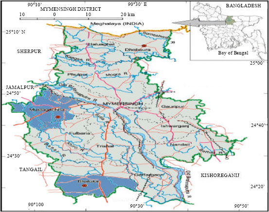 | |
| Fig. 1: | Location of study sites (Banglapedia) |
The sections were then stained with hematoxylin and eosin stains. Then the sections were mounted with Canada balsam and covered by a cover slip. Then the slides were examined under a compound microscope (Olympus). Then photomicrographs from the stained sections taken by using a photomicroscope. Pathological observation were made from the slides and photographs and compared among different months of the study period.
RESULTS AND DISCUSSION
All water quality parameters were within suitable range for fish culture except temperature and alkalinity which were considerably low (18°C and 70 mg L-1) in the months of December and January. Dissolved oxygen (DO) value did not found to vary among the ponds and were within the range from 6 to 8.5 mg L-1. The pH ranged from 6.5-8.5 and ammonia level ranged from 0.001-0.03 mg L-1. Low temperatures have negative impact on fish health. Hossain and Paul (1993) observed that the outbreak of EUS was peak when water temperature was the low, which might be due to the fact that a low temperature fish immune system ceased to function normally. According to Ahmed and Hoque (1999), cooler environment with reduced temperature play major role in the incidence of disease outbreak in the fishes of Bangladesh. Increased symptoms and pathology in December and January would be due to reduced temperature and alkalinity during this period. Hossain (2008) mentioned that, clinical symptoms like scale loss, dermal lesion, loss of caudal fin were seen in December and January. Ahmed and Hoque (1999) also reported that clinical signs like gray white necrotic areas were increased in December, January and February in various carp species in Bangladesh.
Clinically, all the fish of both the upazilas were normal in September, October, February and March as Fig. 2 shows there are no lesion or fin rot.
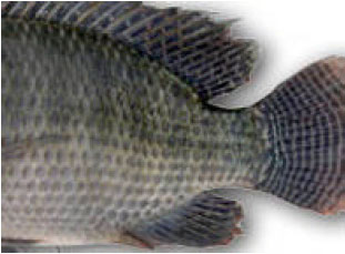 | |
| Fig. 2: | Healthy disease free tilapia |
However, in November mild lesion, rough skin and scale loss were observed in some fishes of Bhaluka. It was observed that, more fish affected from December and January. In December and January, from Bhaluka, fish were observed to have red spots and deep ulcers in the caudal region (Fig. 3). In Muktagacha, pale body colour and swollen anus were observed in December shown in Fig. 4.
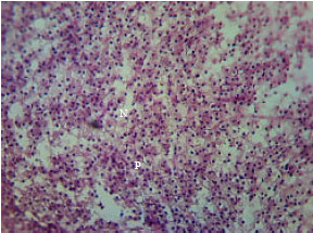
Histopathologically it was observed that in skin and muscle of Oreochromis niloticus, epidermis totally or partly lost, vacuums and necrotic muscle cells were found from fishes of all ponds (Fig. 5, 6) during the September, October and November. In December and January, necrotic muscle with fungal hyphae and fungal granuloma in Bhaluka and trace of fungal granuloma were found in Muktagacha.
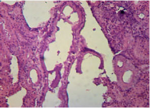
Histopathologically almost normal gill was observed during the months of September and October. Less affected gills were observed in November. Hypertrophy and hyperplasia, gill clubbing, haemorrhage in primary gill lamellae and secondary gill lamellae were lost (Fig. 7, 8) during December and January in Bhaluka and Muktagacha.
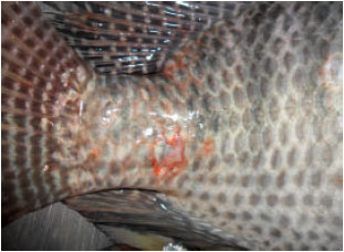 | |
| Fig. 3: | Tilapia with red spots and caudal lesions in January from P1 |
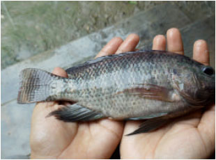 | |
| Fig. 4: | Tilapia with pale body colour and swollen anus in December from P3 |
Liver of investigated fish was normal during September, October and March in both the upazilas. But in November pathologies were found to be started with few abnormalities. Necrosis, fibrosis, haemorrhage and vacuum was observed in December and January (Fig. 9, 10). In case of kidney almost normal structures were observed during the months of September and October. Presence of fungal hyphae and fungal granuloma in fish of both upazilas indicated that fishes were suffered with EUS in December and January (Fig. 11, 12). Ahmed et al. (2007) found that during December and January, epidermis and dermis were totally lost and necrotic, whereas, muscles had fungal granuloma in A. testudineus of investigated farms.
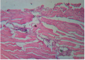 | |
| Fig. 5: | Muscle of O. niloticus from fish of P1 in December, epidermis and dermis were lost; necrosis (N), fungal hyphae (Fh), vacuum (V) and fungal granuloma (fg) were seen. H and E, x125 |
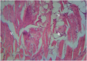 | |
| Fig. 6: | Section of muscle from fish of P4 in January, marked fungal granuloma (fg), necrosis (N) and vacuums (V) were found. H and E, x125 |
Hatai et al. (1994) also reported that fungal hyphae and fungal granulomas in the internal organs and musculature of Colisa lalia suffering from an invasive mycosis in Japan. Thus it could be mentioned that tilapia during the period were infected with EUS. Moniruzzaman (2000) also found almost similar results in skin and muscle of major carps in winter season. In February, vacuums, melanocytes and protozoan cysts were found in Bhaluka.
Liver of investigated fishes were highly necrotic and pyknotic having vacuums, fat droplets, bacterial colony and hemorrhage from the both upazilas during December and January. Ahmed et al. (2009) also found similar result Fig. 9. Liver pathology from fishes of Bhaluka in December. Necrosis (N), fibrosis (fb), Haemorrhage (H) and vacuum (V) found. for freshwater eel. Liver had highly necrotic hepatocytes, pyknotic and inflammatory cell during the months of December and January (Roy et al., 2006).
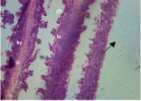 | |
| Fig. 7: | Section of gill of from fish of Bhaluka in December, hypertrophy (ht) and hyperplasia (hy), gill clubbing (gb), hemorrhage (H) in primary gill lamellae and secondary gill lamellae were lost ( |
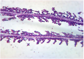 | |
| Fig. 8: | Section of gill obtained from fish of Muktagacha in January, gill clubbing (gb) and pyknosis (p) in primary gill lamellae were found. H and E, x125 |
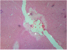 | |
| Fig. 9: | Liver pathology from fishes of Bhaluka in December, necrosis (N), fibrosis (fb), haemorrhage (H) and vacuum (V) found |
 | |
| Fig. 10: | Liver pathology from fishes of Muktagacha in December, necrosis (N) and pyknosis (P) were found |
Hossain et al. (2009) reported that severe necrosis of hepatocytes, pyknosis, vacuoles, fat droplets and hemorrhage were observed in small indigenous species during December and January. In the present study it was observed that liver pathology was recovered during February and March. In the study vacuums, necrosis and pyknosis were found in the month of November. Ahmed et al. (2009) also observed necrosis, vacuums, hemorrhage and blood cells in kidney tubule of Unibus testudineus during the month of November. However, in December and January, degenerated kidney tubules, many pyknotic nuclei, hemorrhage and inflammatory cells were found. However, in March, kidney pathology was recovered to almost normal structure except some vacuums.
From clinical, parasitological and histopathological point of view, fish were less affected during the months of September, October, February and March and increased signs and pathology were recorded during the months of colder season i.e., in December and January.
 | |
| Fig. 11: | Kidney in December from fish of Bhaluka, marked necrosis (N), wide vacuum (V) and pyknosis (P) were present. H and E, x235 |
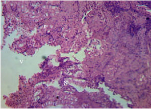 | |
| Fig. 12: | Kidney from fish of Muktagacha, numerous pyknosis (P), necrosis (N) and vacuum (V) were found. H and E, x125 |
This could be due to reduced temperature, alkalinity and dissolved oxygen fish’s metabolic process also reduces and as a result fishes are subjected to be stressed, infected and diseased. Ahmed et al. (2004) who examined through clinical, parasitological and histopathological observation of three small indigenous fishes and found that all fishes were severely affected in the months of December and January. Ahmed and Hoque (1999) also mentioned that in freshwater fish, EUS outbreak was most severe during colder months of the year when water temperature, alkalinity and hardness were reduced at their minimum levels. Parveen et al. (2005) found that during winter season water temperature reduced to nearly 10°C which diminishes antibody in fish. Thus sudden drop of water temperature in winter season was supposed to be an important predisposing cause of diseases. Water exchange, reduced feeding, removal of the dead and moribund fishes as soon as possible are some precautionary measures through which farmers would be benefited in respect of the control of the diseases (Boungou et al., 2008; Mohammed and Sambo, 2008; Ibrahim, 2012).
CONCLUSION
From the present study, it was observed that most of the fish of Bhaluka were more affected and less affected fish were found in Muktagacha. P1 in Bhaluka was modified agricultural land and in dry season its water depth remains very low. There are also possibility of being stressed by different pesticides and insecticides deriving from the agricultural lands. Moreover, reduced water depth, temperature and alkalinity might be reasons for increased infection and pathology of fishes during December and January. Under histopathological observation it was found that a great percentage of fish were affected by various pathogens especially bacteria, protozoan parasites, fungal hyphae and fungal granuloma. Thus it is necessary to take more precautionary measures to prevent and control of such diseases. Some preventive measures should be taken before outbreak of such disease. Farmers should apply 500 g lime and 500 g salt per decimal water body especially before winter, when disease outbreak occurs. It might be continued for 3-4 weeks at 7 days intervals. It is expected farmers would be benefited through increased fish production by controlling serious problems in aquaculture like diseases. Government and NGOs should take initiative to give the local farmers hands-on training on fish health management which can increase local and national production rate, ultimately gain economic benefit.
REFERENCES
- Ahmed, G.U. and M.A. Hoque, 1999. Mycotic involvement in epizootic ulcerative syndrome of freshwater fishes of Bangladesh: A histopathological study. Asian Fish. Sci., 12: 381-390.
Direct Link - Boungou, M., G.B. Kabre, A. Marques and L. Sawadogo, 2008. Dynamics of population of five parasitic monogeneans of Oreochromis niloticus Linne, 1757 in the dam of loumbila and possible interest in intensive pisciculture. Pak. J. Biol. Sci., 11: 1317-1323.
CrossRefPubMedDirect Link - Faruk, M.A.R., M.M.R. Sarker, M.J. Alam and M.B. Kabir, 2004. Economic loss from fish diseases on rural freshwater aquaculture of Bangladesh. Pak. J. Biol. Sci., 7: 2086-2091.
CrossRefDirect Link - Hatai, K., K. Nakamura, S.A. Rha, K. Yuasa and S. Wada, 1994. Aphanomyces infection in Dwarf Gourami (Colisa lalia). Fish Pathol., 29: 95-99.
CrossRef - Ibrahim, M.M., 2012. Variation in Parasite Infracommunies of Tilapia zillii in relation to some biotic and abiotic factors. Int. J. Zool. Res., 8: 59-70.
CrossRef - Mohammed, A.K. and A.B. Sambo, 2008. Haematological assessment of the Nile tilapia Oreochromis niloticus exposed to sublethal concentrations of portland cement powder in solution. Int. J. Zool. Res., 4: 48-52.
CrossRefDirect Link - Parveen, R., G.U. Ahmed and M.L. Ali, 2005. Seasonal variation of diseases of some indigenous fishes from Oxbow Lake fisheries of Bangladesh. Pak. J. Zool., 37: 53-59.
Direct Link