
N.A. Norfarizan-Hanoon
School of Health Sciences, Universiti Sains Malaysia, 16150 Kubang Kerian, Kelantan
R. Asmah
Department of Nutrition and Dietetics,Faculty of Medicine and Health Sciences, Universiti Putra Malaysia, 43400 Serdang, Selangor, Malaysia
M.Y. Rokiah
Department of Nutrition and Dietetics,Faculty of Medicine and Health Sciences, Universiti Putra Malaysia, 43400 Serdang, Selangor, Malaysia
O. Fauziah
Department of Human Anatomy, Faculty of Medicine and Health Sciences, Universiti Putra Malaysia, 43400 Serdang, Selangor, Malaysia
H. Faridah
Malaysian Agricultural Research and Development Institute, Serdang, Selangor, Malaysia
Journal of Biological Sciences
Year: 2009 | Volume: 9 | Issue: 7 | Page No.: 662-668


ABSTRACT
The objective of this study is to determine the effect of Strobilanthes crispus Juice (SCJ) on wound healing and antioxidative enzymes activity in normal and streptozotocin-induced rats. A total of 48 male albino Sprague dawley rats, weight 150-200 g were used. The rats were divided into 8 groups with 6 rats in each group. The healing of 2 cm linear incisions created on the back of each rat was monitored by measuring the length of the wounds daily. The result showed a significant increase (p<0.05) in the percentage of wound healing at day 3 and 7 in the treated group especially treated with 140 mg kg-1 b.wt. of S. crispus juice in diabetic and normal rats compared with the control. Strobilanthes crispus juice increased significantly (p<0.05) of glutathione peroxidase (GPx) and superoxide dismutase (SOD) activity in treated group in diabetic rats. Significant correlation was found between wound healing, GPx and SOD enzymes. In conclusion, S. crispus juice enhanced wound healing in normal and diabetic rats.
PDF Abstract XML References Citation
How to cite this article
N.A. Norfarizan-Hanoon, R. Asmah, M.Y. Rokiah, O. Fauziah and H. Faridah, 2009. Effects of Strobilanthes crispus Juice on Wound Healing and Antioxidant Enzymes in Normal and Streptozotocin-Induced Diabetic Rats. Journal of Biological Sciences, 9: 662-668.
DOI: 10.3923/jbs.2009.662.668
URL: https://scialert.net/abstract/?doi=jbs.2009.662.668
DOI: 10.3923/jbs.2009.662.668
URL: https://scialert.net/abstract/?doi=jbs.2009.662.668
INTRODUCTION
Wound healing is a complex process involving a highly regulated series of biological events. These include a set of co-ordinated interactions between cells in the dermis and the epidermis and important relationships have been found to exist between fibroblasts, keratinocytes and resident dermal cells (Raghow, 1994). Healing of wounds, a fundamental response to tissue injury, occurs by a process of connective tissue repair. A fibrous scar is the end product of this process, the predominant constituent of which is collagen. Collagen and other components of the ground substance are synthesized by the highly vascular granulation tissue that is formed within the wound space. Since, collagen provides strength and integrity to the dermis and all other supporting tissue, the synthesis, secretion and subsequent organisation of collagen plays an integral role in wound healing (Chithra et al., 1998a).
Diabetes mellitus is a condition which is known to be associated with a variety of connective tissue abnormalities. The collagen content of the skin is decreased as a result of reduced biosynthesis and/or accelerated degradation of newly synthesized collagen (Chithra et al., 1998b). These qualitative (Schnider and Kohn, 1981) and quantitative (Spanheimer et al., 1988) abnormalities contribute to the impaired wound healing observed in diabetes.
Diabetic wounds result in significant morbidity, prolonged hospitalization and enormous health-care expenses (Velander et al., 2008). According to the American Diabetes Association, 25% of people with diabetes will suffer from a wound problem during their lifetime and approximately 82,000 limb amputations for nontraumatic wounds were performed in people with diabetes in 2002. The Agency for Health Care Policy and Research reports that wound care for pressure ulcers uses $200 billion a year for hospitalization, durable medical goods, nursing home care, physicians and transportation (Rees, 1999). Surgical treatment of diabetic wounds remains difficult and often insufficient, leading to high morbidity among those patients (Bowler, 2002). Better ways to treat diabetic wounds are needed to develop new therapeutic strategies.
Strobilanthes crispus (S. crispus) is commonly known as daun pecah beling in Jakarta or enyoh kilo, kecibeling, or kejibeling in Java (Sunarto, 1977) as well as pecah kaca or jin batu in Malaysia, one of the herbs that has great potential and is believed to have health-giving properties. It has long been used traditionally to treat diabetes mellitus and related disorders. It is also commonly consumed in the form of herbal tea. The study showed that the leaves of S. crispus possessed anti-AIDS, anti-leukemia (Kusumoto et al., 1992), high antioxidant activity (Ismail et al., 2000; Abu Bakar et al., 2004; Asmah et al., 2006a), anticarcinogenic (Suherman et al., 2004; Fauziah et al., 2005; Asmah et al., 2006b; Mohd Fadzelly et al., 2006a) and antidiabetic (Norfarizan-Hanoon et al., 2009; Mohd Fadzelly et al., 2006b). Therefore, the aim of this study was to determine the effect of S. crispus juice on wound healing and the activity of antioxidant enzymes such as SOD and GPx in normal and streptozotocin-induced diabetic male albino Sprague dawley rats.
MATERIALS AND METHODS
Present study was conducted on November 2007.
Plant material and preparation of S. crispus juice: The leaves of S. crispus were grown and collected from the herbal garden of Faculty of Medicine and Health Science, Universiti Putra Malaysia, Serdang, Selangor. The plant was identified by taxonomist of Department of Botany, Faculty of Science and Technology, Universiti Kebangsaan Malaysia. The voucher number of S. crispus was AZ-6803. Fourteen percent of Strobilanthes crispus leaves was weighed, washed and cut into small pieces. Filtered water containing 0.1% (w/w) sodium metapbisulphite was mixed with the plants and ground into a very fine particle using mechanical grinder. This juice was then mixed with honey and 0.2% (w/w) xantham gum, homogenized using Homogenizer IKA II and pasteurized for 30 min using jacketed heater. The final product was hot-filled into sterilized glass bottle, cooled to a room temperature under running water and kept chilled for analysis.
Study protocol
Experimental rats: Forty-eight Sprague dawley strain male albino white rats, weighing 150 to 200 g were used in this study. The rats were supplied by Institute of Medical Research (IMR), Kuala Lumpur, Malaysia. They were housed in standard cage at an ambient temperature of 25±2°C with 12 h light/12 h dark cycle and placed in Animal House at Faculty Medicine and Health Science, Universiti Putra Malaysia. They were fed with commercial rat feed and tap water ad libitum. Rats were acclimatized to the laboratory conditions for at least 1 week before any experimental work was undertaken. The experiment as designed and conducted according to ethical norms approved by Animal Care and Use Committee (ACUC), Faculty of Medicine and Health Sciences, Universiti Putra Malaysia, Serdang, Selangor. ACUC reference number for this project was UPM/FPSK/PADS/F01-00184.
Induction of experimental diabetes: Streptozotocin (Sigma, USA; 55 mg kg-1 body weight) was freshly dissolved in 0.1 mol L-1 cold sodium citrate buffer, pH 4.5 and injected intraperitoneally to 24 rats. Rats were fasted for 16 h prior to induction of diabetes. Third day after streptozotocin administration, the fasting blood glucose concentration was determined using accutren. Animals having glucose level >198 mg dL-1 were selected in this study for hyperglycaemic or diabetic model.
Wound creation: The experiment was carried out 5 days after the confirmation of diabetes. Hair on the dorsal side of rats was shaved and the skin was cleaned with 70% alcohol. Mid-dorsal linear incisions of 2 cm in length were made on each animal. Wounds were observed and measured daily and photographs taken on days 0 and 7 after treatment. Calculation of percentage of wound healing:
Experimental procedure: In the experiment a total of 48 rats (24 diabetic and 24 normal rats) were used. The rats were divided into eight groups of 6 rats each and the groups were as follows :
| • | Group 1: Diabetic control (Diabetic untreated rats) |
| • | Group 2: Diabetic treated with 70 mg kg-1 b.wt. of S. crispus juice |
| • | Group 3: Diabetic treated with 105 mg kg-1 b.wt. of S. crispus juice |
| • | Group 4: Diabetic treated with 140 mg kg-1 b.wt. of S. crispus juice |
| • | Group 5: Normal control (Normal untreated rats) |
| • | Group 6: Normal treated with 70 mg kg-1 b.wt. of S. crispus juice |
| • | Group 7: Normal treated with 105 mg kg-1 b.wt. of S. crispus juice |
| • | Group 8: Normal treated with 140 mg kg-1 b.wt. of S. crispus juice |
Blood preparation On day 0 (baseline) and seventh day of treatment, the rats were anesthetized with diethyl ether following a 12 h fast. Blood was drawn by cardiac puncture into heparin tube.
Determination of GPx: Dilute 0.05 mL heparinized whole blood with 2 mL diluting agent. Mix well and run the samples using Selectra E Chemical Analyser. GPx catalyses the oxidation of glutathione (GSH) by cumene hydroperoxide. In the presence of Glutathione Reductase (GR) and NADPH the oxidised glutathione (GSSG) was immediately converted to the reduced form with a concomitant oxidation of NADPH to NADP+. The decrease in absorbance at 340 nm was measured.
Determination of SOD: Centifuged 0.5 mL of whole blood for 10 min at 3000 rpm and then aspirate off the plasma. Then washed the erythrocyte four times with 3 mL 0.9% NaCl solution and centrifuged for 10 min at 3000 rpm after each wash. The washed centrifuged erythrocytes should then be made up to 2.0 mL with cold redistilled water, mixed and left to stand at 4°C for 15 min. Dilute 125.0 μL erythrocytes with 3 mL diluent. Samples were run using Selectra E Chemical Analyser.
Statistical analysis: All data were presented as Mean±Standard deviation of mean (SD) using SPSS version 13.0. The data for various biochemical parameters were analysed using ANOVA and the group means were compared by Duncan’s multiple range test. Values were considered statistically significant when p<0.05. Correlation between wound healing, Gpx and SOD enzymes were analyzed by Pearson correlation test.
RESULTS AND DISCUSSION
Table 1 shows the length of wound in untreated and S. crispus juice-treated normal and diabetic rats. There was a significant decrease in length of wound in normal treated with 105 and 140 mg kg-1 b.wt. of S. crispus juice at day 3 and day 7 (Day 3: 1.57 cm ±0.07, Day 7: 0.80 cm ±0.11 ; Day 3: 1.45 cm ±0.03, Day 7: 0.62 cm ±0.11) compared normal control (Day 3: 1.77 cm ±0.12, Day 7: 1.33 cm ±0.12). There also was significant decrease in the length of wound diabetic rats treated with 105 and 140 mg kg-1 b.wt. of S. crispus juice (Day 3: 1.65 cm ±0.07, Day 7: 1.17 cm ±0.07; Day 3: 1.63 cm ±0.05, Day 7: 1.12 cm ±0.07) compared to wound of diabetic control rats (Day 3: 1.85 cm ±0.08, Day 7: 1.50 cm ±0.11) (Fig. 1).
| Table 1: | Length of wound (cm) in diabetic and normal group |
 | |
| Values with different superscript are significantly different | |
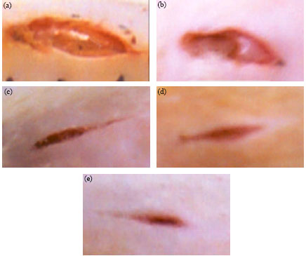 | |
| Fig. 1: | Photo of wound healing in diabetic rats; (a) day 0 and (b-e) day 7 (b: Diabetic control, c: Diabetic + SCJ 70, d: Diabetic + SCJ 105, e: Diabetic + SCJ 140) |
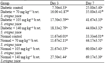
| Table 2: | Percentage of wound healing in diabetic and normal rats |
 | |
| Values with different superscript are significantly different | |
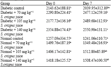
| Table 3: | Glutathione peroxidase activity (u/ml) in diabetic and normal group |
 | |
| Values with different superscript are significantly different | |
There was significant increase the percentage of wound healing in normal treated with 105 and 140 mg kg-1 b.wt. S. crispus juice at day 3 and day 7 (Day 3: 21.67% ±3.33, Day 7: 60.00% ±5.4; Day 3: 27.50% ±1.44, Day 7: 69.17% ±5.39) compared normal control (Day 3: 11.67 %±6.01, Day 7: 33.33% ±6.01) . There also significant increase the percentage of wound healing in diabetic treated with 105 and 140 mg kg-1 b.wt. S. crispus juice at day 3 and day 7 (Day 3: 17.50% ±3.59, Day 7: 41.67% ±3.33; Day 3: 18.33% ±2.79, Day 7: 44.00% ±3.32) (Table 2).
Table 3 shows the GPx activity in erythrocytes of untreated and S. crispus juice-treated normal and diabetic rats. There was increase in treated 140 mg kg-1 b.wt. of S. crispus juice in normal and diabetic rats when compared to the control.
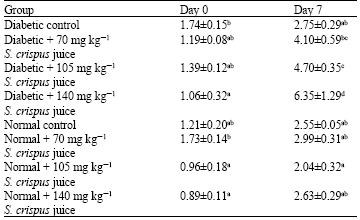
There was significantly increased in treated with 70, 105 and 140 mg kg-1 b.wt. of S. crispus juice in diabetic rats when compared with diabetic control. No significant difference in the SOD activities of untreated and treated normal rats (Table 4).
Significant correlation (p<0.05) were found between wound healing at day 7 and SOD at day 7. Wound healing at day 7 have significant correlation (p<0.01) between wound healing at day 3 and GPx at day 7.
| Table 4: | Superoxide dismutase activity (u/L) in diabetic and normal group |
 | |
| Values with different superscript are significantly different | |
Wound healing is an extreme complex phenomenon involving a number of well-orchestrated processes, including coagulation, inflammation, formation of granulation tissue, synthesis of extracellular matrix protein, remodeling of connective tissue parenchymal components, collagenization and acquisition of wound strength. There are several reports stating that the extracts of several plants, used for wound healing properties (Suguna et al., 1996; Saha et al., 1997; Sunil et al., 1998; Rasik et al., 1996; Mukherjee and Suresh, 2000; Park and Chun, 2001; Nagappa and Cheriyan, 2001).
Cutaneous wound repair is accompanied by an ordered and definable sequence of biological events starting with wound closure and progressing to the repair and remodeling of damaged tissue (Phillips et al., 1991). In spite of tremendous advances in the pharmaceutical drug industry, the availability of drugs capable of stimulating the process of wound repair is still limited (Udupa et al., 1995). Moreover, the management of chronic wounds is another major problem due to the high cost of therapy and the presence of unwanted side effects (Porras-Reyes et al., 1993; Suh et al., 1998). It is consented that Reactive Oxygen Species (ROS) are deleterious to wound healing process due to the harmful effects on cells and tissues. Absorbable synthetic biomaterials are considered to be degraded via ROS (Aliyeva et al., 2004). Free-Radical-Scavenging Enzymes (FRSE) are a cytoprotective enzymatic group that has an essential role in the reduction, de-activation and removal of ROS as well as regulating wound healing process.
Wounding results in the loss of different free radical scavengers, both enzymatic and non-enzymatic, that either partially or completely recover following healing (Shukla et al., 1997). The increase in free radical production and diminished antioxidant activities may worsen the situation and account for the delay in healing in diabetic patients. The present study shows that supplementation of S. crispus juice enhances wound healing in normal and diabetic rats. This probably involves its action as an antioxidant, reducing the level of free radicals and, hence, free radical damage (as indicated in the reduced plasma MDA levels) and by increasing the activity of antioxidant enzymes, especially GPx and SOD. Results obtained in the present study that GPx and SOD were increased in diabetic group treated with S. crispus juice. These antioxidant enzymes are known to quench the superoxide radical and thus prevent the damage of cells caused by free radicals (Shirwaikar et al., 2003). So, scavenging effect might be one of the most important components of wound healing.
Other antioxidants such as ascorbic acid (Ringsdorf and Cheraskin, 1982) and trolox have also been shown to enhance wound healing in diabetic human and rats, respectively (Hallberg et al., 1996). Vitamin C (ascorbic acid) is an essential cofactor for the synthesis of collagen, proteoglycans and other organic components of the intracellular matrix of tissues such as bones, skin, capillary walls and other connective tissues. The combined effect of ascorbic acid on collagen synthesis, antioxidant status and immunomodulation make it an appropriate supplement for wound repair protocols (Mackay and Miller Alan, 2003). Levenson and Demetrio (1992) suggested vitamin A benefits the wound by enhancing the early inflammatory phase, including increasing the number of monocytes and macrophages at the wound site, modulating collagenase activity, supporting epithelial cell differentiation and improving localization and stimulation of the immune response. The effect of vitamin E on wound healing is complex; it may have alternate effects in different types of wounds and in the presence of other nutrients, as well as different functions for water soluble versus lipid soluble preparations of vitamin E. Zinc deficiency has been associated with poor wound healing and decreased breaking strength of animal wounds (Agren and Franzen, 1990). In study (Mohd Fadzelly et al., 2006a, b; Norfarizan-Hanoon et al., 2009) showed that S. crispus, a well-known herb, possesses diverse biological activities and pharmacological functions including antioxidant, antiproliferative, antimicrobial and antihyperglycemic.
Many plants extracts and medicinal herbs have shown potent antioxidant activity (Sleem et al., 1999) may be responsible to support wound healing. Flavonoids, the main components of many plant extracts, acts as powerful free radical scavengers (Bekerecioglu et al., 1998). Research into the role of antioxidants from plant extracts in wound healing has been published widely. The enhanced wound healing may thus be due to the free-radical scavenging action of the plant and the enhanced level of antioxidant enzymes in the tissue. The better collagenation seen under the influence of this plant extract may also be due to the improved antioxidant status.
CONCLUSION
Strobilanthes crispus juice enhances wound healing in normal and diabetic rats. Its mechanism may involve reduction of free radicals; increased glutathione peroxidase and superoxide dismutase; vitamin C, A and E and zinc. Consumption of S. crispus juice could contribute to the additional nutraceutical supplement which had a great potential in healing wounds especially for diabetic patients.
ACKNOWLEDGMENTS
We thank to Dr. Hasdi Mohd Sani, Mr. Kufli, Chemistry Pathology Staffs (Mrs. Safarina, Mrs. Allyna and Mr. Ehsan), Nutrition Lab, staffs and Animal House staffs for their technical expertise and also thanks to Junaidah Mustapha for her help in this project.
REFERENCES
- Bowler, P.G., 2002. Wound pathophysiology, infection and therapeutic options. Ann. Med., 34: 419-427.
CrossRefPubMedDirect Link - Chithra, P., G.B. Sajithlal and G. Chandrakasan, 1998. Influence of Aloe vera on the healing of dermal wounds in diabetic rats. J. Ethnopharmacol., 59: 195-201.
CrossRefPubMedDirect Link - Chithra, P., G.B. Sajithlal and G. Chandrakasan, 1998. Influence of Aloe Vera on collagen characteristics in healing dermal wounds in rats. Mol. Cell. Biochem., 181: 71-76.
PubMedDirect Link - Hallberg, C.K., SD. Troome and N.H. Ansari, 1996. Acceleration of corneal wound healing in diabetic rats by the antioxidant trolox. Res. Commun. Mol. Pathol. Pharmacol., 93: 3-12.
PubMed - Raghow, R., 1994. The role of extracellular matrix in postinflammatory wound healing and fibrosis. J. Federation Am. Soc. Exp. Biol., 8: 823-831.
Direct Link - Rees, R.S. and J.A. Hirshberg, 1999. Wound care centers: Costs, care and strategies. Adv. Skin Wound Care, 12: 4-7.
Direct Link - Schnider, S.L. and R.R. Kohn, 1981. Effects of age and diabetics mellitus on the solubility and nonenzymatic glycosylation of human skin collagen. J. Clin. Inves., 67: 1630-1635.
CrossRefPubMedDirect Link - Shukla, A., A.M. Rasik and G.K. Patnaik, 1997. Depletion of reduced glutathione, ascorbic acid, vitamin E and antioxidant defence enzymes in a healing cutaneous wound. Free Rad. Res., 26: 93-101.
CrossRefPubMedDirect Link - Velander, P., C. Theopold, T. Hirsch, O. Bleiziffer and B. Zuhaili et al., 2008. Impaired wound healing in an acute diabetic pig model and the effects of local hyperglycemia. Wound Repair Regeneration, 16: 288-293.
CrossRefDirect Link - Abu Bakar, M.F., A.H. Teh, A. Rahmat, N. Hashim, F. Othman and S. Fakurazi, 2004. Antioxidant tea from leaves of Strobilanthes crispus. J. Tropical Med. Plants, 5: 199-204.
Direct Link - Rahmat, A., S. Edrini, P. Ismail, T.Y.Y. Hin and M.F. Abu Bakar, 2006. Chemical constituents, antioxidant activity and cytotoxic effects of essential oil from Strobilanthes crispus and Lawsonia inermis. J. Biol. Sci., 6: 1005-1010.
CrossRefDirect Link - Agren, M.S. and L. Franzen, 1990. Influence of zinc deficiency on breaking strength of 3-week-old skin incisions in the rat. Acta Chir. Scand., 156: 667-670.
Direct Link - Bekerecioglu, M., M. Tercan and I. Ozyazan, 1998. The effect of Ginkgo biloba (Egb 761) as a free radical scavenger on the survival of skin flaps in rats. Scand. J. Plastic Reconstructive Surg. Hand Surg., 32: 135-139.
Direct Link - Fauziah, O., P. Hanachi, S. Yogespiriya and R. Asmah, 2005. Evaluation of lesion scoring and aniline hydroxylase activity in hepatocarcinogenesis rats treated with Strobilanthes crispus. J. Med. Sci., 5: 26-30.
CrossRefDirect Link - Ismail, M., E. Manickam, A.M. Danial, A. Rahmat and A. Yahaya, 2000. Chemical composition and antioxidant activity of Strobilanthes crispus leaf extract. J. Nutr. Biochem., 11: 536-542.
CrossRef - Kusumoto, I.T., I. Shimada, N. Kakiuchi, M. Hattori, T. Namba and S. Supriyatna, 1992. Inhibitory effects of Indonesian plant extracts on reverse transcriptase of an RNA tumour virus (I). Phytother. Res., 6: 241-244.
CrossRefDirect Link - MacKay, D.J and A.l. Miller, 2003. Nutritional support for wound healing. Alternative Med. Rev., 8: 359-377.
Direct Link - Abu Bakar, M.F., A.H. Teh, A. Rahmat, F. Othman, N. Hashim and S. Fakurazi, 2006. Antiproliferative properties and antioxidant activity of various types of Strobilanthes crispus tea. Int. J. Cancer Res., 2: 152-158.
CrossRef - Nagappa, A.N. and B. Cheriyan, 2001. Wound healing activity of the aqueous extract of Thespesia populnea fruit. Fitoterapia, 72: 503-506.
Direct Link - Norfarizan-Hanoon, N.A., R. Asmah, M.Y. Rokiah, O. Fauziah and H. Faridah, 2009. Antihyperglycemic, hypolipidemic and antioxidant enzymes effect of Strobilanthes crispus juice in normal and streptozotocin-induced diabetic male and female rats. Int. J. Pharmacol., 5: 200-207.
CrossRefDirect Link - Park, E.H. and M.J. Chun, 2001. Wound healing activity of Opuntia ficus-indica. Fitoterapia, 72: 165-167.
Direct Link - Phillips, G.D., R.A. Whitehead and D.R. Kinghton, 1991. Initiation and pattern of angiogenesis in wound healing in the rat. Am. J. Anat., 192: 257-262.
PubMed - Porras-Reyes, B.H., W.H. Lewis, J. Roman, L. Simchowitz and T.A. Mustoe, 1993. Enhancement of wound healing by the alkaloid taspine defining mechanism of action. Soc. Exp. Biol. Med., 203: 18-25.
PubMed - Ringsdorf, W.M. Jr. and E. Cheraskin, 1982. Vitamin C and human wound healing. Oral Surg. Oral Med. Oral Pathol., 53: 231-236.
PubMed - Saha, K., P.K. Mukherjee, J. Das, M. Pal and B.P. Saha, 1997. Wound healing activity of Leucas lavaulaefolia Rees. J. Ethnopharmacol., 56: 139-144.
PubMed - Shirwaikar, A., A.P. Somashekar, A.L. Udupa, S.L. Udupa and S. Somashekar, 2003. Wound healing studies of Aristolochia bracteolate Lam. with supportive action of antioxidant enzymes. Phytomedicine, 10: 558-562.
CrossRef - Sleem, M., A. Alam, S. Ahmed, M. Iqbal and S. Sultana, 1999. Tephrosia purpurea ameliorates benzoyl peroxide induced cutaneous toxicity in mice: Diminition of oxidative stress. Pharm. Pharmacol. Commun., 5: 455-461.
Direct Link - Suguna, L., P. Sivakumar and G. Chandrakasan, 1996. Effects of Centella asiatica extract on dermal wound healing in rats. Indian J. Exp. Biol., 34: 1208-1211.
PubMedDirect Link - Suh, D.D., I.P. Schwartz, D.A. Canning, H.M. Snyder, S.A. Zderic and A.J. Kirsch, 1998. Comparison of dermal and epithelial approaches to laser tissue soldering for skin flap closure. Lasers Surg. Med., 22: 268-274.
Direct Link - Suherman, J., R. Asmah, O. Fauziah, I. Patimah and A. Nor Haslinda, 2004. Effect of Strobilanthes crispus on tumour marker enzymes and glutathione during chemical hepatocarcinogenesis in the rat. Pak. J. Biol. Sci., 7: 947-951.
Direct Link - Sunilkumar, P.S. and H.G. Shivakumar, 1998. Evaluation of topical formulations of aqueous extract of Centella asiatica on open wounds in rats. Indian J. Exp. Biol., 36: 569-572.
Direct Link - Udupa, A.L., D.R. Kulkarni and S.L. Udupa, 1995. Effect of Tridax procumbens extracts on wound healing. Int. J. Pharmacognosy, 33: 37-40.
CrossRef - Asmah, R., E. Susi, M.A. Abdah, I. Patimah, Y.Y.H. Taufiq and A.B. Mohd Fadzelly, 2006. Anticarcinogenic properties of Strobilanthes crispus extracts and its compounds in vitro. Int. J. Cancer Res., 2: 47-49.
CrossRefDirect Link - Mohd Fadzelly, A.B., R. Asmah and O. Fauziah, 2006. Effects of Strobilanthes crispus tea aqueous extracts on glucose and lipid profile in normal and streptozotocin-induced hyperglycemic rats. Plant Foods Hum. Nutr., 61: 6-11.
Direct Link - Spanheimer, R.G., G.E. Umpierrez and V. Stumpf, 1988. Decreased collagen production in diabetic rats. Diabetes, 37: 371-376.
PubMedDirect Link