
P. Daisy
Department of Biotechnology, Holy Cross College, Tiruchirappalli-620 002, India
Nirmala A. Rayan
Department of Biotechnology, Holy Cross College, Tiruchirappalli-620 002, India
D. Rajathi
Department of Biotechnology, Holy Cross College, Tiruchirappalli-620 002, India
Journal of Biological Sciences
Year: 2007 | Volume: 7 | Issue: 2 | Page No.: 433-437


ABSTRACT
The antidiabetic activity of the aqueous extract of Elephantopus scaber (Linn) root and leaf was evaluated in normal and alloxan-induced hyperglycemic rats. Oral administration of Elephantopus scaber root and leaf extracts (0.3 g kg-1 body weight) for 12 weeks resulted in significant reduction in the glucose levels. The effect of these extracts on triglycerides, HDL, cholesterol, serum urea and creatinine were also assessed to evaluate their activity in controlling diabetes related metabolic alterations. The biochemical estimations were complemented with the immunocytochemical staining to localize pancreatic islets cells secreting insulin. Though treatment with humulin (0.6 g kg-1 body weight) is more effective in normalizing blood glucose levels, regeneration of islet β-cells was mediated only by the extract treatment. The results indicate the effective role of the root and leaf extracts as an hypoglycemic agent against alloxan-induced diabetes in rats.
PDF Abstract XML References Citation
How to cite this article
P. Daisy, Nirmala A. Rayan and D. Rajathi, 2007. Hypoglycemic and Other Related Effects of Elephantopus scaber Extracts on Alloxan Induced Diabetic Rats. Journal of Biological Sciences, 7: 433-437.
DOI: 10.3923/jbs.2007.433.437
URL: https://scialert.net/abstract/?doi=jbs.2007.433.437
DOI: 10.3923/jbs.2007.433.437
URL: https://scialert.net/abstract/?doi=jbs.2007.433.437
INTRODUCTION
Diabetes mellitus is a chronic disease characterized by hyperglycemic effects, alterations in fat and protein metabolisms. Pharmacotherapy involves insulin injection and oral chemical drugs, but their use is complemented with side effects including hypoglycemic coma and disturbances of liver and kidney functions. In addition drugs are not suitable during pregnancy, on the other hand herbal drugs are considered less toxic and free from side effects than synthetic drugs (Bailey and Day, 1989).
Medicinal plants are found to possess certain active principles which can cure many diseases, also the World Health Organisation approves the use of plant based drugs in the treatment of ailments including diabetes mellitus (WHO, 1980).
Elephantopus scaber (Linn), of Compositae family commonly known as‘Anoshovadhi’ is a small herb which grows wild throughout the warm parts of India. The plant has been used in the Indian system of medicine as an analgesic, diuretic, astringent and antiemetic. The leaves of the plants are used for conditions like bronchitis, smallpox, diarrhea and as a brain tonic (Rastogi and Mehrotra, 1990). The major phytochemical constituents of the plant are elephantopin, triterpenes, stigmasterol, epifriedelinol and lupeol (Rajkapoor et al., 2002). It has also been shown to possess anti inflammatory and antitumour activity in animal models (Sankar et al., 2001). Reports of this plant on antidiabetic activity are scant. In view of the above information and folklore use of the root and leaves of this plant as an antidiabetic agent, the present study was undertaken to assess the hypoglycemic and related effects of aqueous extracts of Elephantopus scaber root and leaf.
MATERIALS AND METHODS
Preparation of plant extract: The leaves and roots of Elephantopus scaber were collected from Kerala and authenticated at the Department of Botany, Holy Cross College, Trichy. The air dried leaves and roots were powdered, the coarse powder was passed through No. 10 mesh and used for extraction. The powders were boiled in water separately (100 g/1000 mL distilled water), the decoction was filtered, using Whatman filter paper and the filtrates were evaporated to dryness under reduced pressure in rotary evaporator. The dry residues obtained were stored for further use. The residues prepared were quantitatively dissolved in distilled water for oral administration. For convenience the aqueous extract of E. scaber root and leaf are named ESR and ESL, respectively.
Animals used: Wistar male albino rats weighing 150-220 g, bred and maintained in our animal house were used for these studies. They were allowed a standard pellet diet (Kamadhenu agencies, Bangalore, India) and water ad libitum.
Induction of diabetes in rats: Stable diabetes was induced by the administration of alloxan monohydrate (Sigma Chemicals, USA) to the rats starved for 16 h. Intraperitoneal injection of 150 mg kg-1 body weight of alloxan dissolved in physiological saline produced persistent hyperglycemia after 7 days, as revealed by determination of blood sugar levels.
Experimental design: Experiments were carried out in normoglycemic and hyperglycemic rats assigned into five treatment groups (n = 8); Group I (untreated control rats) and Group II (Diabetic control rats) received distilled water; aqueous extract of Elephantopus scaber root (0.3 g kg-1) was given to group III diabetic rats whereas group IV had diabetic rats treated with (0.3 g kg-1) E. scaber leaf extract. Group V diabetic rats were given humulin (Ranbaxy Ltd., India) intraperitoneally (0.6 g kg-1). Distilled water and aqueous extract were administered orally for 12 weeks using gastric tubes. At the end of the experiments, blood samples (1 mL) were collected from the tail vein under mild ether anesthesia in tubes containing 50 μL of anticoagulant. Plasma was obtained by centrifuging and stored in refrigerator until analysed. Pancreas was excised and used for immunocytchemical studies.
Biological assays. Blood glucose was determined using Glucose Oxidase/Peroxidase method (Trinder, 1969). Serum insulin was assayed using Radio immunoassay kit purchased from Disasorin, Italy. Glycosylated haemoglobin was estimated using the diagnostic kit from Biosystems, Spain and liver glycogen was also estimated by the Grover (2000) method. Serum total cholesterol and HDL cholesterol were estimated using Cholesterol Oxidase/Peroxidase method (Allain et al., 1974) while the Phosphate oxidase/Peroxidase method (Fossati and Prencipe, 1982) was used to estimate triglycerides. Serum urea was assayed using Mod. Berthelot method (Fawcett and Scott, 1960) and creatinine by the Kinetic method (Bowers, 1980).
Statistical analysis: Students t-test and a probability level of p<0.05 were chosen as the criterion of statistical significance. Values reported are mean±SD.
Immunocytochemical studies: Small slices of pancreas were fixed in Bouin’s fluid (Humason, 1979), dehydrated in graded alcohol, embedded in paraffin and sectioned at 5 micron thickness for immunostaining. All immunochemicals were purchased from Biomeda, (CA) USA. All section were de-paraffinized in xylene bath to remove excess wax and then treated with the blocking reagent to block the endogenous peroxidase activity. After washing away the excess alcohol by immersing in Tris buffer, pH-7.5, prediluted primary polyclonal antiguinea pig antibody to insulin (1:1000) were added to the sections and incubated for 1 h. The secondary antibodies for insulin were anti-rabit polyclonal antibodies, which after incubation for half an hour, were rinsed with tris buffer, following which peroxidase solution was added and incubated for 30 min. AEC (3-amino, 9-ethyl carbazole) chromogen substrate was added to the sections, incubated for 15 min and observed under a Leitz diaplan microscope.
RESULTS
Administration of alloxan monohydrate produced diabetes in rats after a week. The effect of ESR, ESL extracts and insulin on blood glucose, serum insulin, glycosylated haemoglobin and liver glycogen were elevated in diabetic rats as compared to normal rats (Table 1). Oral administration of ESR and ESL extracts and insulin significantly lowered the high levels of blood glucose and glycosylated hemoglobin in diabetic rats. Liver glycogen depleted and serum insulin level decreased progressively in diabetic control animals, whereas, both parameters recorded a significant improvement after extract treatment and were normalized in insulin treated group (Table 1).
A marked increase in cholesterol and triglyceride were observed in diabetic control rats, whereas there was a decline in HDL cholesterol. Lipid parameters viz., cholesterol and triglyceride showed a decline and HDL cholesterol showed an improvement after treatment with ESR and ESL in diabetic rats. Serum and creatinine showed considerable increase in the diabetic control animals, in contrast, treatment with extracts corrected this aberration (Table 2).
| Table 1: | Blood glucose, glycosylated hemoglobin, lives glycogen and serum insulin levels of untreated and treated rats after repeated oral administration of ESR, ESL and insulin, after 12 weeks |
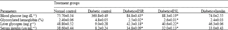 | |
*p<0.05 compared with diabetic control | |
| Table 2: | Serum cholesterol, HDL cholesterol, triglycerides, urea and creatinine levels of untreated and treated rats after repeated oral administration of ESR and ESL and insulin, after 12 weeks |
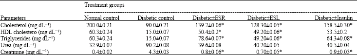 | |
*p<0.05 compared with diabetic control group | |
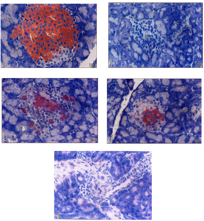 | |
| Fig. 1: | Immunocytochemical staining of β-cells of the islet (A, acinar cells; I, islet cells (x 400) |
Immunocytochemistry: Immunocytochemical staining of the islets from untreated rat revealed a normal population of well granulated β-cells with a majority being insulin positive (Fig. 1a). The pancreas of the alloxan-induced diabetic group showed no positive staining for insulin positive beta cells (Fig. 1b). Similarly islets of the drug (insulin) injected group too, completely lacked insulin positive beta cells (Fig. 1e). The ESR and ESL treatment in diabetic rats (Fig. 1c and d, respectively), restored more than half of the normal population of insulin producing β-cells.
DISCUSSION
It is well known that Alloxan-a diabetogenic agent easily penetrates cells, causing changes that result in the formation of hydrogen peroxides as well as superoxides and hydroxy radicals (Zhang et al., 1992). The avalanche formation of these active oxygen species destructs the DNA structure leading to the impairment of insulin secreting β-cells (Takasu, 1991).This decreases endogenous insulin release and ultimately depresses the tissue glucose utilization.
A significant hypoglycemic activity was associated with ESR, ESL and drug (insulin) treated groups. This may be due to the beta cell stimulation resulting in increased insulin levels. This idea of decrease in blood glucose due to an increase in insulin production was well supplemented by the increased serum insulin levels of extract and drug treated diabetic rats. However the rejuvenation and regeneration of beta cells in the pancreas were witnessed only in the ESR and ESL treated diabetic rats whose pancreas was processed for immunocytochemical staining. This result emphasizes the recuperative property of the ESR and ESL extracts.
In diabetic conditions, the erythrocytes were more prone to oxidative stress and hence exhibit high glycosylated haemoglobin levels (Andallu and Varadacharyulu, 2002), also the normal capacity of the liver to synthesize glycogen is impaired (Welihinda and Karunanayake, 1986) ESR and ESL treatment to diabetic rats decreased the glycosylated haemoglobin levels and ensured the normal synthesis of liver glycogen. Hyperglycemia is often accompanied by an increase in total cholesterol, triglycerides and a decrease in HDL (Markku, 1995). In extracts and drug treated animals, the decrease in blood glucose level is accompanied by the decrease in the serum cholesterol and triglyceride levels. Distinct metabolic renal alterations are seen in experimental diabetes, which is marked by high creatinine and serum urea levels. The plant extract (ESR and ESL) treatment has brought down the elevated levels more effectively than the drug treatment, indicating a positive effect on renal function.
The presents study reveals that the root and leaf extracts of Elephantopus scaber has hypoglycemicaction and besides its antidiabetic activity they also show a positive control on the secondary complications of diabetes. Even though the commercially available drug shows the same trend, regeneration of the damaged β-cells is unique to the extract treatment.
Comparing the root and leaf extract of Elephantopus scaber, the root extract shows a better activity. Further studies to isolate the fractions responsible for this mechanism of action are in progress in our laboratory.
ACKNOWLEDGMENT
The author wishes to thank the Principal, Holy Cross College, Trichy, India for the support provided.
REFERENCES
- Allain, C.C., L.S. Poon, C.S.G. Chan, W. Richmond and P.C. Fu, 1974. Enzymatic determination of total serum cholesterol. Clin. Chem., 20: 470-475.
CrossRefPubMedDirect Link - Andallu, B. and N. Varadacharyulu, 2002. Control of hyperglycemia and retardation of cataract by mulberry (Morus indica L.) leaves in streptozotocin diabetic rats. Ind. J. Exp. Biol., 40: 791-795.
PubMedDirect Link - Bailey, C.J. and C. Day, 1989. Traditional plant medicines as treatments for diabetes. Diabetes Care, 12: 553-564.
CrossRefDirect Link - Bowers, L.D., 1980. Kinetic serum creatinine assay I. The role of various factors in determining specificity. Clin. Chem., 26: 551-554.
Direct Link - Fawcett, J.K. and J.E. Scott, 1960. A rapid and precise method for the determination of urea. J. Clin. Pathol., 13: 156-159.
CrossRefPubMedDirect Link - Fossati, P. and L. Prencipe, 1982. Serum triglycerides determined colorimetrically with an enzyme that produces hydrogen peroxide. Clin. Chem., 28: 2077-2080.
CrossRefPubMedDirect Link - Grover, J.K., V. Vats and S.S. Rathi, 2000. Anti-hypergl ycemic effect of Eugenia jambolana and Tinospora cordifolia in experimental diabetes and their effects on key metabolic enzymes involved in carbohydrate metabolism. J. Ethnopharmacol., 73: 461-470.
Direct Link - Rajkapoor, B., B. Jayakar, R. Ananadan, 2002. Antitumour activity of Elephantopus scaber Linn. against Dalton's Ascitic Lymphoma. Ind. J. Pharm. Sci., 64: 71-73.
Direct Link