
Idris Sherifat Banke
Department of Veterinary Preclinical Studies, Faculty of Veterinary Medicine, Universiti Putra Malaysia, 43400 UPM, Serdang, Selangor, Malaysia
LiveDNA: 234.35358
Arifah Abdul Kadir
Department of Veterinary Preclinical Studies, Faculty of Veterinary Medicine, Universiti Putra Malaysia, 43400 UPM, Serdang, Selangor, Malaysia
LiveDNA: 60.2132
Faez Firdaus Abdullah Jesse
Department of Veterinary Clinical Studies, Faculty of Veterinary Medicine, Universiti Putra Malaysia, 43400 UPM, Serdang, Selangor, Malaysia
LiveDNA: 60.13767
Siti Zubaidah Ramanoon
Department of Farm and Exotic Animal Medicine and Surgery, Faculty of Veterinary Medicine, Universiti Putra Malaysia, 43400 UPM, Serdang, Selangor, Malaysia
Muhammad Abdul Basit
Department of Veterinary Preclinical Studies, Faculty of Veterinary Medicine, Universiti Putra Malaysia, 43400 UPM, Serdang, Selangor, Malaysia
Md Zuki Abu Bakar Zakaria
Department of Veterinary Preclinical Studies, Faculty of Veterinary Medicine, Universiti Putra Malaysia, 43400 UPM, Serdang, Selangor, Malaysia
International Journal of Pharmacology
Year: 2021 | Volume: 17 | Issue: 2 | Page No.: 73-83


ABSTRACT
Background and Objective: Corynebacterium pseudotuberculosis is the causative agent of caseous lymphadenitis, a zoonotic bacterial disease of small ruminants. The disease is unresponsive to antibiotic treatment due to the intracellular nature of the bacteria, thick purulent exudates, antibiotic resistance and formation of biofilms. The objective of this study was to evaluate the antibiofilm activity of oxytetracycline in free (OTC) and cockle shell loaded forms (OTC-CS-CaCO3NP) against C. pseudotuberculosis. Materials and Methods: The Minimum Inhibitory Concentration (MIC) and Minimum Biofilm Eradication Concentration (MBEC) of oxytetracycline in free and cockle shell loaded forms against C. pseudotuberculosis were determined by the microtiter plate assay method. Biofilm formation and inhibition assays were studied spectrophotometrically using the crystal violet assay method. TEM, SEM and fluorescent microscopy were used to assess the antibacterial and antibiofilm effect against C. pseudotuberculosis. Results: The MIC of OTC and OTC-CS-CaCO3NP against C. pseudotuberculosis were 125 and 500 μg L–1, respectively while the MBEC was 250 and >2000 μg L–1. TEM analysis showed OTC-CS-CaCO3NP disrupted cell membrane and formation of vacuoles in C. pseudotuberculosis. Conclusion: Microscopic analysis of the biofilms revealed OTC-CS-CaCO3NP caused significant morphological changes compared to OTC. These results suggest the potential applicability of OTC-CS-CaCO3NP for the treatment of caseous lymphadenitis.
PDF Abstract XML References Citation
Copyright: © 2021. This is an open access article distributed under the terms of the creative commons attribution License, which permits unrestricted use, distribution and reproduction in any medium, provided the original author and source are credited.
How to cite this article
Idris Sherifat Banke, Arifah Abdul Kadir, Faez Firdaus Abdullah Jesse, Siti Zubaidah Ramanoon, Muhammad Abdul Basit and Md Zuki Abu Bakar Zakaria, 2021. Antibiofilm Activity of Oxytetracycline Loaded Calcium Carbonate Aragonite Loaded Nanoparticle Against Corynebacterium pseudotuberculosis. International Journal of Pharmacology, 17: 73-83.
DOI: 10.3923/ijp.2021.73.83
URL: https://scialert.net/abstract/?doi=ijp.2021.73.83
DOI: 10.3923/ijp.2021.73.83
URL: https://scialert.net/abstract/?doi=ijp.2021.73.83
INTRODUCTION
Corynebacterium pseudotuberculosis is a gram-positive, pleomorphic and facultative intracellular bacterium that causes Caseous Lymphadenitis (CLA) in ruminants1. CLA in goats is a zoonotic infection that is accompanied by high treatment cost2. The disease is common in goats world-wide3. The difficulty in treatment encountered in this disease is because antibiotics do not reach the lesions as C. pseudotuberculosis is usually located deep within the lesions surrounded by pus and encapsulated by a thick fibrous capsule4. Also, C. pseudotuberculosis is resistant to antibiotics because it forms biofilms in the host5,6. OTC, a naturally occurring tetracycline with a broad spectrum of antibacterial action is used in veterinary medicine for the treatment of diseases caused by Gram-positive and Gram-negative bacteria7. It is one of the antibiotics used by clinicians in the treatment of CLA in small ruminants8,9. Unfortunately, its widespread use and misuse have resulted in generational microbial resistance against this drug10-12. Therefore, it is pertinent to develop newer methods of improving efficacy and reducing resistance to OTC by using alternative methods of delivery13. Recently, the efficacy of gold and silver nanoparticles against C. pseudotuberculosis has been reported13-15. The use of nanoparticles to treat otherwise a resistant infection is receiving great attention because of the physicochemical characteristic of nanoparticles which allows them to act as therapeutic agents or carriers of antibiotics16. To solve the problem of resistance as well as improve bioavailability and increase the antibacterial activity of OTC, we developed a nano-antibiotic formulation against C. pseudotuberculosis. Loading antibiotics into nanoparticles for drug delivery is a new area in the field of nanomedicine17. The use of CS-CaCO3NP to deliver antibiotics offers several advantages over other inorganic nanocarriers18. This is because CS-CaCO3NP are fabricated from cheap and readily available raw materials as well as maintain loaded drug crystallinity and functionality18,19. The delivery of antibiotics like ciprofloxacin, vancomycin, gentamicin and tetracycline for targeted delivery against Salmonella spp., Staphylococcus aureus, Bacillus subtillis and Shigella flexineri using CS-CaCO3NP yielded promising results compared to the free forms of the drugs12,18-21. Also, CS-CaCO3NP allows for surface functionalization to improve cellular uptake and sustained drug release22-24. Lastly, CS-CaCO3NP is considered safe and non-toxic22-25. Although OTC has been investigated encapsulated in nanoparticles7,11.
This study aimed to find out the effect of OTC-CS-CaCO3NP against C. pseudotuberculosis that remains largely unexplored in trying to find effective treatment against CLA in small ruminants.
MATERIALS AND METHODS
Study area: The study was carried out at Microbiology laboratory, Faculty of Veterinary Medicine, Universiti Putra Malaysia, Malaysia from November, 2018-June, 2019.
Fabrication of CS-CaCO3NP and OTC-CS-CaCO3NP: Fabrication and characterization of CS-CaCO3NP and Oxytetracycline loaded calcium carbonate aragonite nanoparticle (OTC-CS-CaCO3NP) as well as in vitro release of OTC from CS-CaCO3NPwere done as described in our previous study26.
Bacterial culture and staining characteristics: Corynebacterium pseudotuberculosis used in this work was obtained from the Bacteriology Laboratory of Faculty of Veterinary Medicine, Universiti Putra Malaysia, Malaysia. Blood agar was used to culture the bacteria at 37°C for 48 hrs. Gram staining was done to confirm the staining characteristic of the organism.
Antibacterial mode of action: High-Resolution Transmission Electron Microscopy (HR-TEM) was performed to study the effect of cockle shell nanoparticles on bacterial cell integrity and to further explain the mechanism of action of the drug. Control and C. pseudotuberculosis treated with OTC-CS-CaCO3 NP, CS-CaCO3NP and OTC for 24 hrs were fixed and prepared for viewing by HR-TEM. Briefly, bacteria were fixed in fixation solution (2% glutaraldehyde+2% paraformaldehyde dissolved in 0.12 M Sorensen’s buffer, pH 7.4) and then stained by (2%) Osmium Tetroxide solution for 1 hr at 4°C. Embedding sections were cut on an MT6000 ultramicrotome (Sorvall, New Castle, DE). Thin sections (0.1-0.05 mm) of the embedded bacterial cell ultrastructural changes induced in C. pseudotuberculosis treated with OTC-CS-CaCO3 NP, CS-CaCO3NP and OTC were examined under the electron microscope (Hitachi H-7100, Hitachi Ltd., Tokyo, Japan).
Corynebacterium pseudotuberculosis biofilm formation: Bacterial colonies were collected from the blood agar plate and dissolved in Tryptic Soy Broth (TSB) with 2% FBS. The density of the bacterial suspension was standardized by comparison with 1.0 McFarland standard (3.0×10⁸ CFU mL–1). This was incubated at 37°C overnight. Biofilm growth, staining and quantification were done as described by. Overnight C. pseudotuberculosis culture was diluted (1:30) into fresh TSB to give an approximate bacterial count of 1.0×10⁷ CFU mL–1. Total 100 μL of the diluted culture was added into each well of the 96 well non-tissue culture treated plates. This was then incubated at 37°C on a rocker at 10 rpm for 6, 12 and 24 hrs at 10% CO2 to evaluate biofilm formation. Following incubation, the unattached C. pseudotuberculosis cells were discarded by turning the plate over and shaking out the liquid. Then the plate was gently submerged in a small tub of water and the water was gently shaken out to remove the remaining media and unattached planktonic cells. About 125 μL of a 0.1% solution of crystal violet was added to each well of the 96 well plates for 10-15 min and incubated at room temperature. After incubation, the plate was submerged in a clean plate of water to completely wash out the crystal violet. This was then blotted dry on a paper tissue and the plates turned upside down to dry overnight. To quantify the number of biofilms formed, 125 μL of 30% acetic acid in water to each well of the microtiter plate to solubilize the crystal violet. The 96 well plates were then incubated at room temperature for 10-15 min after which 125 μL of the solubilized biofilm stained with crystal violet was then transferred to a new microtiter plate. The biofilms were quantified by reading the absorbance in a microplate reader (TEKAN Infinite M200PRO) (Tekan, Seestrasse, Mannedorf Switzerland) at 600 nm using 30% acetic acid in water as the blank. The experiment was done 3 times on different days and the average Optical Density (OD) at 600 nm was determined. The results were interpreted as follows: Positive (>0.24), weak (>0.12 and <0.24), or negative: (<0.12)27.
MIC and MBEC of CS-CaCO3NP, OTC-CS-CaCO3NP and OTC Against C. pseudotuberculosis: The MIC of CS-CaCO3NP, OTC-CS-CaCO3 NP and OTC was determined using the microtiter plate assay method. Total 100 μL of sterile Mueller Hinton Broth (MHB) was added to 96 well microtiter plates then 100 μL of CS-CaCO3NP, OTC-CS-CaCO3 NP and OTC prepared in MHB were added to the first well and 2-fold serial dilutions were done (2000-15.6 μg mL–1). Treatment was not added to the last row to serve as the control. A 5 μL of diluted overnight cultures (1:100) of C. pseudotuberculosis (1.0×10⁸ CFU mL–1) in TSB supplemented with 2% FBS was added to the treatment rows except for the control. This was incubated overnight at 37°C for 24 hrs. MIC was determined, visually assessing the turbidity in the CS-CaCO3NP, OTC-CS-CaCO3 NP and OTC 96 well plates and further confirmed using a micro ELISA auto reader (TEKAN Infinite M200PRO) (Tekan, Seestrasse, Mannedorf, Switzerland) at a wavelength of 600 nm. The lowest concentration that inhibited bacterial growth (showed no visible growth after 24hrs incubation) was assigned as the MIC.
The MBEC was done as described by Mulla et al.28 with modifications. Overnight C. pseudotuberculosis culture (From TSB with 2% FBS) was diluted (1:30) into fresh MHB (with 2% FBS) broth for biofilm assay. This is to give an approximate bacterial count of 1.0×10⁷ CFU mL–1 in sterile glass tubes. A 150 μL of the diluted culture was added into each well of the 96 well microtiter plate and incubated at 37°C on a rocker at 10 rpm for 24 hrs at 10% CO2.
Biofilm formation was confirmed by staining the 96 well plates with 0.1% crystal violet for 10-15 min. The plates were then submerged in a tube with water, shaken out to remove the excess dye and blotted dry on a stack of paper towels to remove the excess cells and dye.
Mechanical disruption of biofilms was done by ultrasonic disruption of the biofilms in the 96 well microtiter plates after adding 150 μL of MHB with 2% FBS for 5 min and then incubated at 37°C for 24 hrs with OTC-CS-CaCO3 NP, CS-CaCO3NP and OTC (2000-15.6 μg mL–1). MBEC was determined by inspecting the plates visually for turbidity and confirmed by reading the absorbance at 600 nm in a 96-well plate reader (TEKAN Infinite M200PRO) (Tekan, Seestrasse, Mannedorf Switzerland) as described by Harrison et al.29.
Biofilm inhibition assay: Inhibition of C. pseudotuberculosis biofilm formation by OTC-CS-CaCO3NP and OTC was studied spectrophotometrically using crystal violet assay as described previously30. Overnight C. pseudotuberculosis culture was diluted (1:30) into fresh TSB to give an approximate bacterial count of 1.0×10⁷ CFU mL–1. A 100 μL of the diluted culture was added into each well of the 96 well non-tissue culture treated plates containing different concentrations of OTC-CS-CaCO3NP NP and OTC ranging from 2500-78.13 μg mL–1 obtained by 2-fold serial dilution in MHB and incubated at 37°C for 24 hrs on a rocker. The wells were then washed with sterile PBS to ensure the removal of unattached planktonic C. pseudotuberculosis. A 125 μL of a 0.1% solution of crystal violet solution was added to each well for 15 min. After incubation, the crystal violet solution in the wells was removed and excess crystal violet was washed out with sterile PBS. Total 125 μL of 30% acetic acid in water was added to each well of the microtiter plate to solubilize the crystal violet stain for 10-15 min, then the solubilized biofilm was transferred to a new microtiter plate and absorbance was measured spectrophotometrically at 600 nm. The percentage of biofilm inhibition was calculated as follows:
 |
Morphological changes of C. pseudotuberculosis biofilms exposed to OTC-CS-CaCO3NP
Scanning electron microscopy: Total 96 well plates with the bacterial biofilm were incubated for 24 hrs at 37°C with MBEC of CS-CaCO3NP, OTC-CS-CaCO3 NP and OTC. The supernatant was removed and rinsed with PBS, the biofilm was then disrupted by sonication then collected with pipettes into centrifuge tubes. The control and treated biofilms were centrifuged at 3000 rpm for 5 min after which the supernatant was removed and was fixed with glutaraldehyde for 6 hrs and further prepared for analysis using SEM. Briefly, the bacterial biofilm was fixed in fixation solution (2% glutaraldehyde+2% paraformaldehyde dissolved in 0.12 M Sorensen’s buffer, pH 7.4), dehydrated with graded levels of acetone 35, 50, 75, 90 and 100% sequentially. Then stained by (2%) Osmium Tetroxide solution for 2 hrs at 4°C and was examined and photographed using a scanning electron microscope (JSM-6400 Scanning Microscope) (Jeol, USA).
Fluorescent microscopy: Plates with control biofilm and biofilm treated with MBEC of CS-CaCO3NP, OTC-CS-CaCO3NP and OTC were incubated for 24 hrs at 37°C. The plates were rinsed with PBS then treated with 6 μL of Acridine orange/Ethidium bromide (1 and 3 mg mL–1, respectively) was added to each well, then incubated in the dark for 20 min. Excess dye was removed from each well and the wells were viewed under a fluorescent microscope before the dyes leak out of the biofilm. The wells were photographed by a fluorescence microscope (Motic Asia, Hong Kong) with a FITC filter (green) for live cells a TRITC filter (red) for dead biofilms as described by Skogman et al.31.
RESULTS
Cultural and staining characteristics of C. pseudotuberculosis: The result of Fig. 1a revealed the colonial morphology of C. pseudotuberculosis on 5% sheep blood agar. It presented as small (about 1 mm in diameter) with whitish and translucent colonies which did not produce hemolysis following 48 hrs of incubation at 37°C. Gram staining the smear made from the cultured C. pseudotuberculosis showed a pleomorphic Chinese Letter-like presentation which stained Gram-positive (Fig. 1b).
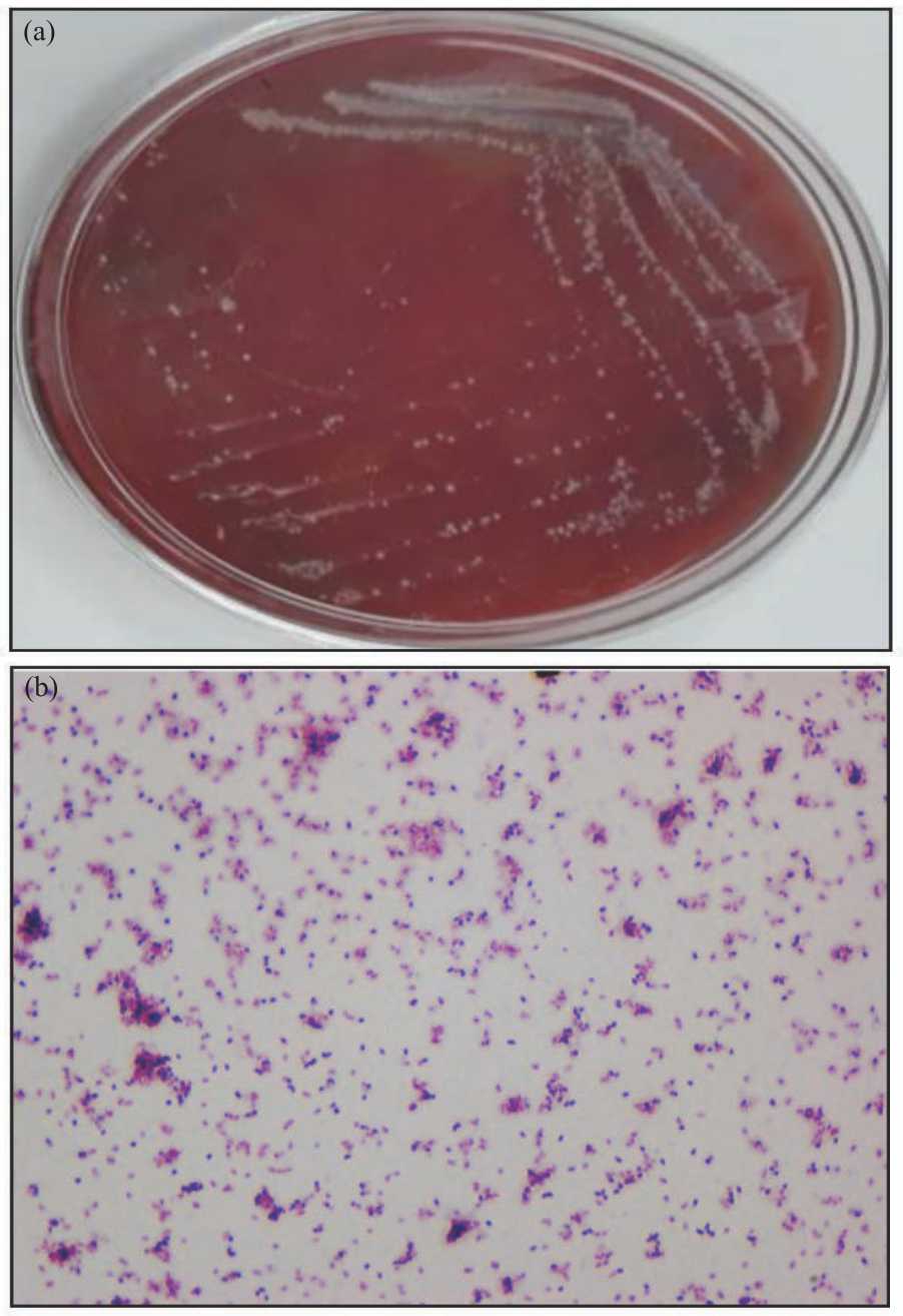 |
| Fig. 1(a-b): | Cultural and staining characteristics of C. pseudotuberculosis (a) C. pseudotuberculosis after 48 hrs of incubation at 37°C grew as small (1 mm diameter) white, opaque and dry colonies with no haemolysis and (b) C. pseudotuberculosis stained Gram-positive displaying the characteristic Chinese-letter presentation (magnification ×100) |
| Table 1: MIC and MBEC of CS-CaCO3NP, OTC-CS-CaCO3NP and OTC against C. pseudotuberculosis | |||
| Treatment (μg mL–1) | CS-CaCO3NP | OTC-CS-CaCO3NP | OTC |
| MIC | NE | 125 | 500 |
| MBEC | NE | 250 | >2000 | NE: No antibacterial effect, MIC: Minimum inhibitory concentration, MBEC: Minimum biofilm eradication concentration |
MIC and MBEC of CS-CaCO3NP, OTC-CS-CaCO3NP and OTC against C. pseudotuberculosis: The MIC values of OTC-CS-CaCO3NP and OTC that inhibited the growth of C. pseudotuberculosis in Mueller Hinton broth was 125 and 500 μg mL–1, respectively. Whereas the MBEC was 250 and >2000 μg mL–1 (Table 1).
Antibacterial mode of action of OTC-CS-CaCO3NP: TEM studies were done to observe the antibacterial effect of OTC-CS-CaCO3NP on C. pseudotuberculosis cell walls. The images obtained indicated OTC-CS-CaCO3NP that disrupted the integrity of the cell wall as shown in Fig. 2a-d.
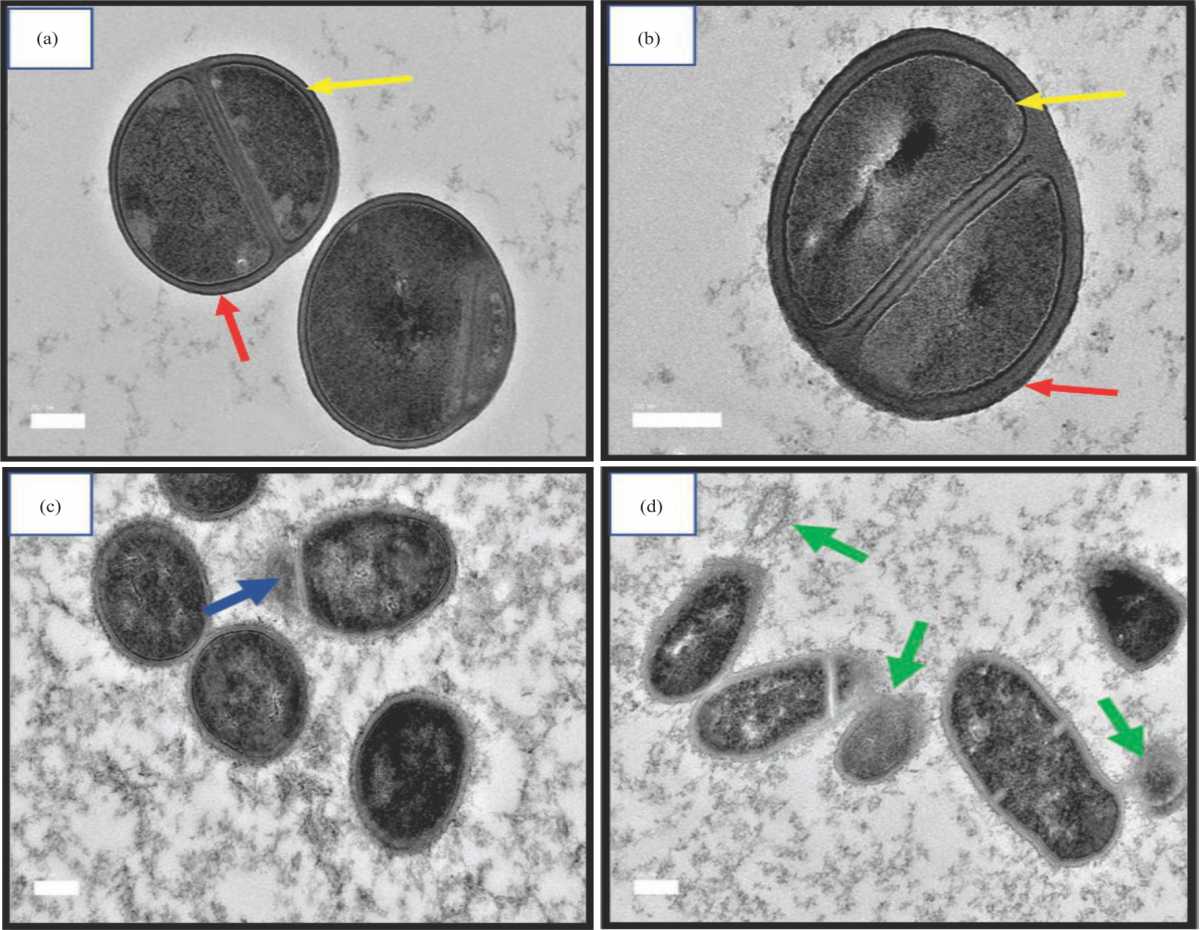 |
| Fig. 2(a-d): | TEM images of C. pseudotuberculosis (a) Control, (b) CS-CaCO3NP treated bacterial showing the normal characteristic electro dense (red arrow) and a plasma membrane layer (yellow arrow) of Corynebacterium spp., (c) OTC treated bacteria showing the mild dissolution of membrane layers (blue arrow) and (d) OTC-CS-CaCO3NP) showing dissolution of cells (green arrow) (scale bar 200 μm) |
The control C. pseudotuberculosis cell (Fig. 2a) appeared with a complete cell wall and plasma membrane showing the normal structure of C. pseudotuberculosis having a close association between the thick electron-dense layer (yellow arrow) and the plasma membrane (blue arrow). All the layers were also present in the CS-CaCO3NP treated group (Fig. 2b) implying that CS-CaCO3NP had no antibacterial effect on C. pseudotuberculosis.
Treatment with OTC caused the bacteria cells to have slightly reduced thickness of the electron-dense layer with the majority of the cells remaining undamaged (Fig. 2c).
Furthermore, treatment of C. pseudotuberculosis with OTC-CS-CaCO3NP resulted in changes in the morphology of the bacterial surface. After 24 hrs, OTC-CS-CaCO3NP caused the outer layers of the cells to be flattened out with reduced thickness, membrane dissolution, broken cell membrane and presence of vacuoles within the bacterial cells (Fig. 2d). TEM analysis further revealed that OTC treated C. pseudotuberculosis showed intact bacterial cells (Fig. 3a) whereas treatment with OTC-CS-CaCO3NP resulted in leakage of the cytoplasmic components to the outer environment through the disruption of the cell wall (Fig. 3b).
Corynebacterium pseudotuberculosis biofilms formation: C. pseudotuberculosis biofilms significantly increased from 0.32±0.05 at 6 hrs to 2.17±0.72 and 2.94±0.25 at 12 and 24 hrs respectively, at 600 nm optical density (Fig. 4).
Biofilm inhibition assay: OTC-CS-CaCO3NP and OTC at concentrations from >MBEC to the sub-MBEC (2500-78.13 μg mL–1) were utilized to study the antibiofilm activity against C. pseudotuberculosis using crystal violet biofilm assay as shown in Fig. 2b-c. Biofilm formations by C. pseudotuberculosis were significantly reduced by OTC-CS-CaCO3NP at concentrations of 156.3 and 78.13 μg mL–1 (p<0.05) (Fig. 5a).
 |
| Fig. 3(a-b): | TEM images of C. pseudotuberculosis (a) After treatment with OTC showing intact bacterial cells and (b) OTC-CS-CaCO3NP showing the dissolution of cells and presence of vacuoles (red arrow), broken cell membrane (blue arrow) and detachment of cytoplasmic membrane from the bacterial cell wall (green arrow) (scale bar 1 μm) |
 |
| Fig. 4: | Biofilm formation (Optical density600) of C. pseudotuberculosis by crystal violet assay |
Whereas the OTC treated biofilms were significantly reduced starting from 625-78.13 μg mL–1 (p<0.05) (Fig. 5b).
Morphological changes assessment
Scanning electron microscopy: Visualization of C. pseudotuberculosis biofilms with scanning electron microscopy and fluorescence microscopy showed morphological differences in the OTC-CS-CaCO3NP and OTC treated biofilm architectures. The control and CS-CaCO3NP treated C. pseudotuberculosis displayed the characteristic 3-dimensional morphology showing rod-shaped, smooth-surfaced bacteria cells closely packed together. Also, the cell membranes of the bacterial cells were intact (Fig. 6a-b) whereas C. pseudotuberculosis treated with OTC revealed bacterial cells in the biofilm less compactly held together, with blebbing and roughness on the outer surfaces of the cells (Fig. 6c). OTC-CS-CaCO3NP treated biofilms showed roughness on the outer surface of the bacteria seen in the biofilm. Most of the bacterial cells in the biofilm were digested and having fewer bacterial cells with swollen, rough, irregular and non-uniform shapes (Fig. 6d). This result shows that OTC-CS-CaCO3NP destroyed the cell membrane, with a roughness of the outer surface of the bacteria seen in the biofilm. Most of the bacterial cells in the biofilm were digested and having fewer bacterial cells with swollen, rough, irregular and non-uniform shapes.
Fluorescence microscopy: Visual examination of fluorescence images of untreated biofilms and CS-CaCO3NP, OTC-CS-CaCO3NP and OTC treated biofilms showed colours depicting the different stages of cell death (Fig. 7a-d). Control and biofilms treated with CS-CaCO3NP showed live bacterial cells (bright green) (Fig. 7a-b). Whereas OTC-CS-CaCO3NP treated biofilms had dead bacterial cells (Red), early apoptotic bacterial cells (greenish-yellow), late apoptotic bacterial cells (Orange-red) and dying cells (faint green) (Fig. 7c).
 |
| Fig. 5(a-b): | Antibiofilm formation activity by crystal violet assay (a) OTC-CS-CaCO3NP antibiofilm activity on C. pseudotuberculosis and (b) OTC antibiofilm activity on C. pseudotuberculosis |
 |
| Fig. 6(a-d): | Scanning electron microscope image of C. pseudotuberculosis biofilm (a) intact cell membrane and smooth cell surface, (b) Treatment with CS-CaCO3NP showing intact cell membrane and smooth cell surface, (c) Treatment with OTC showing bacterial cells with pleomorphic shapes, roughness and blebbing on the cell surfaces and (d) Treatment with OTC-CS-CaCO3NP showing digested bacterial cells with irregular shapes, rough cell surface and membrane |
However, treatment of biofilms with OTC resulted in viable bacterial cells (bright green), early apoptotic cells (greenish-yellow) and a few late apoptotic cells (orange-red) (Fig. 7d).
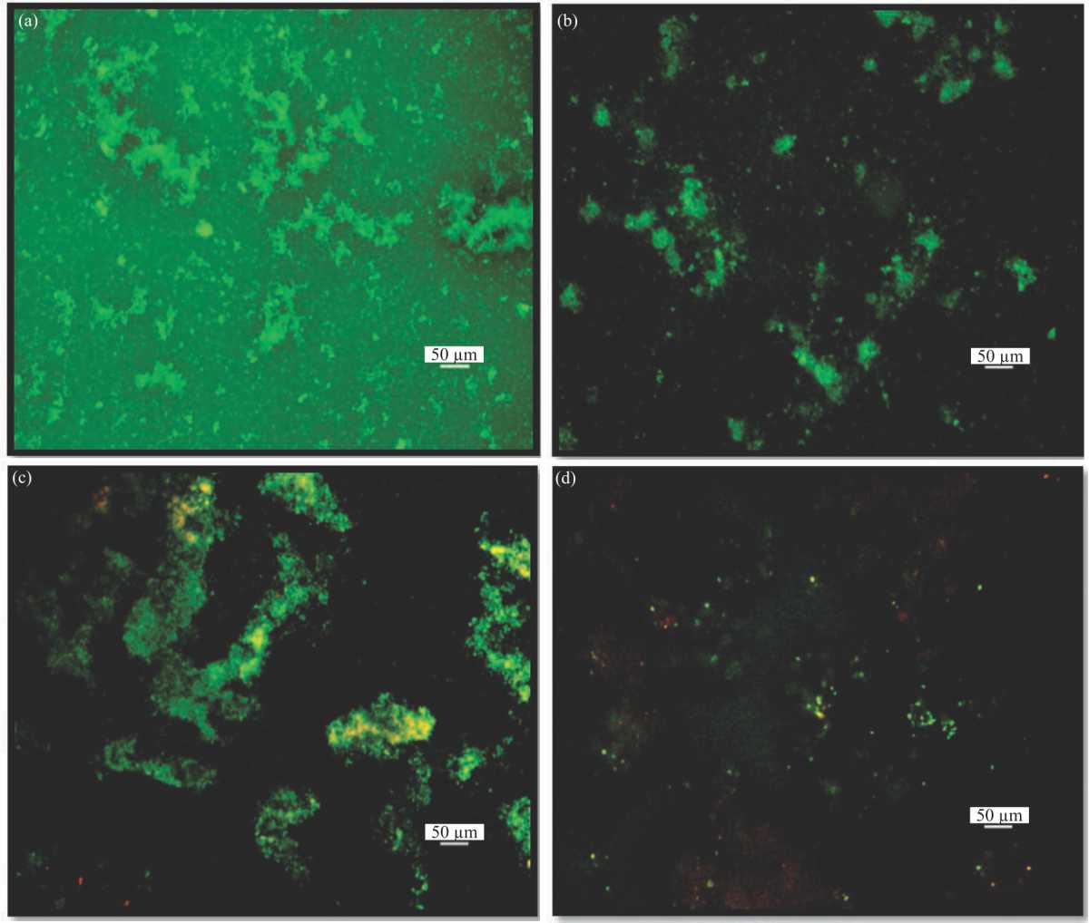 |
| Fig. 7(a-d): | Fluorescence microscope image of C. pseudotuberculosis biofilm (a) Viable cells (The bright green coloured biofilms indicate live biofilms), (b) Treatment with CS-CaCO3NP and stained by acridine orange/ethidium bromide (The bright green coloured biofilms indicate live biofilms, (c) Treatment with OTC (The green colour depicts viable biofilms, the greenish-yellow = early apoptotic biofilms and orange-red = late apoptotic biofilms) and (d) Treatment with OTC-CS-CaCO3NP Showing dead biofilms (red), early apoptotic biofilms (greenish-yellow) and late apoptotic biofilms (orange-red) |
DISCUSSION
In the present study, the planktonic form of C. pseudotuberculosis was highly susceptible to OTC compared to the biofilm form which agrees with the results of Olson et al.5. The lower MIC of OTC-CS-CaCO3NP compared to that of OTC in the present study means that encapsulation of OTC in CS-CaCO3NP nanoparticles enhanced the antibacterial sensitivity of OTC against C. pseudotuberculosis. The antibacterial effect of OTC-CS-CaCO3NP may be due to the large surface area of the CS-CaCO3NP conjugated with a considerable number of OTC molecules that bind to the outer surface of C. pseudotuberculosis which enables easy entry into the bacterium. Furthermore, the lower MIC of OTC-CS-CaCO3NP as against that of OTC may have been caused by a synergistic mechanism of action of both OTC and CS-CaCO3NP in the OTC-CS-CaCO3NP. Nanoparticles exert synergistic antibacterial effect by disruption of the cell wall integrity because of adsorption of the negatively charged CS-CaCO3NP on the surface of C. pseudotuberculosis, combined with the regular mechanism of action of the antibiotic-loaded nanoparticle19 and in this case OTC which is via protein synthesis inhibition. MBEC values in the present study for both OTC-CS-CaCO3NP and OTC suggest that the formation of biofilms by C. pseudotuberculosis is the major reason why CLA in animals is unresponsive to antibiotics because biofilms can withstand very high concentrations of antibiotics that would readily destroy the planktonic form of bacteria32.
The close association of the thick electron-dense layer and the plasma membrane seen in the C. pseudotuberculosis cells on HR-TEM is characteristic of Corynebacterium species33. An earlier study by Burkovski,33 revealed that the electro dense layer is made up of peptidoglycans while the electro light area is composed of mycolic acid is which plays a major role in the pathogenicity and permeability of Corynebacterium species. All the layers were also present in the CS-CaCO3NP treated group implying that CS-CaCO3NP had no antibacterial effect on C. pseudotuberculosis. Furthermore, in the OTC-CS-CaCO3NP group, there was no clear demarcation of these layers as there is a spread out of the outer envelope of the bacteria. Therefore, we suggest that the antibacterial mechanism of action of OTC-CS-CaCO3NP could be via 2 pathways: (i) Damage to the outer envelope of C. pseudotuberculosis disrupting the gradient of the electrochemical potential of ions across the membrane (Fig. 3b). (ii) OTC-CS-CaCO3NP may have increased the rate of Reactive Oxygen Species (ROS) within the organism as evidenced by the high amounts of vacuoles seen in C. pseudotuberculosis. This agrees with Mohamed et al.14 who reported that the antibacterial mechanism of action of gold nanoparticles against C. pseudotuberculosis was due to induction of oxidative stress and cell membrane damage. Mukherjee et al.12 reported that loading tetracyclines into calcium phosphate nanoparticles block the tetracycline efflux pump in Shigella flexneri 2a which resulted in high amounts of tetracycline in the cell and hence improved antibacterial activity. OTC-CS-CaCO3NP may also block the tetracycline efflux pump in the bacteria cells which results in increased concentration of the OTC within the bacteria cell as explained by Mukherjee et al.12. However, in the OTC treated C. pseudotuberculosis although some cells were damaged, the cell envelopes were intact with no obvious disorganization. This is similar to what was observed by Pan et al.19 on HR-TEM after treating Bacillus subtillis with free gentamicin and gentamicin-loaded CaCO3 nanoparticles. The similarities may be because CS-CaCO3NP enhanced the penetration of loaded into it, therefore, increasing damage to the bacteria envelope.
The increasing biofilm formation between 6-24 hrs is expected because C. pseudotuberculosis readily forms biofilms following culture in TSB with 2% FBS at 10% CO2 as reported by Yaacob et al.5. High in vitro biofilm formation capacity is an indication that C. pseudotuberculosis readily forms biofilms following culture in TSB with 2% FBS at 10% CO2 as reported by researchers5, Since TSB with 2% FBS at 10% CO2 mimics the environment of the organism in host tissue5,then it can be said that similar conditions are necessary for the growth of C. pseudotuberculosis biofilms in vitro and in vivo. Moreover, the absorbance at 600 nm is proportional to the number of biofilms formed. The C. pseudotuberculosis biofilm inhibition assay revealed that greater than 50% of the C. pseudotuberculosis was viable after 24 hrs following OTC treatment. The results suggest that OTC-CS-CaCO3NP maintained high biofilm inhibition up to 156.3 μg mL–1 whereas biofilm inhibition was concentration-dependent for OTC.
The C. pseudotuberculosis in the control and CS-CaCO3NP treated groups were predominantly green implying that the bacterial cells within the biofilms were viable as the stain (acridine orange) used enters the cells of intact viable cells and stain the nucleus green34,35. For the OTC-CS-CaCO3NP treated biofilms, more red colouration was present and this may have been due to destruction to the cytoplasmic membrane of C. pseudotuberculosis done by OTC-CS-CaCO3NP which caused the influx of the ethidium bromide dye into the cytoplasm to stain the nucleus red. Furthermore, in the OTC-CS-CaCO3NP treated group, the early and late apoptotic cells (greenish-yellow and orange-red, respectively) show that OTC-CS-CaCO3NP exerted its effect on C. pseudotuberculosis biofilms since most of the cells were in the different stages of cell death. On the other hand, OTC treated biofilms exerted a mild antibiofilm effect as there were predominantly viable bacterial cells within the biofilm with few early apoptotic cells. Thus, from this study, it is clear that OTC was successfully released from OTC-CS-CaCO3NP to the bacterial cells after penetrating the biofilm which prevented the proliferation of new biofilms as well as the destruction of existing biofilm. Similar reports were noted by Mu et al.35 Consequently, the SEM and fluorescent microscopy results from this work results agree with the MIC, MBEC and TEM results.
CONCLUSION
This study reveals that OTC-CS-CaCO3NP exerts a better antibacterial and antibiofilm effect against C. pseudotuberculosis compared to free OTC based on the MIC, MBEC and biofilm inhibition studies. The improved antibiofilm effect is attributed to the damage to the outer envelope of C. pseudotuberculosis caused by OTC-CS-CaCO3NP as seen on HR-TEM. Thus, OTC-CS-CaCO3NP may provide a promising new strategy for the treatment of C. pseudotuberculosis infection. Therefore, further studies investigating the potential of OTC-CS-CaCO3NP for the effective treatment of C. pseudotuberculosis in vivo is necessary.
SIGNIFICANCE STATEMENT
This study discovered that the antibiofilm effect of OTC-CS-CaCO3NP is more pronounced than that of free OTC which can be beneficial in C. pseudotuberculosis treatment. Our findings coupled with the increasing use of calcium-based nanoparticles opens uncover promising perspectives for OTC-CS-CaCO3NP in the treatment of caseous lymphadenitis.
ACKNOWLEDGMENTS
This project was financed by Geran Putra, Universiti Putra Malaysia (Grant Code: GP/2018/9616700).
REFERENCES
- Li, H., H. Yang, Z. Zhou, X. Li and W. Yi et al., 2018. Isolation, antibiotic resistance, virulence traits and phylogenetic analysis of Corynebacterium pseudotuberculosis from goats in southwestern China. Small Rum. Res., 168: 69-75.
CrossRefDirect Link - Oreiby, A.F., 2015. Diagnosis of caseous lymphadenitis in sheep and goat. Small Rum. Res., 123: 160-166.
CrossRefDirect Link - Minozzi, G., S. Mattiello, L. Grosso, P. Crepaldi, S. Chessa and G. Pagnacco, 2017. First insights in the genetics of caseous lymphadenitis in goats. Ital. J. Anim. Sci., 16: 31-38.
CrossRefDirect Link - Oreiby, A.F. and Y.M. Hegazy, 2016. Diagnosis of ovine caseous lymphadenitis by blood and milk gamma interferon assays. Small Rumin. Res., 144: 109-112.
CrossRefDirect Link - Yaacob, M.F., A. Murata, N.H.M. Nor, F.F.A. Jesse and M.F.Z.R. Yahya, 2021. Biochemical composition, morphology and antimicrobial susceptibility pattern of Corynebacterium pseudotuberculosis biofilm. J. King Saud Univ. Sci., Vol. 33.
CrossRefDirect Link - Sá, M.C.A., J.L.A. Veschi, G.B. Santos, E.S. Amanso and S.A.S. Oliveira et al., 2013. Activity of disinfectants and biofilm production of Corynebacterium pseudotuberculosis. Pesq. Vet. Bras., 33: 1319-1324.
CrossRefDirect Link - Mishra, D., P. Khare, K. Shanker, D.K. Singh and S. Luqman, 2016. Controlled delivery systems of cellulose matrix for oxytetracycline: In vitro dissolution. New. Horiz. Transl. Med., 3: 66-72.
CrossRefDirect Link - Senturk, S. and M. Temizel, 2006. Clinical efficacy of rifamycin SV combined with oxytetracycline in the treatment of caseous lymphadenitis in sheep. Vet. Rec., 159: 216-217.
CrossRefDirect Link - Osman, A.Y., F.F.J. Abdullah, E.L.T. Chung, Y. Abba, M.A. Sadiq and K. Mohammed et al., 2015. Caseous lymphadenitis in a Goat: A case report. Int. J. Livest. Res., 5: 128-132.
CrossRefDirect Link - Guerra, W., P.P. Silva-Caldeira, H. Terenzi and E.C. Pereira-Maia, 2016. Impact of metal coordination on the antibiotic and non-antibiotic activities of tetracycline-based drugs. Coord. Chem. Rev., 328: 188-199.
CrossRefDirect Link - Georgescu, D., A.M. Brezoiu, R.A. Mitran, D. Berger, C. Matei and B. Negreanu-Pirjol, 2017. Mesostructured silica–titania composites for improved oxytetracycline delivery systems. C.R. Chim., 20: 1017-1025.
CrossRefDirect Link - Mukherjee, R., D. Dutta, M. Patra, B. Chatterjee and T. Basu, 2019. Nanonized tetracycline cures deadly diarrheal disease ‘shigellosis’ in mice, caused by multidrug-resistant Shigella flexneri 2a bacterial infection. Nanomed. Nanotechnol. Biol. Med., 18: 402-413.
CrossRefDirect Link - Santos, L.M., D. Stanisic, U.J. Menezes, M.A. Mendonça and T.D. Barral et al., 2019. Biogenic silver nanoparticles as a post-surgical treatment for Corynebacterium pseudotuberculosis infection in small ruminants. Front. Microbiol., Vol. 10.
CrossRefDirect Link - Mohamed, M.M., S.A. Fouad, H.A. Elshoky, G.M. Mohammed and T.A. Salaheldin, 2017. Antibacterial effect of gold nanoparticles against Corynebacterium pseudotuberculosis. Int. J. Vet. Sci. Med., 5: 23-29.
CrossRefDirect Link - Stanisic, D., N.L. Fregonesi, C.H.N. Barros, J.G.M. Pontes and S. Fulaz et al., 2018. NMR insights on nano silver post-surgical treatment of superficial caseous lymphadenitis in small ruminants. RSC Adv., 8: 40778-40786.
CrossRefDirect Link - Zazo, H., C.I. Colino and J.M. Lanao, 2016. Current applications of nanoparticles in infectious diseases. J. Controlled Release, 224: 86-102.
CrossRefDirect Link - Aderibigbe, B.A., 2017. Metal-based nanoparticles for the treatment of infectious diseases. Molecules, Vol. 22.
CrossRefDirect Link - Mukherjee, R., M. Patra, D. Dutta, M. Banik and T. Basu, 2016. Tetracycline-loaded calcium phosphate nanoparticle (Tet-CPNP): Rejuvenation of an obsolete antibiotic to further action. Biochim. Biophys. Acta, Gen. Subj., 1860: 1929-1941.
CrossRefDirect Link - Pan, X., S. Chen, D. Li, W. Rao, Y. Zheng and Z. Yang, 2018. The synergistic antibacterial mechanism of gentamicin-loaded CaCO3 nanoparticles. Front. Chem., Vol. 5.
CrossRefDirect Link - Isa, T., Z.A.B. Zakaria, Y. Rukayadi, M.N.M. Hezmee, A.Z. Jaji and M.U. Imam et al., 2016. Antibacterial activity of ciprofloxacin-encapsulated cockle shells calcium carbonate (aragonite) nanoparticles and its biocompatibility in macrophage J774A.1. Int. J. Mol. Sci., Vol. 17.
CrossRefDirect Link - Saidykhan, L., Z. Zakaria, Y. Rukayadi, A.U. Kura and L.S. Yazan, 2016. Development of nanoantibiotic delivery system using cockle shell-derived aragonite nanoparticles for treatment of osteomyelitis. Int. J. Nanomed., 11: 661-673.
CrossRefDirect Link - Fu, W., M.H.M. Noor, L.M. Yusof, T.A.T. Ibrahim, Y.S. Keong, A.Z. Jaji and M.Z.A.B. Zakaria, 2017. In vitro evaluation of a novel pH sensitive drug delivery system based cockle shell-derived aragonite nanoparticles against osteosarcoma. J. Exp. Nanosci., 12: 166-187.
CrossRefDirect Link - Hammadi, N.I., Y. Abba, M.N.M. Hezmee, I.S.A. Razak and A.Z. Jaji et al., 2017. Formulation of a sustained release docetaxel loaded cockle shell-derived calcium carbonate nanoparticles against breast cancer. Pharm. Res., 34: 1193-1203.
CrossRefDirect Link - Min, K.H., E.Y. Jang, H.J. Lee, Y.S. Hwang, J.I. Ryu, J.H. Moon and S.C. Lee, 2019. pH-responsive mineralized nanoparticles for bacteria-triggered topical release of antibiotics. J. Ind. Eng. Chem., 71: 210-219.
CrossRefDirect Link - Jaji, A.Z., Z.A.B. Zakaria, R. Mahmud, M.Y. Loqman and M.N.M. Hezmee et al., 2017. Safety assessments of subcutaneous doses of aragonite calcium carbonate nanocrystals in rats. J. Nanopart. Res., Vol. 19.
CrossRefDirect Link - Idris, S.B., A.A. Kadir, F.F.A. Jesse, S.Z. Ramanoon, M.A. Basit, Z.A. Zakaria and M.Z.A.B. Zakaria, 2019. Synthesis, characterization and in vitro release of oxytetracycline loaded in pH-responsive CaCO3 nanoparticles. J. Appl. Pharm. Sci., Vol. 9.
CrossRefDirect Link - Qin, H., H. Cao, Y. Zhao, C. Zhu and T. Cheng et al., 2014. In vitro and in vivo anti-biofilm effects of silver nanoparticles immobilized on titanium. Biomaterials, 35: 9114-9125.
CrossRefDirect Link - Mulla, S., A. Kumar and S. Rajdey, 2016. Comparison of MIC with MBEC assay for in vitro antimicrobial susceptibility testing in biofilm forming clinical bacterial isolates. Adv. Microbiol., 6: 73-78.
CrossRefDirect Link - Harrison, J.J., H. Ceri, J. Yerly, C.A. Stremick, Y. Hu, R. Martinuzzi and R.J. Turner, 2006. The use of microscopy and three-dimensional visualization to evaluate the structure of microbial biofilms cultivated in the calgary biofilm device. Biol. Proced. Online, 8: 194-215.
CrossRefDirect Link - Goel, S. and P. Mishra, 2018. Thymoquinone inhibits biofilm formation and has selective antibacterial activity due to ROS generation. Appl. Microbiol. Biotechnol., 102: 1955-1967.
CrossRefDirect Link - Skogman, M.E., P.M. Vuorela and A. Fallarero, 2016. A platform of anti-biofilm assays suited to the exploration of natural compound libraries. J. Vis. Exp.
CrossRefDirect Link - Bjarnsholt, T., 2013. The role of bacterial biofilms in chronic infections. Apmis, 121: 1-58.
CrossRefPubMedDirect Link - Burkovski, A., 2013. Cell envelope of corynebacteria: Structure and influence on pathogenicity. ISRN Microbiol., 2013: 1-11.
CrossRefDirect Link - Smith, S.M., D. Ribble, N.B. Goldstein, D.A. Norris and Y.G. Shellman, 2012. A Simple Technique for Quantifying Apoptosis in 96-Well Plates. In: Methods in Cell Biology, Buque, A. and L. Galluzzi (Eds.)., Elsevier, New York, pp: 361-368.
CrossRefDirect Link - Mu, H., J. Tang, Q. Liu, C. Sun, T. Wang and J. Duan, 2016. Potent antibacterial nanoparticles against biofilm and intracellular bacteria. Sci. Rep., Vol. 6.
CrossRefDirect Link