

Mobasher Ahmad
Department of Pharmacology, University College of Pharmacy, University of the Punjab, 54000 Lahore, Pakistan
Iqra Masood
Department of Pharmacology, Lahore College of Women University, Lahore Pakistan
Hafeez Ikram
Department of Pharmacology, Lahore College of Women University, Lahore Pakistan
Rukhsana Anwar
Department of Pharmacology, University College of Pharmacy, University of the Punjab, 54000 Lahore, Pakistan
LiveDNA: 92.16634
Afroze Mobasher
Department of Pharmacology, University College of Pharmacy, University of the Punjab, 54000 Lahore, Pakistan
Sairah Hafeez Kamran
Department of Pharmacology, University College of Pharmacy, University of the Punjab, 54000 Lahore, Pakistan
Usman Akhtar
Department of Pharmacology, University College of Pharmacy, University of the Punjab, 54000 Lahore, Pakistan
International Journal of Pharmacology
Year: 2018 | Volume: 14 | Issue: 1 | Page No.: 61-67





ABSTRACT
Background and Objective: Phoenix dactylifera L. (Ajwa dates) is a primeval plant possessing special characteristic in therapeutics. The aim of the current study was to investigate the pharmacological effects of aqueous and methanolic extracts of Phoenix dactylifera L. on liver, lipid and renal parameters in azithromycin treated rabbits. Materials and Methods: Experimental animals were divided into 4 groups (n = 4) group I (Normal rabbits), group II Azithromycin (AZT) (30 mg kg–1 for 7 days), group III Azithromycin+Aqueous extract (AZT-Aqu) AZT 30 mg kg–1 for 7 days daily then aqueous extract of Phoenix dactylifera L. (300 mg kg–1 for 7 days), group IV Azithromycin+Methanolic extract (AZT-Met) AZT 30 mg kg–1 for 7 days daily, then methanolic extract of Phoenix dactylifera L. (300 mg kg–1 for 7 days). The data were analyzed as Mean±SEM. Student’s-test was applied for statistical evaluation by using SPSS version 19. Results: The serum enzyme levels of liver (AST, ALT, LDH and ALP), lipid profile, creatinine and uric acid levels were analyzed for determining treatment effects of Phoenix dactylifera L. Liver function tests, lipid profile, creatinine and uric acid showed (p<0.05) significant decrease and improvement when compared with the group II. The histopathological analysis of liver showed less damage in treated animals. Conclusion: The result of current study concludes that Phoenix dactylifera L. significantly reduces the toxicity induced by azithromycin.
PDF Abstract XML References Citation
Article History
Received: June 13, 2017;
Accepted: September 06, 2017;
Published: December 15, 2017
Copyright: © 2018. This is an open access article distributed under the terms of the Creative Commons Attribution License , which permits unrestricted use, distribution and reproduction in any medium, provided the original author and source are credited.
How to cite this article
Mobasher Ahmad, Iqra Masood, Hafeez Ikram, Rukhsana Anwar, Afroze Mobasher, Sairah Hafeez Kamran and Usman Akhtar, 2018. Pharmacological Investigation of Phoenix dactylifera L. in Azithromycin Induced Toxicity. International Journal of Pharmacology, 14: 61-67.
DOI: 10.3923/ijp.2018.61.67
URL: https://scialert.net/abstract/?doi=ijp.2018.61.67
DOI: 10.3923/ijp.2018.61.67
URL: https://scialert.net/abstract/?doi=ijp.2018.61.67
INTRODUCTION
Plants have been used as medicines by human beings from the very beginning of time. Various experimentations revealed plants implications as a vital source of medicines. The muslims have been using Phoenix dactylifera L. fruit as well as seeds for treatment of multiple ailments traditionally and religiously. Later, with the advent of science and technology, investigations on several medicinal plants provided evidence for the existing theoretical knowledge1.
Phoenix dactylifera L. (Ajwa dates) are one of the most repetitively and extensively mentioned fruits in the Quran. Date palm have a long history of cultivation and utilization which dates back to atleast 5000 years. It is diploid, perennial, monocotyledonous and dioecious plant belonging to family Arecaceae 2.
Phoenix dactylifera L. fruit contains high percentage of carbohydrates, dietary fiber, fats and various types of amino acids and vitamins are also present3,4. They possess free radical scavenging, antioxidant5, antimutagenic, antimicrobial6, hepatoprotective, nephroprotective, anticancer7 and immunostimulant activities8. The antioxidant activity is attributed to the extensive range of phenolic compounds, alkaloids, volatile oils, sterols and flavonoids5,9,10.
Azithromycin (AZT, macrolide antibiotics) used in treatment of upper and lower respiratory tract, pharyngitis, tonsillitis, dermatological infections, streptococcal pharyngitis, urogenital infections, otitis media and sexually transmitted disease11-13. It is rapidly absorbed orally and extensively distributed in tissue. Hepatic metabolism of AZT results into inactive metabolites and its elimination is biphasic, with half-life of 5 days14. AZT can cause serious adverse effects including cardiomyopathy, angioedema, cholestatic jaundice and gastrointestinal effects. Severe hepatocellular toxicity and hepatic encephalopathy have been reported15. Various case study also reported AZT induced liver injury16,17. The purpose of the current study was to investigate the pharmacological potential of Phoenix dactylifera L. in treatment and prophylactic effect in azithromycin induced animal model.
MATERIALS AND METHODS
Place of work: The study was carried out in 2013-2016, with the collaboration of two oldest institution of Pakistan University College of Pharmacy University of the Punjab, Lahore and Lahore College of Women University, Jail Road Lahore, Pakistan.
Chemicals: All the chemicals used in the experimental procedures were of analytical grade.
Plant used: Fresh pure Ajwa dates (Phoenix dactylifera L.) were brought from gardens of Madina Munawara, Saudi Arabia and identified by a botanist in November, 2014. The flesh was separated manually from pits (seeds), sun dried and cut into small pieces. Aqueous and methanolic extract was prepared as previously described by Ragab et al.2. The extract was prepared at Lahore College for Women University and the experimental procedures were carried out except for phytochemial and histopathological analysis, which were performed at Punjab University College of Pharmacy, University of the Punjab.
Aqueous extract of Phoenix dactylifera L.: Two hundred fifty grams dried Phoenix dactylifera L. were soaked in distilled water (1500 mL) and kept on orbital shaker for constant stirring (75 rpm) for 24 h. The mixture was filtered to get cold macerate and marc left behind. The marc was subjected to hot percolation using Soxhlet apparatus (Roshanico, Pakistan) with distilled water (750 mL). Hot extract (percolate) and cold extract (macerate) were mixed and concentrated for obtaining thick condensate in rotary evaporator subjected to reduced pressure (70 KPa) and controlled temperature (50°C) at constant speed (70 rpm). The resulting condensate was kept in incubator (Memmert IN30plus, Germany) at 37°C for further drying till formation of viscous paste. Percentage yield of this dark brown extract was calculated to be 45% and stored in moisture free container at room temperature until use.
Methanolic extract of Phoenix dactylifera L.: A similar process was adopted for preparation of methanol extract as mentioned above using absolute methanol (99%) instead of distilled water. The percentage yield of the extract was 55% .
Drug used: Azomax tablets 500 mg (manufactured by Novartis, marketed by Sandoz), purchased from local market and crushed in pestle and mortar to form powder.
Animals: Guide for the care and use of laboratory animals by the US National Institutes of Health (NIH publication No. 85-23, revised1985) was followed and according to the procedures and treatment protocols approved by the Institutional Ethical Committee of Lahore College for Women University. Male rabbits (Oryctolagus cuniculus) not less than 1 Kg and purchased from local animal market, inhabited in cages in animal house of university and fed in accordance with the standard housing laboratory conditions (fed with fresh green fodder and water) and acclimatized for 1 week before experiment.
Experimental design: For treatment study, rabbits were divided into 4 groups of six rabbits each group, group I Nor (normal rabbits), group II AZT (AZT 30 mg kg–1 for 7 days), group III AZT-Aqu (AZT 30 mg kg–1 for 7 days daily then aqueous extract of Phoenix dactylifera L. (300 mg kg–1 for 7 days), group IV AZT-Met [(AZT 30 mg kg–1 for 7 days daily then methanolic extract of Phoenix dactylifera L. (300 mg kg–1 for 7 days)].
For prophylactic study, rabbits were divided into 4 groups of six rabbits each group, group I Nor (normal rabbits), group II AZT (AZT 30 mg kg–1 for 7 days), group III AZT-Aqu (Aqueous extract of Phoenix dactylifera L. (300 mg kg–1 for 7 days then AZT 30 mg kg–1), group IV AZT-Met (methanolic extract 300 mg kg–1 for 7 days then AZT 30 mg kg–1).
Phytochemical investigations: Various qualitative phytochemical tests were performed for confirming the presence of different chemical groups i.e., alkaloids, tannins, glycosides and saponins etc.18, in both aqueous and methanol extracts of Phoenix dactylifera L. The investigations are in accordance with the developed protocols as described in previous study19 included:
Alkaloids, Mayer's test: The 0.5 g extract diluted in 10 mL of 1.0% of hydrochloric acid, boiled for 2 min on water bath and filtered. Mayer's reagent’s few drops added to 1 mL filtrate. Pale precipitates formed indicating presence of alkaloids19.
Saponins: The extract (0.2 g) was added to 5 mL distilled water and shook vigorously. Formation of stable persistent solution for 5 min indicated presence of saponins19.
Reducing sugars, fehling's test: The extract (0.2 g) added to equal volumes of boiling Fehling's solution A & B. Brick red precipitates confirmed reducing sugars19.
Tannins: The extract, 0.1 g was diluted in 5 mL distilled water and few dropsof 1% lead acetate solution was added. Formation of red precipitates indicated presence of tannins19.
Phenols, ferric chloride test: The extract 0.2 g was mixed with 1 mL absolute ethanol and added few drops of 10% solution of ferric chloride. Formation of bluish color showed the presence of phenols19.
Resins, water acetone test: Two mL extract was treated with ethanol and added small amount of distilled water. Shake solution well. Turbidity indicated the presence of resins19.
Terpenoids, salkowaski test: The extract (0.5 g) was added to 2 mL chloroform and mixed. Three mL ‘concentrated sulphuric acid’ added. Formation of layer and reddish brown coloration showed the presence of terpenoids19.
Flavonoids: Twenty mg of extract was added to 1 mL of ethanol and added 8 drops of dilute hydrochloric acid followed by small amount of zinc. Solution boiled for 6 min. Reddish pink color appeared indicating the presence of flavonoids19.
Anthraquinones: The extract (0.5 g) was added to 10 mL of boiled sulphuric acid, filtered when hot. Filtrate was shaken with 5 mL benzene. Layered benzene was separated and added 1 mL of 10% ammonia solution in it. Pink color appearance in the lower layer of ammonia showed the presence of anthraquinones19.
Biochemical investigations: Blood samples were collected from marginal ear vein of all experimental rabbits and clotted for 45 min at room temperature. Serum was separated by centrifugation at 1500 rpm for 20 min and used for the estimation of biochemical parameters like alanine aminotransferases (ALT), aspartate aminotransferases (AST), alkaline phosphatases (ALP), creatinine, uric acid, cholesterol, triglycerides, lactate dehydrogenase (LDH), high density lipoproteins (HDL) and low density lipoproteins (LDL) using commercial kits according to the manufacturers protocol.
Histopathological investigations: For histopathological studies of rabbits liver, modified Luna technique was applied20. Three rabbits from each group of prophylactic study were randomly selected. On last day of sampling, anesthetized with chloroform and dissected. Liver obtained from each rabbit was cleaned using saline solution and was fixed with formalin (37%). Paraffin embedded blocks were prepared and 5 μm thick microtome sections were cut and stained with eosin and hematoxylin (Courtesy of Post graduate medical institute). High resolution images were obtained from Chughtai Laboratories, Lhr. and were identified by pathologist (PGMI).
Statistical analysis: The data were analyzed carefully and represented as Mean±SEM. Student’s-test was applied for statistical evaluation of biochemical parameters using SPSS version 19. The value of p<0.01 was considered as statistical significant and p<0.001 was considered highly significant19.
RESULTS
Phytochemical investigations of Phoenix dactylifera L. extract: The phytochemical testing of aqueous and methanolic extracts of Phoenix dactylifera L. showed the presence of various phytochemicals. The aqueous extract contained alkaloids, saponins, reducing sugars, tannins, phenols, terpenoids, flavonoids, anthraquinones except for resins. The methanolic extract contained all the phytochemicals as mentioned above including resins.
Biochemical investigations of various groups (treatment study) of rabbits treated with azithromycin and Phoenix dactylifera L. (aqueous and methanolic) extracts: Liver function test (ALT, AST, ALP) showed significant (p<0.001) increase when treated with AZT for 7 days when compared to normal group. There was significant (p<0.001) decrease after administration of aqueous and methanolic extract for 7 days in AZT-Aqu and AZT-Met groups in serum ALT, AST when compared with AZT group but non-significant decrease was observed in serum ALP in AZT-Met group and significant (p<0.001) decrease in AZT-Aqu group when compared with AZT group as shown in Table 1. Table 1 described the biochemical analysis of treatment group.
Biochemical investigations of various groups (prophylactic study) of rabbits treated with Phoenix dactylifera L. (aqueous and methanolic) extracts then azithromycin: In prophylactic study, serum ALT, AST, ALP showed highly significant (p<0.001) increase in AZT group when compared with normal group, respectively as shown in Table 2. AZT-Aqu groups showed significant (p<0.05) decrease in serum ALT in AZT-Aqu group and very significant (p<0.01) decrease in Aqu-Met group when compared with AZT group. Serum AST, ALP showed highly significant (p<0.001) decrease in AZT-Aqu and AZT-Met groups, respectively when compared with AZT group. Table 2 described the biochemical analysis of prophylactic group.
Histopathological investigation of prophylactic study: Histopathological examination of prophylactic group revealed that rabbits treated with AZT shows marked signs of congestion of blood vessels, inflammation and necrosis of hepatocytes whereas after treatment with aqueous and methanolic extract of Phoenix dactylifera L. restored and prevent liver from further damage and oxidative stress as shown in Fig. 1.
| Table 1: | Biochemical investigations of various groups (treatment study) of rabbits treated with azithromycin and Phoenix dactylifera (Aqueous and Methanolic) extracts |
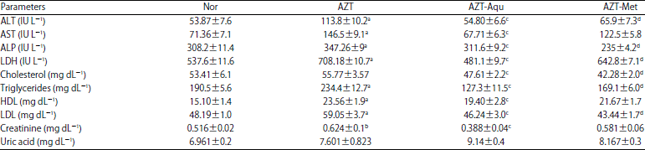 | |
ap<0.001 Nor Vs AZT , bp<0.01 Nor Vs AZT, cp<0.001 AZT Vs AZT-Aqu, dp<0.001 AZT Vs AZT-Met. Values expressed in terms of Mean±SEM | |
| Table 2: | Biochemical investigations of various groups (prophylactic study) of rabbits treated with Phoenix dactylifera (Aqueous and Methanolic) extracts then azithromycin |
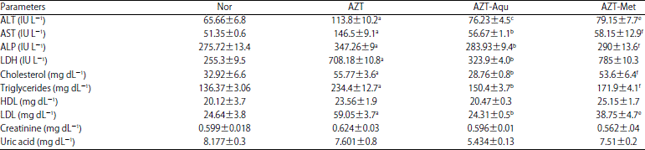 | |
ap<0.001 Nor Vs AZT, bp<0.001 AZT Vs AZT-Aqu, Cp<0.05 AZT Vs AZT-Aqu, dp<0.01 AZT Vs AZT-Aqu, ep<0.01 AZT Vs AZT-Met, fp<0.001 AZT Vs AZT-Met. Values expressed in terms of Mean±SEM | |
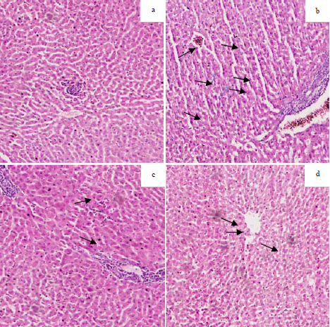 | |
| Fig. 1(a-d): | Histopathological investigation of prophylactic study, (a): Normal group, (b): AZT group (AZT 30 mg kg–1 for 7 days) black arrows shows marked congestion of blood vessels, Focci of inflammation, Marks of fibrosis, early necrosis like changes. Hepatocytes appear normal (c): AZT-Aqu and (d): AZT-Met groups showed marked reduction of inflammation as compared to b |
DISCUSSION
Phoenix dactylifera L. was evaluated in the current study for its pharmacological effects Againstazithromycin, an azalide subgroup of the macrolides. Although it is considered to be safe but the advent of adverse effects relating to macrolides cannot be ruled out completely. Potential side effects include cardiomyopathy, cholestatic jaundice and severe hepatocellular toxicity15. Macrolide antibiotics are among the most commonly prescribed antibiotics for the treatment of various systemic infections (most commonly respiratory tract infections21).
The current study showed significant increase in serum ALT, AST and ALP (p<0.001)in the azithromycin treated group. These results are due to the cholestatic effects on liver cells. The data shows highly significant reduction in the serum hepatic enzymes level (p<0.05) by Phoenix dactylifera L. extracts could be attributed to a decrease in the lipid peroxidation of hepatocellular membrane induced by azithromycin in both treated and prophylactic studies. Also, it might be due to the accelerated regeneration/repairing of damaged hepatocytes2.
The presence of ALT,AST,ALP in serum is basically associated with hepatic cell necrosis22. They are present in cytoplasm of the cell and released after cellular damage into the systemic circulation23. As demonstrated in current study, elevated level of ALT,AST,ALP plus comparative histopathological study of liver (Fig. 1) from different groups further revealed the hepatoprotective effect of Phoenix dactylifera L. extracts. Phoenix dactylifera L. has been found to quench the hydroxyl and superoxide anion radicals6. And explains the role of such antioxidants in inducing the observed decline in serum levels of ALT, AST and ALP (p<0.005), as well as reduction in the histopathlogical alterations of the liver tissues in rabbits pretreated with Phoenix dactylifera L. extracts (Fig. 1). AZT group shows congestion of portal vessels, necrosis of liver cells and dilatation of sinusoids. Like other macrolide, AZT toxicity destroy the hepatic cells and other cells as previously reported24. From the results of the present study, it can also be concluded that Phoenix dactylifera L. may boost up the antioxidant enzymes that stabilize biological molecules in the liver tissue, rendering them more resistant to oxidative stress and can be considered as a functional food for liver toxicity25.
Lactate dehydrogenase, a cardiac as well as hepatic enzyme showed a significant (p<0.05) elevation in azithromycin treated group in the current study. The difference was also statistically significant (p<0.05) in comparison to combined azithromycin and Phoenix dactylifera L. treated group.
In the present study, a significant (p<0.05) decrease has been observed in the lipid profile i.e., cholesterol, triglycerides, HDL and LDL-cholesterol in the subject animals treated with Phoenix dactylifera L. Rise in cholesterol level after azithromycin treatment was not significant but very significant increase was observed in triglycerides, HDL and LDL cholesterol levels (p<0.005).The decline in the lipid biochemical parameters was more significant with Phoenix dactylifera L. aqueous extract (p<0.005) as compare to that with methanolic extract (p>0.05).
Treatment with azithromycin (alone) alters genetic profile of cells and increases predominantly the lipid/cholesterol genes expression26. Rise in the lipid profile in the treated control group in present study well correlates this fact. On the other hand fixed oils and plant sterols present in Phoenix dactylifera L. may account for its antihyperlipidemic activity. Saponins, polyphenolics and flavonoids might also be responsible for the lipid lowering effects27.
Antioxidant components present in Phoenix dactylifera L. i.e. vitamin E, melatonin and ascorbic acid were suggested to form the basis of nephroprotection27. In the present study increased levels of serum creatinine were observed in the azithromycin treated group. In Phoenix dactylifera L. extracts treated group, a significant decline in creatinine level was noted (p<0.05) when compared with treated control. These results clarify a positive role of Phoenix dactylifera L. in kidney protection from azithromycin toxicity.
The outcome of the present study reported the ameliorative and protective effects of Phoenix dactylifera L. (Ajwa dates) against the toxic effects of azithromycin.
CONCLUSION AND FUTURE RECOMMENDATIONS
In the present study Phoenix dactylifera L. aqueous and methanolic extracts showed significant activity in treating and preventing the azithromycin induced hepatic, lipid and renal toxicities. The histopathological studies also showed less necrosis and morphological changes in treated groups. The presence of phenols, terpenoids, flavonoids might be responsible for the pharmacological actions of Phoenix dactylifera L. Further investigations are required to identify the active molecules in Phoenix dactylifera L. and elucidation of their molecular mechanisms.
SIGNIFICANCE STATEMENT
This study discovers the beneficial pharmacological effects of Phoenix dactylifera L. on liver, lipid and renal parameters in acute and chronic diseases. These effects will help the researchers to further evaluate the pharmacological mechanisms of Phoenix dactylifera L. in liver, renal and cardiovascular ailments.
REFERENCES
- Marwat, S.K., M.A. Khan, M.A. Khan, M. Ahmad, M. Zafar, Fazal-ur-Rehman and S. Sultana, 2009. Fruit plant species mentioned in the Holy Qura'n and Ahadith and their ethnomedicinal importance. Am.-Eurasian J. Agric. Environ. Sci., 5: 284-295.
Direct Link - Ragab, A.R., M.A. Elkablawy, B.Y. Sheik and H.N. Baraka, 2013. Antioxidant and tissue-protective studies on Ajwa extract: Dates from Al Madinah Al-Monwarah, Saudia Arabia. J. Environ. Anal. Toxicol. Vol., 3.
CrossRefDirect Link - Ali, A. and S. Abdu, 2011. Antioxidant protection against pathological mycotoxins alterations on proximal tubules in rat kidney. Funct. Foods Health Dis., 1: 118-134.
Direct Link - Zhang, C.R., S.A. Aldosari, P.S.P.V. Vidyasagar, K.M. Nair and M.G. Nair, 2013. Antioxidant and anti-inflammatory assays confirm bioactive compounds in Ajwa date fruit. J. Agric. Food Chem., 61: 5834-5840.
CrossRefDirect Link - Al-Farsi, M., C. Alasalvar, A. Morris, M. Baron and F. Shahidi, 2005. Comparison of antioxidant activity, anthocyanins, carotenoids and phenolics of three native fresh and sun-dried date (Phoenix dactylifera L.) varieties grown in Oman. J. Agric. Food Chem., 53: 7592-7599.
CrossRefDirect Link - Vayalil, P.K., 2002. Antioxidant and antimutagenic properties of aqueous extract of date fruit (Phoenix dactylifera L. Arecaceae). J. Agric. Food Chem., 50: 610-617.
CrossRefPubMedDirect Link - Ishurd, O., F. Zgheel, A. Kermagi, M. Flefla and M. Elmabruk, 2004. Antitumor activity of β-D-glucan from Libyan dates. J. Med. Food, 7: 252-255.
CrossRefDirect Link - Al-Farsi, M.A. and C.Y. Lee, 2008. Nutritional and functional properties of dates: A review. Crit. Rev. Food Sci. Nutr., 48: 877-887.
CrossRefDirect Link - Baliga, M.S., B.R.V. Baliga, S.M. Kandathilc, H.P. Bhatd and P.K. Vayalil, 2011. A review of the chemistry and pharmacology of the date fruits (Phoenix dactylifera L.). Food Res. Int., 44: 1812-1822.
CrossRefDirect Link - Hong, Y.J., F.A. Tomas-Barberan, A.A. Kader and A.E. Mitchell, 2006. The flavonoid glycosides and procyanidin composition of Deglet Noor dates (Phoenix dactylifera). J. Agric. Food Chem., 54: 2405-2411.
CrossRefPubMedDirect Link - Holmes, K.K., 2005. Azithromycin versus penicillin G benzathine for early syphilis. N. Engl. J. Med., 353: 1291-1293.
CrossRefDirect Link - Duran, J.M. and G.W. Amsden, 2000. Azithromycin: Indications for the future? Expert Opin. Pharmacother., 1: 489-505.
CrossRefDirect Link - Drew, R.H. and H.A. Gallis, 1992. Azithromycin-spectrum of activity, pharmacokinetics and clinical applications. Pharmacother.: J. Hum. Pharmacol. Drug Ther., 12: 161-173.
Direct Link - Das, B.K., 2011. Azithromycin induced hepatocellular toxicity and hepatic encephalopathy in asymptomatic dilated cardiomyopathy. Indian J. Pharmacol., 43: 736-737.
Direct Link - Lockwood, A.M., S. Cole and M. Rabinovich, 2010. Azithromycin-induced liver injury. Am. J. Health-Syst. Pharm., 67: 810-814.
CrossRefDirect Link - Chandrupatla, S., A.J. Demetris and M. Rabinovitz, 2002. Case report: Azithromycin-induced intrahepatic cholestasis. Dig. Dis. Sci., 47: 2186-2188.
Direct Link - Ishtiaq, S., M. Ahmad, U. Hanif, S. Akbar and S.H. Kamran, 2014. Phytochemical and in vitro antioxidant evaluation of different fractions of Amaranthus graecizans subsp. silvestris (Vill.) Brenan. Asian Pac. J. Trop. Med., 7: S342-S347.
CrossRefDirect Link - Blasi, F., 2004. Atypical pathogens and respiratory tract infections. Eur. Respir. J., 24: 171-182.
CrossRefDirect Link - Rodriguez-Garay, E.A., 2002. Cholestasis: Human disease and experimental animal models. Ann. Hepatol., 2: 150-158.
PubMedDirect Link - Sallie, R., J.M. Tredger and R. Williams, 1991. Drugs and the liver part 1: Testing liver function. Biopharm. Drug Dispos., 12: 251-259.
CrossRefPubMedDirect Link - Pessayre, D., D. Larrey, C. Funck-Brentano and J.P. Benhamou, 1985. Drug interactions and hepatitis produced by some macrolide antibiotics. J. Antimicrob. Chemother., 16: 181-194.
CrossRefDirect Link - Rock, W., M. Rosenblat, H. Borochov-Neori, N. Volkova, S. Judeinstein, M. Elias and M. Aviram, 2009. Effects of date (Phoenix dactylifera L., Medjool or Hallawi Variety) consumption by healthy subjects on serum glucose and lipid levels and on serum oxidative status: A pilot study. J. Agric. Food Chem., 57: 8010-8017.
CrossRefPubMedDirect Link