
Shreesh K. Ojha
Department of Pharmacology,New Delhi-110 029, India
Mukesh Nandave
Department of Pharmacology,New Delhi-110 029, India
Sachin Arora
Department of Pharmacology,New Delhi-110 029, India
Rajeev Narang
Department of Cardiology,New Delhi-110 029, India
Amit K. Dinda
Department of Pathology, All India Institute of Medical Sciences,New Delhi-110 029, India
Dharamvir Singh Arya
Department of Pharmacology,All India Institute of Medical Sciences,New Delhi-110 029, India
International Journal of Pharmacology
Year: 2008 | Volume: 4 | Issue: 1 | Page No.: 1-10


ABSTRACT
The present study was undertaken to evaluate the cardioprotective potential of hydro-alcoholic extract of Tribulus terrestris Linn. (Family; Zygophyllaceae), a traditional medicine used in Indian and Chinese systems of medicine. Wistar male albino rats weighing 150-200 g were randomly divided into three main experimental groups; sham (saline treated only), isoproterenol (ISP) control (saline and ISP) and Tribulus terrestris treatment groups (T. terrestris and ISP). Saline or T. terrestris extract 250 mg kg-1 once daily were orally administered for 30 days. Isoproterenol was administered in rats to induce myocardial infarction. On days 29 and 30, the animals of ISP control and T. terrestris treatment group were administered ISP (85 mg kg-1, subcutaneously) at an interval of 24 h. On the day 31, 48 h after first dose of ISP, hemodynamic parameters were recorded. After sacrificing the animals the hearts were excised and subjected to biochemical, histopathological and ultrastructural studies. ISP-administration produced a significant decrease in the activities of endogenous antioxidant defence enzymes viz. superoxide dismutase (SOD), catalase (CAT), glutathione peroxidase (GSHPx) and tissue antioxidant, reduced glutathione (GSH) along with a concomitant increase in the lipid peroxidation product malonaldehyde (MDA). In addition, a significant decrease in the activities of myocardial injury markers i.e., creatine phosphokinase-MB (CK-MB isoenzyme) and lactate dehydrogenase (LDH) was also observed in the heart of ISP control group as compared to sham control. Cardiac dysfunction was observed as a decrease in mean arterial pressure (MAP), heart rate (HR), left ventricular rate of peak positive and negative pressure change {(+) and (-) LV dP/dt} and elevated left ventricular end diastolic pressure (LVEDP) following ISP administration. These functional alterations were supported by severe modifications in histopathological and ultrastructural assessment. Pretreatment with T. terrestris resulted in the increased activities of SOD, CAT, GSHPx and prevention of depletion of tissue glutathione along with inhibition of lipid peroxidation. In addition treatment with T. terrestris decreased the leakage of CK-MB and LDH enzymes from myocardium, there was a significant improvement in cardiac function as evidenced by correction of MAP, HR, LVEDP and contractility and relaxation. The possible underlying mechanism of the cardioprotective effect of T. terrestris could be due to restoration of endogenous myocardial antioxidant status or free radical scavenging activity along with correction of the altered hemodynamic parameters and preservation of histoarchitectural and ultrastructural alterations.
PDF Abstract XML References Citation
How to cite this article
Shreesh K. Ojha, Mukesh Nandave, Sachin Arora, Rajeev Narang, Amit K. Dinda and Dharamvir Singh Arya, 2008. Chronic Administration of Tribulus terrestris Linn. Extract Improves Cardiac Function and Attenuates Myocardial Infarction in Rats. International Journal of Pharmacology, 4: 1-10.
DOI: 10.3923/ijp.2008.1.10
URL: https://scialert.net/abstract/?doi=ijp.2008.1.10
DOI: 10.3923/ijp.2008.1.10
URL: https://scialert.net/abstract/?doi=ijp.2008.1.10
INTRODUCTION
Among cardiovascular diseases (CVDs), myocardial infarction (MI) accounts for majority of death and disability and has high incidence rate in both developed and developing countries as seen over the past quarter century (Agrawal et al., 2006). Reduction of death incidences and prevention of myocardial infarction are of utmost importance. In spite of development in modern drugs, there is a growing interest in the use of herb based drugs from alternative medicine for long-term prevention of myocardial ischemia in high risk patients along with a realization that the use of herbal products can influence the course of heart diseases and may provides anintegrated approach of nutritional substances, which help in restoring and maintaining, the balanced body systems (Hertog et al., 1993; Dhar et al., 1968).
The plant Tribulus terrestris Linn, a deciduous tree of the Zygophyllaceae family, is an important herb from Indian and Chinese traditional medicine literature for the treatment of various diseases especially ischemic heart diseases (Warrier, 1994). Modern pharmacological studies have reported its hypoglycemic (Amin et al., 2006), nephroprotective (Kavitha and Jagdeesan, 2006), diuretic (Anand et al., 1994), aphrodisiac (Gauthaman et al., 2002) and immunomodulator (Bhattacharya et al., 2000) activities. T. terrestris extract standardized to contain saponins have been reported to dilate coronary arteries, increase coronary blood flow and scavenge free radicals in patients with ischemic heart diseases (Sharifi et al., 2003; Guo et al., 2005) and indicated for angina pectoris, myocardial infarction, congestive heart failure and stroke (Phillips et al., 2006; Guo et al., 2005). In addition to the antianginal activity, their antihypertensive effect has been demonstrated owing to its angiotensin converting enzyme (ACE) inhibitor property (Sharifi et al., 2003). Experimental studies also showed that ACE inhibitors administered chronically before acute MI might limit myocardial infarct size, improve cardiac function and prevent cardiac hypertrophy (Stauss et al., 1994). The extract of the plant contains a large amount of flavonoids, saponins, alkaloids and tannins, which possess significant antioxidant (Wang et al., 1997) and cardiac (Achenbach et al., 1996) activity. In addition to antioxidant constituents, it has been reported to consist of adaptogenic components which provide resistance against stress (Gauthaman et al., 2002). Several herbal antioxidants and adaptogens rich herbs have been shown to possess cardioprotective activity. However, so far very few scientific studies has been undertaken to evaluate the cardioprotective potential of T. terrestris. In addition, the exact mechanism of its cardioprotective effects with respect to the present knowledge of the pathophysiology of myocardial infarction has not been investigated. In view of the above, the present study was designed to evaluate the cardioprotective potential of lyophilized extract T. terrestris in isoproterenol (ISP)-induced myocardial infarction in rats.
We aimed to investigate the effects of chronic administration of the hydro-alcoholic extract of T. terrestris on: (i) hemodynamics e.g., heart rate (HR), systolic, diastolic and mean arterial blood pressure (SAP, DAP and MAP, respectively) (ii) left ventricular function e.g., left ventricular peak positive pressure change (rate of pressure development), left ventricular rate of peak negative pressure change (rate of pressure decline) {(+) and (-) LV dP/dt} indicating the inotropic and lusitropic state of heart respectively and left ventricular end-diastolic pressure (LVEDP), a surrogate marker of preload (iii) endogenous antioxidant status e.g., reduced glutathione (GSH) and antioxidant enzymes; superoxide dismutase (SOD), catalase (CAT) and glutathione peroxidase (GSHPx) (iv) lipid peroxidation marker e.g., malonaldialdehyde (MDA) (v) myocyte injury marker enzymes e.g., creatine phosphokinase-MB isoenzyme (CK-MB) and lactate dehydrogenase (LDH) (vi) histoarchitectural changes and (vii) ultramicroscopic changes in rats subjected to acute ischemic injury of heart following ISP-administration.
MATERIALS AND METHODS
Plant extract: Standardized hydro-alcoholic lyophilized extract of whole plant of Tribulus terrestris was procured from Sanat Products Ltd., New Delhi, India. The identity of Tribulus terrestris was authenticated on the basis of routine pharmacognistical studies including organoleptic tests and macroscopic and microscopic observations. The voucher specimen of lyophilized extract of T. terrestris (No. TTS 104) has been deposited in Cardiovascular Laboratory, Department of Pharmacology, All India Institute of Medical Sciences, New Delhi, India for further reference. Phytochemical analysis of the extract was performed for saponin content determination. The total saponin content was 43.77% w/w and the extractive values of 1 g sample of Tribulus terrestris in water and 50% v/v methanol were 81.36 and 90.31%, respectively. The dose of T. terrestris (250 mg kg-1) in the present study was selected on the basis of a pilot study assessing the antioxidant activity of T. terrestris in rat hearts (Ojha et al., 2006). The doses screened were 75, 150, 250 and 350 mg kg-1 day-1 and 250 mg kg-1 of T. terrestris exhibited maximum antioxidant and anti-peroxidative activity in hearts. Therefore, this dose was selected for further evaluation in the ISP-induced myocardial infarction incorporating biochemical, hemodynamic, histological and ultra-structural assessments.
Chemicals: All chemicals used in the study were of analytical grade and obtained from standard drug houses. Isoproterenol hydrochloride was obtained from Sigma Chemicals Co. (St. Louis, MO), USA.
Animals: Laboratory bred Wistar male albino rats weighing 150-200 g were obtained from the Central Animal House Facility of All India Institute of Medical Sciences, New Delhi, India. The study protocol was reviewed and approved by the Institutional Animal Ethics Committee (227/IAEC/03) and conducted in accordance with the Indian National Science Academy Guidelines for the use and care of experimental animals in research. The animals were acclimatized with the atmosphere of departmental animal house and housed under standard laboratory conditions of temperature at 25±2°C, relative humidity of 50±10% and light: dark cycle of 12 h photoperiod. All experiments were performed between 9.00 and 16.00 h. They were group housed in polypropylene cages (38x23x10 cm) with not more than four animals per cage and had free access to food pellets and tap water.
Study design: A total of thirty-nine rats were used in the study and equally divided into three main experimental groups as follows:
Control group: Rats of this group received oral saline once daily for 30 days and on days 29 and 30 administered 0.5 mL saline s.c. at an interval of 24 h.
ISP group: Rats of this group received oral saline once daily for 30 days and on days 29 and 30 administered ISP (85 mg kg-1, s.c.) at an interval of 24 h.
T. terrestris plus ISP group: Rats of this group received T. terrestris extract 250 mg kg-1 day-1 for 30 days and on days 29 and 30 administered ISP (85 mg kg-1, s.c.) at an interval of 24 h.
On day 31, 48 h after injection of ISP administration or saline, hemodynamic parameters were recorded.
Induction of myocardial infarction and hemodynamic assessment: Animals of all the experimental groups were anesthetized intraperitoneally with pentobarbitone sodium (60 mg kg-1). Atropine (4 mg kg-1) was administered along with the anesthetic agent to maintain the heart rate especially during the surgery and to reduce tracheo-bronchial secretions. The body temperature was monitored and maintained at 37°C during the surgical period. The neck was opened with a ventral midline incision to perform tracheostomy and rats were ventilated with room air from a positive pressure ventilator (Inco, India) using compressed air at a rate of 90 strokes/min and a tidal volume of 10 mL kg-1. Ventilator setting and PO2 were adjusted as needed to maintain the arterial blood gas parameters within the physiological range. The left jugular vein was cannulated with polyethylene tube for continuous infusion of 0.9% saline solution. The right carotid artery was cannulated and the cannula was filled with heparinized saline. The cannula was connected with Cardiosys CO-101 (Experimentria, Hungary) using a pressure transducer and the signal was amplified by means of an amplifier for the measurement of SAP, DAP, MAP and HR. The left thoracotomy was preformed at the fifth intercostal space on left side and the heart was exposed. A sterile metal cannula (1.5 mm bore) was introduced into the cavity of the left ventricle from the posterior apical region of the heart for measuring left ventricular dynamics such as (+) LV dP/dt, (-) LV dP/dt and LVEDP. The canula was connected to a pressure transducer (Gould Statham P23ID, USA) through a pressure-recording catheter on the Polygraph (Grass 7D, USA). After the stabilization time of 10 min the tracings has been recorded polygraph paper following baseline measurements at different sensitivity and speed.
After recording hemodynamic parameters, the animals were sacrificed under an overdose of anesthesia (pentobarbitone, 100 mg kg-1; i.v.) and the heart was excised. The excised hearts were rinsed with ice cold saline and snap frozen in liquid nitrogen for biochemical analysis. However, the hearts subjected for histopathological studies were fixed in 10% buffered formalin and for ultrastructural studies in Karnowsky reagent.
Biochemical studies: Hearts stored in liquid nitrogen were brought to room temperature and weighed. A 10% homogenate of whole heart was prepared in 50 mM phosphate buffer (pH 7.4) and an aliquot of 0.5 mL was used for the assay of MDA (Ohkawa et al., 1979) and reduced GSH (Moron et al., 1979). Remaining the homogenate was centrifuged at 7000 rpm for 15 min and the supernatant was used for estimation of the following biochemical parameters: SOD (Misra and Fridovich, 1976), CAT (Aebi, 1974), GSHPx (Paglia and Valentine, 1967) and protein (Lowry et al., 1951). Myocardial injury markers, CK-MB isoenzyme (Lamprecht et al., 1974) and LDH (Cabaud and Wroblewski, 1958) were estimated spectrophotometrically.
Histopathological studies (Light microscopy): For histopathological studies, the hearts fixed in 10% buffered neutral formalin solution were cut in to four segments from apex to bottom to visualize myocardial lesions at different levels. The segments of the tissue fixed tissues were embedded in paraffin wax and serial semi thin sections of 4 μm thickness were cut. After hematoxylin and eosin staining, for histoarchitectural evaluation, these sections were examined under light microscope (Nikon, Tokyo, Japan) and photomicrographs were taken. Representative area images were captured in an image analysis system. The Image Analyzer consisted of BX-50 Research Microscope (Olympus, Japan), Coolsnap 10 bit Digital Camera (Media Cyberneticus, USA) with an image analysis software Image Plus Pro (Media Cyberneticus, USA). The slides were evaluated for myonecrosis, inflammatory cell infiltration and edema.
Ultrastructural studies (Transmission electron microscopy): For ultrastructural study, at the end of experiment small pieces of myocardial tissue (approximately 1-2 mm in thickness) were immediately fixed in ice-cold Karnovsky`s fixative. The tissues were then washed in phosphate buffer (0.1 M, pH 7.4) and post fixed for 2 h in 1% osmium tetroxide in the same buffer at 4°C. The specimens were then washed in phosphate buffer, dehydrated with graded acetone and then embedded in araldite CY 212 to make tissue blocks. The semi thin as well as ultra thin sections (80-100 nm) were cut by an ultramicrotome (Ultracut E, Reichert, Austria). The sections were stained with uranyl acetate and lead acetate and examined under transmission electron microscope (Morgagni 268D, Fei Co., The Netherlands) operated at 60 KV.
Statistical analysis: The statistical analysis was carried out by SPPSS statistical package version 10. In tables, figures and text the data are expressed as mean±SD. Differences between groups were examined for statistical significance using one-way analysis of variance (ANOVA) followed by Bonferroni Multiple Range post-hoc analysis. p<0.05 was considered as statistical significance level.
RESULTS
There was no significant difference in body weight of the treated rats, when compared with sham control, either at the beginning or at end of the study period.
ISP induced myocardial necrosis produced a significant reduction in activities of SOD, CAT and GSHPx (Table 1) as compared to sham control. Treatment with T. terrestris significantly prevented fall in enzyme activities and increased the activities of SOD, CAT and GSHPx as compared to ISP control.
A significant decrease in myocardial GSH level and increase in lipid peroxidation product MDA, was observed in the ISP control group in comparison to sham control (Table 2). However, T. terrestris extract significantly prevented lipid peroxidation and decline of GSH level and as compared to ISP control.
We observed a significant decrease in the myocardial enzyme activities, CK-MB isoenzyme and LDH (Table 3) in the rat myocardium of ISP control group as compared to sham control. Treatment with T. terrestris extract significantly prevented loss of CK-MB isoenzyme and LDH enzymes and restored both in the myocardium.
A significant fall in SAP, DAP and MAP was observed in the ISP control group as compared to sham control (Fig. 1). T. terrestris treatment significantly increased MAP with a slight but insignificant improvement in SAP and DAP in comparison to sham control. Subsequent to ISP administration, a significant fall in HR was observed as compared to sham control (Fig. 2). T. terrestris treatment significantly improved HR in comparison with ISP control.
| Table 1: | Changes in activities of antioxidant enzymes in heart homogenates |
 | |
| *: p<0.05 vs sham control; #: p<0.05 vs ISP control. Each value represents mean±SD of six readings (n = 6) | |
| Table 2: | Changes in lipid peroxidation and GSH level in heart homogenates |
 | |
| *: p<0.05 vs sham control; #: p<0.05 vs ISP control. Each value represents mean±SD of six readings (n = 6) | |
| Table 3: | Changes in the activities of CK-MB and LDH in heart homogenates |
 | |
| *: p<0.05 vs sham control; #: p<0.05 vs ISP control. Each value represents mean±SD of six readings (n = 6) | |
 | |
| Fig. 1: | Changes in SAP, DAP and MAP in different experimental groups, *: p<0.05 vs sham control; #: p<0.05 vs ISP control. Each value represents mean±SD of six readings (n = 6) |
ISP administration resulted in left ventricular dysfunction, as indicated by a significant fall in (+) and (-) LVdP/dt (Fig. 3) and increase in LVEDP as compared to sham control (Fig. 4). Myocardial relaxation (lusitropic state) denoted by (-) LVdP/dt was significantly restored by T. terrestris treatment as compared with ISP control (Fig. 3). Additionally, T. terrestris treatment markedly reduced LVEDP as compared to ISP control (Fig. 4).
 | |
| Fig. 2: | Changes in heart rate in different experimental groups, *: p<0.05 vs sham control; #: p<0.05 vs ISP control. Each value represents mean±SD of six readings (n = 6) |
 | |
| Fig. 3: | Changes in (+) and (-) LV dP/dt in different experimental groups, *: p<0.05 vs sham control; #: p<0.05 vs ISP control. Each value represents mean±SD of six readings (n = 6) |
 | |
| Fig. 4: | Changes in left ventricular end-diastolic pressure in experimental groups, *: p<0.05 vs sham control; #: p<0.05 vs ISP control. Each value represents mean±SD of six readings (n = 6) |
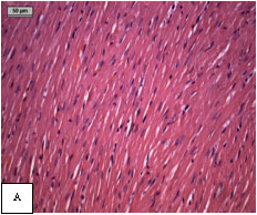
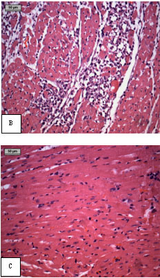
On histopathological examination depicted in Fig. 5A-C, we observed normal architecture of the heart in sham control (Fig. 5A). However, significant myocyte membrane damage with extensive myonecrosis, fibroblastic proliferation and infiltration of inflammatory cells was observed in ISP control hearts (Fig. 5B) in comparison with sham control. T. terrestris treatment extract prevented myonecrosis (Fig. 5C) as demonstrated by significant reduction in the infiltration of inflammatory cells, vacuolar changes as well as edema as compared to the ISP control group.
| |||
| Fig. 5: | Photomicrograph of heart tissue (H and E X 100) (A) Sham group showing normal architecture. Endocardium and pericardium are seen within normal limits without infiltration of inflammatory cells (B) ISP group showing focal myonecrosis with myophagocytosis and lymphocytic infiltration. Vacuolar changes and prominent edema along with chronic inflammatory cells were seen in subendocardium (C) Tribulus terrestris treated group administered showing lesser infiltration of inflammatory cells and myonecrosis | ||
| |||
| Fig. 6: | Electron micrograph of heart tissue (A) Normal ultra structure of heart tissue of sham group (1250X) (B) Extensive muscle necrosis with significant disruption of myofibrils, intracytoplasmic vacuoles and lipid droplets with irregular mitochondria in ISP control group (4400X) (C) Lesser muscle necrosis with slight disruption of myofibrils and mitochondria in Tribulus terrestris treated group (2800X) | ||

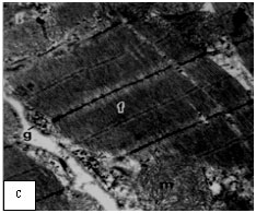
Electron microscopic examination of the myocardium revealed normal appearance of cell organelles in sham control rats showing sarcomere and abundant mitochondria (Fig. 6A). In ISP control group, the myocardial damage was marked by significant disruption of myofibrils (Fig. 6B). In addition, the myocardial tissue of ISP control group revealed several intracytoplasmic vacuoles and lipid droplets. Other ultrastructural changes include the appearance of small and irregular mitochondria with loss of cristae (Fig. 6B). However, treatment of animals with T. terrestris showed structural protection of the myocardium with respect to ISP control group (Fig. 6C).
DISCUSSION
Tribulus terrestris has significant cardioprotective activity as shown by its mitigating effects on ISP-induced hemodynamic, biochemical, histopathological and ultrastructural perturbations. In the present study, ISP, a β-adrenergic agonist was used to produce myocardial necrosis and interstitial fibrosis at its supra-maximal dosages in a multi-step manner. In addition, isoproterenol causes substrate loss, depletion of energy reserves, hypoxia due to myocardial hyperactivity, increased inotropic activity, coronary hypotension, a high-oxygen demand and production of free radicals resulting from oxidative metabolism of catecholamine (Tappia et al., 2001). Free radical mediated peroxidation of membrane phospholipids and consequent changes in membrane permeability are the primary reasons for cardiotoxicity induced by ISP (Rathore et al., 1998; Blasig et al., 1984). ISP-induced oxidative stress also depresses the sarcolemmal Ca2+ transport and results in the development of intracellular Ca2+ overload and ventricular dysfunction (Tappia et al., 2001). The oxidative stress may be exerted through quinine metabolites of ISP which react with oxygen to produce superoxide anions and others reactive oxygen species and interfere with SOD, glutathione reductase and ATP pumps (Rathore et al., 1998; Remiao et al., 2000). Experimental and clinical studies have demonstrated that therapeutic interventions having antioxidant or free radical scavenging activity may exert cardioprotective effects against oxidative stress associated with various cardiovascular diseases, including ischemic heart disease (Bandyopadhyay et al., 2004). Hence, medicinal herbs or natural products possessing antioxidant constituents has recently gained a great deal of scientific interest (Gupta et al., 2004; Mohanty et al., 2004; Karthikeyan et al., 2003) as it is expected to offer better protection against oxidative stress associated diseases through cellular adaptive mechanism and may provide a lead for promotion of therapeutic agent especially of natural origin.
In the present study, chronic administration of T. terrestris extract maintained the antioxidant activity of the endogenous antioxidants as evidenced by increase in the levels of reduced GSH and activities of endogenous antioxidant enzymes such as SOD, CAT and GSHPx along with decrease in the level of lipid peroxidation product, MDA. The depletion in GSH levels along with increased plasma lipid peroxides (measured as TBARS or thiobarbituric acid-reactive substances) has been observed during myocardial ischemia and associated with the consumption of endogenous antioxidant and enzymes such as SOD, CAT and GSHPx. These antioxidant enzymes are known to remove the ISP-induced generation of toxic oxygen species by their peroxidative and free radical scavenging activity. In addition, an increase in MDA levels was observed in the heart tissue following ISP administration. The myocardial necrosis observed in the animals receiving ISP can also be attributed to per-oxidative damage as it has been previously reported that ISP generates lipid peroxide (Blasig et al., 1984).
The fall in activity of GSHPx in the ISP group might be co-related with decreased availability of its substrate, GSH. Moreover, due to impairment in both enzymatic and non-enzymatic antioxidant defense mechanisms, it is possible that the free radicals may not be effectively neutralized, thereby rendering the myocardium susceptible to lipid peroxidation. The increased GSH levels observed in the T. terrestris groups in the present study may be due to its enhanced synthesis, as antioxidant compounds have been shown to increase glutathione reductase activity, which, maintains GSH in its reduced state (Mohanty et al., 2004). The observations of our study are in conformity with earlier reports that have demonstrated the modulation of synthesis of cellular antioxidants by treatment with natural products (Gupta et al., 2004; Mohanty et al., 2004; Karthikeyan et al., 2003).
In addition to the antioxidant parameters, the estimation of CK-MB isoenzyme and LDH levels, both of which are useful parameters for assessing cardiomyocyte damage (Jennings et al., 1990). These enzymes normally exist in cellular compartment and leak out into the plasma during myocardial injury due to disintegration of contractile elements and sarcoplasmic reticulum. ISP challenge in the present study produced significant depletion of myocardial CK-MB isoenzyme and LDH. T. terrestris treatment prevented the leakage of these enzymes and restored their activities as compared to the ISP control. The observation that T. terrestris treatment significantly restored CK-MB isoenzyme and LDH levels is suggestive of its cardioprotective effect.
As described earlier, hemodynamic parameters were also incorporated into the experimental design for a better understanding of the co-relation between biochemical and functional changes in the myocardium subjected to ISP-induced damage. Previous studies have shown that ISP mediated oxidative stress could progresses to myocardial necrosis which leads to cardiac dysfunction characterized by increased end-diastolic volume and pressure and left ventricular wall thickness (Grimm et al., 1998; Teerlink et al., 1994) and these changes are significantly prevented by antioxidants. In the present study, the favorable modulation of hemodynamic and left ventricular function by T. terrestris treatment also supports the previous observations it may modify the course of action and help in retaining the function of heart to normal level.
In the ISP control group, myocardial dysfunction was clearly evident by a significant fall in SAP, DAP and MAP, HR, LV (+) and (-) dP/dt along with a rise in LVEDP, which might be ascribed to its ISP-induced myocardial infarction. The left ventricular rate of negative pressure change was more markedly depressed indicating a more diastolic dysfunction per se which may result in the persistence of elevated LVEDP. It is speculated that deteriorating myocardial contractile status following ISP-induced necrosis might be responsible for the significant fall in MAP. Normally a fall in MAP reflexly increases HR and myocardial contractility. However, none of these effects were observed in the present study, suggesting impairment in the conducting system of the heart following ISP-induced myocardial necrosis. T. terrestris treatment in our study significantly increases MAP and HR as compared to the isoproterenol control group. Moreover, T. terrestris appeared to improve left ventricular function as evidenced by significant preservation of both inotropic and lusitropic functions of the heart along with correction of elevated LVEDP. Preservation of cardiac reflexes resulting in increased heart rate and ventricular function to maintain cardiac output may be on account of myocardial salvage, produced by T. terrestris.
The histopathological examination of myocardium in ISP control animals showed presence of focal myonecrosis with myophagocytosis and lymphocytic infiltration in sub-endocardial region indicative of infarct like lesions similar to previous studies (Grimm et al., 1998; Teerlink et al., 1994). Treatment with T. terrestris extract preserved the normal histoarchitecture and ultrastructure of the myocardium as evidenced by reduced myonecrosis and lesser infiltration of inflammatory cells. Taken together, the biochemical and histopathological results of the present study demonstrate the cardioprotective effects of T. terrestris. Additionally, ultrastructural perturbations in ISP administered rat hearts further confirmed the injured state of myocardium. Restoration of myocardial CK-MB activity, along with preservation of myofibrils and mitochondrial morphology is indicative of cytoprotective activity of T. terrestris.
Recently, adaptogens are demonstrated to confer resistance against a variety of stresses and aid in myocardial adaptation. Various experimental studies have demonstrated the adaptogenecity i.e., increased non-specific resistance to stress of medicinal herbs is considered as a function of antioxidant components which may produce myocardial adaptation (Bhattacharya and Muruganandam, 2003; Lei and Chiou, 1986). The major active constituents of T. terrestris are flavonoids and steroidal saponins, lignanamides, protodioscin and tribulosins (Wang et al., 1997; Achenbach et al., 1996). It has been reported that protodioscin and tribulosins are the adaptogenic components present in T. terrestris (Gauthaman et al., 2002). The myocardial adaptation against ischemic stress may be mediated through augmentation of cellular tissue antioxidant enzymes such as SOD, CAT and GSHPx and stress proteins. Although, the exact mechanism of such myocardial adaptation is not known. Further, flavonoids have been shown to exhibit wide spectrum of biological activities including free radical scavenging, antioxidant and anti-ischemic activities (Hertog et al., 1993). Therefore, it could be possible that T. terrestris may have exerted its cardioprotective effects partly due to its antioxidant as well as adaptogenic constituents.
In summary, the present study demonstrated that multiple mechanisms may be responsible for the cardioprotective effect of Tribulus terrestris. Improved hemodynamics and left ventricular function along with improved endogenous myocardial antioxidant status and myocardial adaptability assure its cardioprotective potential in ISP-induced myocardial necrosis. The histopathological and ultrastructural examination, further confirmed its cardioprotective effects through preservation of cell organelle and myocyte membrane integrity. In conclusions, the observations of present study provide a scientific basis for cardioprotective effect of Tribulus terrestris during myocardial ischemia and demonstrate its therapeutic potential in the treatment of ischemic heart disease.
ACKNOWLEDGMENTS
The financial aid from UGC, New Delhi is gratefully acknowledged. The authors also like to express their thanks to Mr. B.M. Sharma, Department of Pharmacology, AllMS, New Delhi, India for his valuable technical assistance in conducting the study.
REFERENCES
- Achenbach, H., H. Hubner and M. Reiter, 1996. New cardioactive steroid saponins and other glycosides from Mexican Tribulus cistoides. Adv. Exp. Med. Biol., 404: 357-370.
Direct Link - Agrawal, V.K., D.R. Basannar, R.P. Singh, M. Dutt, D. Abraham and M.S. Mustafa, 2006. Coronary risk factors in a rural community. Indian J. Public Health, 50: 19-23.
PubMedDirect Link - Amin, A., M. Lotfy, M. Shafiullah and E. Adeghate, 2006. The protective effect of Tribulus terrestris in diabetes. Ann. N. Y. Acad. Sci., 1084: 391-401.
Direct Link - Anand, R., G.K. Patnaik, D.K. Kulshreshtha and B.N. Dhawan, 1994. Activity of certain fractions of Tribulus terrestris fruits against experimentally induced urolithiasis in rats. Indian J. Exp. Biol., 32: 548-552.
Direct Link - Bandyopadhyay, D., A. Chattopadhyay, G. Ghosh and A.G. Datta, 2004. Oxidative stress-induced ischemic heart disease: Protection by antioxidants. Curr. Med. Chem., 11: 369-387.
Direct Link - Bhattacharya, S.K., A. Bhattacharya and A. Chakrabarti, 2000. Adaptogenic activity of Siotone, a polyherbal formulation of Ayurvedic rasayanas. Indian J. Exp. Biol., 38: 119-128.
PubMedDirect Link - Bhattacharya, S.K. and A.V. Muruganandam, 2003. Adaptogenic activity of Withania somnifera: An experimental study using a rat model of chronic stress. Pharmacol. Biochem. Behav., 75: 547-555.
CrossRefDirect Link - Blasig, I.E., R. Blasig and H. Lowe, 1984. Myocardial lipid peroxidation during isoproterenol-induced blood flow reduction in rat myocardium. Biomed. Biochim. Acta, 43: S171-S174.
Direct Link - Cabaud, P.G. and F. Wroblewski, 1958. Colorimetric measurements of lactic dehydrogenase activity of body fluids. Am. J. Clin. Pathol., 30: 234-236.
Direct Link - Dhar, M.L., M.M. Dhar, B.N. Dhawan, B.N. Mehrotra and C. Ray, 1968. Screening of Indian plants for biological activity: I. Indian J. Exp. Biol., 6: 232-247.
PubMedDirect Link - Gauthaman, K., P.G. Adaikan and R.N.V. Prasad, 2002. Aphrodisiac properties of Tribulus terrestris extract (Protodioscin) in normal and castrated rats. Life Sci., 71: 1385-1396.
CrossRefDirect Link - Grimm, D., D. Elsner, H. Schunkert, M. Pfeifer and D. Griese et al., 1998. Development of heart failure following isoproterenol administration in the rat: Role of the renin-angiotensin system. Cardiovas. Res., 37: 91-100.
PubMedDirect Link - Guo, Y., H.J. Yin, D.Z. Shi and K.J. Chen, 2005. Effects of Tribuli saponins on left ventricular remodeling after acute myocardial infarction in rats with hyperlipidemia. Chin. J. Integr. Med., 11: 142-146.
CrossRefDirect Link - Gupta, S.K., I. Mohanty, K.K. Talwar, A. Dinda and S. Joshi et al., 2004. Cardioprotection from ischemia and reperfusion injury by Withania somnifera: A hemodynamic, biochemical and histopathological assessment. Mol. Cell. Biochem., 260: 39-47.
CrossRef - Hertog, M.G.L., E.J.M. Feskens, D. Kromhout, M.G.L. Hertog, P.C.H. Hollman, M.G.L. Hertog and M.B. Katan, 1993. Dietary antioxidant flavonoids and risk of coronary heart disease: The Zutphen elderly study. Lancet, 342: 1007-1011.
CrossRefPubMedDirect Link - Jennings, R.B., C.E. Murry, C.J.R. Steenbergen and K.A. Reimer, 1990. Acute myocardial ischemia: Development of cell injury in sustained ischemia. Circulation, 82: 112-122.
PubMedDirect Link - Karthikeyan, K., B.R.S. Bai, K. Gauthaman, K.S. Sathish and S.N. Devaraj, 2003. Cardioprotective effect of the alcoholic extract of Terminalia arjuna bark in an in vivo model of myocardial ischemic reperfusion injury. Life Sci., 73: 2727-2739.
CrossRefPubMedDirect Link - Kavitha, A.V. and G. Jagadeesan, 2006. Role of Tribulus terrestris (Linn.) (Zygophyllacea) against mercuric chloride induced nephrotoxicity in mice, Mus musculus (Linn.). J. Environ. Biol., 27: 397-400.
PubMed - Lei, X.L. and G.C. Chiou, 1986. Cardiovascular pharmacology of Panax notoginseng (Burk) F.H. Chen and Salvia miltiorrhiza. Am. J. Chin. Med., 14: 145-152.
Direct Link - Lowry, O.H., N.J. Rosebrough, A.L. Farr and R.J. Randall, 1951. Protein measurement with the folin phenol reagent. J. Biol. Chem., 193: 265-275.
CrossRefPubMedDirect Link - Misra, H.P. and I. Fridovich, 1976. The oxidation of phenylhydrazine: Superoxide and mechanisms. Biochemistry, 15: 681-687.
CrossRefDirect Link - Mohanty, I., D.S. Arya, A. Dinda, K.K. Talwar, S. Joshi and S.K. Gupta, 2004. Mechanisms of cardioprotective effect of Withania somnifera in experimentally induced myocardial infarction. Basic Clin. Pharmacol. Toxicol., 94: 184-190.
CrossRefPubMedDirect Link - Moron, M.S., J.W. Depierre and B. Mannervik, 1979. Levels of glutathione, glutathione reductase and glutathione S-transferase activities in rat lung and liver. Biochim. Biophys. Gen. Subj., 582: 67-78.
CrossRefPubMedDirect Link - Ohkawa, H., N. Ohishi and K. Yagi, 1979. Assay for lipid peroxides in animal tissues by thiobarbituric acid reaction. Anal. Biochem., 95: 351-358.
CrossRefPubMedDirect Link - Paglia, D.E. and W.N. Valentine, 1967. Studies on the quantitative and qualitative characterization of erythrocyte glutathione peroxidase. J. Lab. Clin. Med., 70: 158-169.
CrossRefPubMedDirect Link - Phillips, O.A., K.T. Mathew and M.A. Oriowo, 2006. Antihypertensive and vasodilator effects of methanolic and aqueous extracts of Tribulus terrestris in rats. J. Ethnopharmacol., 104: 351-355.
CrossRefDirect Link - Rathore, N., S. John, M. Kale and D. Bhatnagar, 1998. Lipid peroxidation and antioxidant enzymes in isoproterenol induced oxidative stress in rat tissues. Pharmacol. Res., 38: 297-303.
CrossRefDirect Link - Remiao, F., H. Carmo, F.D. Carvalho and M.L. Bastos, 2000. Inhibition of glutathione reductase by isoproterenol oxidation products. J. Enzyme Inhibition Med. Chem., 15: 47-61.
CrossRefDirect Link - Sharifi, A.M., R. Darabi and N. Akbarloo, 2003. Study of antihypertensive mechanism of Tribulus terrestris in 2K1C hypertensive rats: Role of tissue ACE activity. Life Sci., 73: 2963-2971.
Direct Link - Stauss, H.M., Y.C. Zhu, T. Redlich, D. Adamiak, A. Mott, K.C. Kregel and T. Unger, 1994. Angiotensin-converting enzyme inhibition in infarct-induced heart failure in rats: Bradykinin versus angiotensin II. J. Cardiovasc. Risk, 1: 255-262.
Direct Link - Tappia, P.S., T. Heta and N.S. Dhalla, 2001. Role of oxidative stress in catecholamine-induced changes in cardiac sarcolemmal Ca2+ transport. Arch. Biochem. Biophys., 377: 85-92.
Direct Link - Teerlink, J.R., J.M. Pfeffer and M.A. Pfeffer, 1994. Progressive ventricular remodeling in response to diffuse isoproterenol-induced myocardial necrosis in rats. Circ. Res., 75: 105-113.
Direct Link - Wang, Y., K. Ohtani, R. Kasai and K. Yamasaki, 1997. Steroidal saponins from fruits of Tribulus terrestris. Phytochemistry, 45: 811-817.
Direct Link