
A.Y. Obaid
Department of Chemistry, Faculty of Science, King Abdulaziz University, P.O. Box 80203 Jeddah 21589, Saudi Arabia
O.A. Abu-Zinadah
Department of Biological Sciences, Faculty of Science, King Abdulaziz University, P.O. Box 80203 Jeddah 21589, Saudi Arabia
H.K. Hussein
Department of Biological Sciences, Faculty of Science, King Abdulaziz University, P.O. Box 80203 Jeddah 21589, Saudi Arabia
International Journal of Biological Chemistry
Year: 2011 | Volume: 5 | Issue: 2 | Page No.: 103-115


ABSTRACT
This research was carried out to evaluate the potential and beneficial effect of green tea (Camellia sinensis) and its fractions in direct fire burns in comparison with antibiotic manufactured for this purpose using basic morphological and histological methods. Twenty-five animals were divided into five groups (5 animals each). These groups were control untreated group; crude extraction treated group, polyphenol fraction group, terpenes extract group and antibiotic ointment treated group. All types of treatments reduce the wounds to minimum volume at the beginning of the fifth week. Most wounds were recovered completely, giving normal epidermis, dermis and hair growing, whoever some burns showed no hair growth and gave abnormal appearance of newly formed skin. Furthermore, histological findings revealed normal cutaneous architecture of the extract treated wounds more than that appeared in the untreated wounds. The healing capability of Camellia sinensis was evident when compared to the impaired healing process of the wounds administered to the Baneocin antibiotic ointment treated rabbits that illustrated a delay in coagulation and inflammation phase of healing after five weeks of wounding. However, the healing effect of crude extract was more effective than that observed with the Baneocin treated or essential oil derivatives (terpenes and polyphenoles), respectively. The contract ability and closure times of wounds were very high by application of crude extract and antibiotic than those through application of terpenes and polyphenoles. Terpenes gave another way of treatment, it was good antimicrobial material and so did polyphenoles. They prevent microbial growing above the wounds after the first hours of application.
PDF Abstract XML References Citation
Received: August 14, 2010;
Accepted: December 14, 2010;
Published: February 22, 2011
How to cite this article
A.Y. Obaid, O.A. Abu-Zinadah and H.K. Hussein, 2011. The Beneficial Effects of Green Tea Extract and its Main Derivatives in Repairing Skin Burns of Rabbit. International Journal of Biological Chemistry, 5: 103-115.
URL: https://scialert.net/abstract/?doi=ijbc.2011.103.115
URL: https://scialert.net/abstract/?doi=ijbc.2011.103.115
INTRODUCTION
Plant extracts have been widely used as topical application for wound healing and disease treatments. These plants share a common character; they all produce flavonoid compounds with phenolic structures. These phytochemicals are highly reactive with other compounds, such as reactive oxygen species and biologic macromolecules, to neutralize free radicals or initiate biological effects. A short list of phenolic phytochemicals with promising properties to benefit human health includes a group of polyphenol compounds, called catechins, found in green tea (Katiyar et al., 2001).
The chemical composition of green tea varies with climate, season, horticultural practices and position of the leaf on the harvested shoot. The major components of interest are the Terpenes and polyphenols. Terpenes and terpenoids are the primary constituents of the essential oils of many types of plants and flowers. Essential oils are used widely as natural flavor additives for food, as fragrances in perfumery and in traditional and alternative medicines such as aromatherapy. Synthetic variations and derivatives of natural terpenes and terpenoids also greatly expand the variety of aromas used in perfumery and flavors used in food additives. Vitamin A is an example of a terpene (Liu et al., 2008).
The major polyphenols in green tea are flavonoids. The four major flavonoids in green tea are the Catechins Epicatechin (EC), Epigallocatechin (EGC), Epicatechin Gallate (ECG) and Epigallocatechin Gallate (EGCG). Epigallocatechin gallate is viewed as the most significant active component. The leaf bud and first leaves are richest in EGCG. The usual concentration of total polyphenols in dried green tea leaves is about 8 to 12%. Other compound of interest in dried green tea leaves include terpenes, gallic acid, quercetin, kaempferol, myricetin, caffeic acid and chlorogenic acid (Cabrera et al., 2006).
A large amount of information has been published concerning the effects of green tea and its major constituents on human health. This beverage has been consumed in many countries for a very long time and today interest is growing because scientific reports indicate that tea could bring benefits for health and may help prevent chronic diseases. Tea was first introduced into European countries from China by Portuguese and Dutch explorers as a medicinal herb. It was taken in China 5000 years ago for its stimulating and detoxifying properties in the elimination of alcohol and toxins, to improve blood and urine, to relieve joint pains and to improve resistance to diseases (Moreno et al., 2006; Jazani et al., 2007a; Kilicalp et al., 2009; AL-Rejaie, 2009).
Tea grew rapidly in importance and was incorporated into many social rituals notably in China, Japan and England. Today, tea is the second most popular beverage in the world after water (Wang et al., 1992). Interest in the growing green tea stimulated many studies for protection against several alterations including cancer. These fractions were found to be antioxidant through their peroxides activity, lipoxygenases and cyclo-oxygenases are capable of co-oxidizing molecules other than their regular subtracts, with the potential for increasing oxidative damage in some tissues. Green tea polyphenols were found to inhibit Cyclooxygenase (COX)-2 and 5-, 12 and 15-lipoxygenase activities in human colon mucosa cells and human colon cancer cells. Feeding green tea polyphenols to mice inhibited ultraviolet light-induced increases in epidermal COX activity (Lou et al., 1999) whereas topical application of green tea (Chen et al., 1998) and black tea polyphenoles (Liu et al., 2008) inhibited phorbol ester-induced increases in epidermal COX and lipoxygenase activities. The inhibition of phase I enzymes which are responsible for activation of many carcinogens in animals and induction of phase II enzymes, which are associated with enhanced excretion of carcinogens considered the mode of action of green tea fractions (Duansak et al., 2003).
In fact, Burns are a major problem in many developing countries. Eupolin ointment is a tropical agent used in treatment of soft tissue wounds and burns. Many studies using green tea polyphenols as chemo-preventive, natural healing and anti-aging agents of burn wounds. Studies using this extract have illustrated antimicrobial and anticoagulation effects as well as the promotion of tissue remodeling in the wound healing process (Lee et al., 2005; Sakarcan et al., 2005; Liu et al., 2008; Oboh, 2009). However, the mechanism by which this agent affects cell involved in the wound healing process is unknown. In many researches, fibroblasts and endothelial cells, two cell types that play a crucial role in wound healing enhanced growth of fibroblasts and endothelial cells.
The aim of this study was to evaluate the potential and beneficial effect of green tea and its fractions in burns in comparison with antibiotic manufactured for this purpose using basic morphological methods. The studies which used green tea extracts to treat inflammation, oxidation, bacteria and cancer as well as enhanced the immune system, stimulated us to test whether such green tea and its fractions can be used to treat burn wounds through its multipotential pharmaceutical activities and to what extend this plant can repair fire induced burns of rabbit skin through the macro-observation of wound using the local rabbit as a model experimental animal.
MATERIALS AND METHODS
Plant material and extraction: Green tea is tea made solely with the plant leaves of Camellia sinensis that have undergone minimal oxidation during processing. The used crude extract (water extract) of the green tea was produced by a published extraction method (Wall et al., 1996) by using A Soxhlet extractor. The method moves bringing the material to be extracted into contact with the extraction water solvent for a period of time followed by separation of solution from solid debris.
Animal's model: Premature local rabbits, Oryctolagus caniculus were obtained from unburied strain of private farm in Jeddah District, Saudi Arabia. The average weight of animals was between 0.50-0.75 kg. The sex was not taken into consideration in this study, however, males and females were separated to avoid pregnancy. The animals were housed in good conditions. Commercial foods, vegetables were the types of food through this experiment and water was put ad labitum. The animals were housed in stainless steel cages in air conditioning and sterile room. Twenty-five animals were divided into five groups (5 animals each). The five groups were: Control, crude fraction, polyphenol fraction, terpenes extract and antibiotic ointment treated group. This study was conducted from September to December 2010.
Experimental treatments: Hairs of the dorsal skin were shaved mechanically 24 h before burn. The burns were done by direct fire by putting hot glass rod 80°C on the dorsal skin for 1 min three times on different areas upon the shaved skin of the 5 above mentioned groups on 5x5 cm2 area. After induction of burns, the animals were housed separately to avoid contact and contamination. Twelve hours dark/light cycle by sterilization lamps and the treatment started at the next day of burn induction.
Topical treatment of burns: The control group was left without treatment. One group was treated by crude extract by spreading a layer above the burn (0.05 mL) two times a day. Minute drops of terpenes and polyphenols were allowed to cover burns (0.02 mL) per rabbit a day in the other two groups. Spreading thin layer of Baneocin (Bacitracin zinc 250 IU and neomycin 5000 IU) twice daily topically treated the antibiotic groups. Baneocin ointment, broad-spectrum antibiotic for topical therapy produced by Pharco Pharmaceutically, Egypt, under the license of Biochemie GmbH, Vienna, Austria.
Chromatographic fractionation procedures: It is the most widely technique used in fractionation of extracts and has made possible in the isolation of many naturally occurring compounds. This is based on the fact that a dynamic equilibrium is established between the concentrations of solute into phases (distribution coefficient). One phase is the mobile fluid and the other phase is being solid consisting of fine particles (Braithwaite and Smith, 1985).
Layer chromatography and detection of bands formed: The thin-layer plates may be available commercially as ready-made products. Nine groups of solved are considered each having solvent strength (related to its polarity). The most common layer system phase silica derivatives. This method involves applying the plant extract in the form of band on the thin layer plate (0.05-0.25 mm thick). Care must be taken to avoid the sample being applied in a diffused band. The plate is developed in a solvent system known to separate the components.
It is often achieved by spray the layer with a fine moist of water so that it is wetted enough to become transparent. Non-water soluble compounds will appear as darker areas with transmitted light. Another method is that by iodine vapor the supernatant is evaporated to yield required compound.
Test for phenolic and terpenoids compounds: Ferric ions when added to solution of phenols produce dark green or blue-black complex. Terpenoids give positive reaction (colors ranging from red to blue) when warmed with acetic anhydride and treated with a few drops of concentrated sulphuric acid (Liebermann-Burchard reaction). So that bands appear as yellow or brown bands. The marked bands are carefully scraped off and mixed with an excess volume of suitable solvent (ethanol) and then the suspension is filtered (Braithwaite and Smith, 1985).
Macroscopic measurements: Outlook of the burns, the periphery, epithialation, contamination, closure time, contracting ability growing hair time were observed and recorded. The peripheries of burns were observed: the erythma, thickness and inflammation were recorded; the contamination of burn wounds. However, the types of microbes were not taken into consideration but taken as a separated work; the closure time was estimated through the daily measurements of the volumes of the burn wounds. On the other hand, the contracting ability was estimated according to the repairing of wound to the normal skin architecture through topical treatment of burn wounds and the growing hair time is the first day of seeing hairs upon the repaired skin.
Microscopic observation: Three rabbits of each group was sacrificed after 5 weeks and the burned skin was fixed in neutral formalin solution (10%) till 24 h, then washed to another 24 h, in running water, the skin was dehydrated in ascending ethyl alcohol series, embedded in paraffin wax, sectioned and stained by hematoxylin and eosin stains . The significance of the tabulated values was tested by Student t-test between the treated and untreated values.
Biological activities: To test the effect of green tea extract and its derivatives on some microorganisms, cultures of certain types of bacteria were topically treated by three treatments: Crude extract, polyphenoles and terpenes. These bacteria were Enterococcus faecalis, Staphyloccous aureus and Escherichia coli. These types of bacteria significant differently response with different types of treatment applications and doses (1, 2, 3, 5, 10 and 20 mm) of different treatments. The microbial infection with these bacteria is the first reason of inflammation and contamination of wounds.
RESULTS
The mean volume of burns, which induced by fire, was slowly reduced by time when different types of treatments were applied. The burns were recovered by different ways; some burns wounds were covered by thick coat of keratinized layer, which shedding off leaving new skin layer, however, the newly formed skin was very thin. Some other burn wounds still covered by such hard coat through the whole experimental period. The response of other rabbits to burns was very bad with no defense. In spite of all rabbits felt ill and their activities were badly affected, most rabbits were recovered very soon. Their hairs also were grown sooner. The skin became very well without any burns signs.
Macroscopic examination of burn-wounds: It was found that antibiotic and crude extract gave highly significant difference (p<0.001) if compared with control (untreated) group, while polyphenols and terpenes gave significant results if compared with control ones (p<0.01, Table 1). The burns wounds of the control group reduced from 4.22±0.92-0.92±0.15 cm2 at the end of the fifth week of observation (p<0.001). In crude extract group the wounds were reduced from 3.10±0.55-0.09±0.03 cm2 (p<0.001). In some burns were left abnormally skin structure, others gave on growth and some became normal with growth. All types of treatments reduced the wounds to the minimum volume at the beginning of the fifth weeks, whoever some burns showed no hair growth. At the third week of treatment the burns reduced to half volume of burns treated by antibiotic and crude extract (from 3.66 and 3.10 cm2 to 1.81 and 1.67 cm, respectively, Table, 1). Polyphenoles and terpenes reduced the volumes of burns wounds at lower rate than antibiotic and crude extract. The contractability of wounds was high by application of polyphenoles and terpenes and very high by application of antibiotic and crude extract (p<0.001). The closure times were ranged between 15 and 18 days in control group. These times reduced significantly to 8 and 10 days; 7 and 9 days; 9 and 11 days and 10 and 12 days by application of antibiotic, crude extract, polyphenoles and terpenes, respectively. The recovery days were between 16 and 20 days after topical application of mentioned control treatment, while the recovery days were between 7 to 9 days of crude extract treatment (Table 2).
| Table 1: | Effect of topical application of treatments on fire induced burns of local rabbits throughout five weeks interval |
 | |
| * = p<0.01, ** = p<0.001, Mean areas of wounds are mentioned as cm2±SD | |
| Table 2: | Changes of burns parameters after different types of treatments |
 | |
| ** = p<0.001, + low rate, ++ median rate, +++ high rate, ++++ very high rate | |
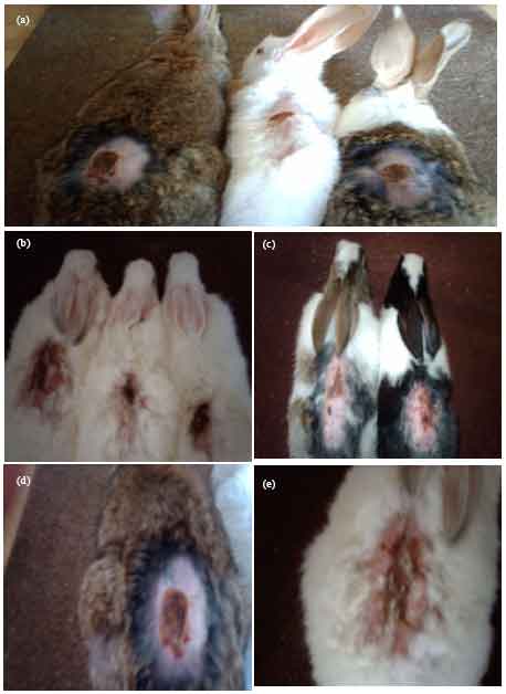 | |
| Fig. 1: | Topical dorsal skin burn wound site obtained from the controls and green tea (Camellia sinensis) treated rabbit groups on the first day of wounding. (a) Control not treated wound; (b) Only Baneocin treated wound; (c) Wound treated with Camellia sinensis crude extract; (d) Wound treated with polyphenoles and (e) wound treated with terpenes |
The growing hairs were between 22 and 25 days after topical application of control treatment, while there were between 15 to 18 days of crude extract treatment. Topical dorsal skin burn wound site obtained from the control and green tea (Camellia sinensis) in the first day of wounding are illustrated in Fig. 1(a-e) and after 35 days of wounding are illustrated in Fig. 2(a-e). Both Fig. 1 and 2 represented control not treated wound; only Baneocin treated wound; wound treated with (Camellia sinensis crude extract; wound treated with polyphenoles and wound treated with terpenes as the captions from A to E, respectively.
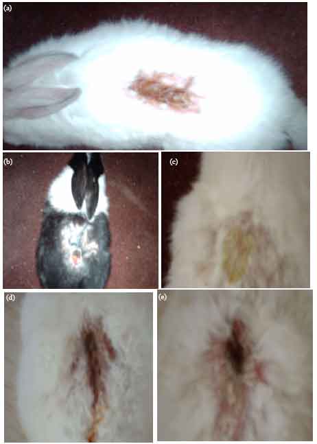 | |
| Fig. 2: | Topical dorsal skin burn wound site obtained from the controls and green tea (Camellia sinensis) treated rabbit groups after 35 days of wounding. (a) Control not treated wound; (b) Only Baneocin treated wound; (c) Wound treated with Camellia sinensis crude extract; (d) Wound treated with polyphenoles and (e) Wound treated with terpenes |
Microscopic examination of burn-wounds: Green tea crude extract was the best treatment applied on the burn wound if compared with the antibiotic (Baneocin zinc), polyphenoles and terpenes fractions of this extract. However, some burns wounds were recovered leaving abnormal skin appearance and no hair growth. The newly formed epidermis were few thin layers, neither dermal papillae nor horny layer were formed. The hair grew at first around the edges of the wounds, which showed certain hyperplasia. The hair roots developed from these edges and extent underneath the newly formed skin.
Histological sections of cutaneous wound site obtained from the control and green tea (Camellia sinensis) treated groups are illustrated in Fig. 3(a-e).
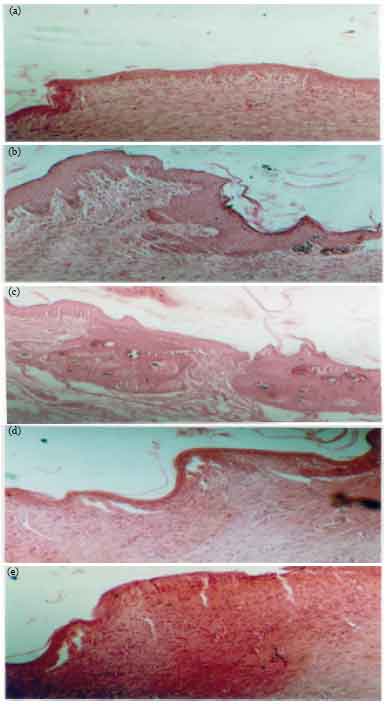 | |
| Fig. 3: | Hematoxylin and eosin staining histological sections of cutaneous wound site obtained from the controls and green tea (Camellia sinensis) treated rabbits, revealing epidermal and dermal architecture of the burn wounds on day 35 after wounding. (a) Control (not treated) wound; (b) only Baneocin treated wound; (c) wound treated with Camellia sinensis crude extract; (d) wound treated with polyphenoles and (e) wound treated with terpenes |
While the antibiotic caused the quick recovery of wounds, the crude extract improved the growing of newly formed skin layers. Large numbers of burns wounds were looked like the normal skin with the best and normal hair growth. The epidermis was darkly stained which reflected the activities of these layers to give complete epidermal structure.
Polyphenoles application gave great attention to use such fraction in certain topical treatment, burn wounds included. While response of it was in lower rate if compared with above-mentioned treatment no and horny layer was formed, the wounds were recovered completely.
Terpenes gave another way of treatment, it was good antimicrobial material and so did polyphenoles. They prevent microbial growing above the wounds after the first hours of application, however, the wounds recovery was in lower rate, some burns gave abnormal appearance of the newly formed skin and absent of hair roots.
Biological activities: By application of twenty millimeters from three types of treatments (crude extract, polyphenoles and terpenes), the three types of bacteria (Enterococcus faecalis, Staphylococcus aureus and Escherichia coli) were significantly destructed leaving certain small spots which give the same response when they cultured and treated again. Ten millimeters dose gave good response with Staphylococcus aureus only and median response with the other types of bacteria. By decreasing the dose of treatments the different types of bacteria differentially response. Five millimeters dose of crude extract gave the significant response with all types of bacteria. The same dose of polyphenoles gave significant response with Staphylococcus aureus. In the mean time, terpenes gave median response with all tested types of bacteria. 1, 2 and 3 mm doses gave insignificant response.
DISCUSSION
The possible mechanism of wound healing observed in both crude extraction of green tea Camellia sinensis and its main derivatives (polyphenoles and terpenes) in treated rabbit (Table 1, 2), may be attributed to compound(s) responsible for the wound healing. Wound healing is a complex natural regeneration process of skin cells to minimize or eliminate scarring as well to help heal and repair damage. The normal subsequent events of cutaneous healing occur in three overlapping phases: coagulation and inflammation, proliferation and remodeling (Eming et al., 2007). Coagulation and inflammation begin immediately after injury and are characterized by platelet aggregation to control excessive blood loss from the damaged vessels and the influx of inflammatory cells into the wound site. These cells play multiple roles in wound healing, including release of protease for wound debridement, phagocytosis of debris and bacteria and secretion of various cytokines and growth factors. Which, in turn, cause migration and division of cells involved in the proliferative phase and support the results reported earlier by Stipcevic et al. (2006). During this phase, angiogenesis, collagen deposition, epithelialization and newly formed granulation tissue, consisting of endothelial cells, macrophages, fibroblasts and the components of a new provisional extracellular matrix begin to cover and fill the wound area to restore tissue integrity (Midwood et al., 2004; Eming et al., 2007; Jazani et al., 2007b). However, the remodeling phase of repair involves collagen cross linking and reorganization, evolution of granulation tissue into scar tissue and cells no longer needed are removed by apoptosis (Rai et al., 2005). Present findings here come in agreement with the above-cited findings and contribute the development of wound healing after treatments.
Green tea (Camellia sinensis) crude extract and its essential derivatives accelerate the wound healing in rabbit by influencing different aspects of the healing process at various days after wounding, under the conditions of the present investigation (Table 1, 2 and Fig. 1-3). However, the healing effect of essential oil derivatives (polyphenoles and terpenes and respectively) was less effective than that observed with the crude extract treated or Baneocin treated, respectively. Potential healing of Camellia sinensis was high-lighted by reduced inflammation and wound debridement (Fig. 2), closure of the wound area and recovery time due to rapid wound contraction (Table 2), full-thickness epidermal regeneration and organization (Fig. 3c), increase in the wet weight of granulation tissue indicating rapid maturation due to high vascularization and deposition of extracellular matrix elements involving collagen and fibroblasts (Fig. 3). Furthermore, histological findings revealed normal cutaneous architecture of the extract treated wounds more than that appeared in the control (untreated wounds). The healing capability of Camellia sinensis was evident when compared to the impaired healing process of the wounds administered to the Baneocin treated rabbits that showed a delay in coagulation and inflammation phase of healing after wounding (Fig. 3a, b). Previous studies have reported that this may be associated with an increase in the number of acute inflammatory cells, impaired leukocytes function, inadequate migration of neutrophils and macrophages to the wound along with reduced chemotaxis, failure in the resolution of inflammation and a defect in neutrophil apoptosis (Gutierrez-Fernandez et al., 2007; Hirsch et al., 2008; AL-Sowyan, 2009) come in agreement with our results. This is also in accordance with results that indicated that the reduced microcirculation and diminished expression of growth factors contribute to the disruption of wound healing, such as increasing wound volume and impairment in wound closure (Table 1 and 2), fibroblasts proliferation leading to inadequate deposition of fibrous collagen tissue and scarcely formed granulation tissue (Sivan-Loukianova et al., 2003; Cianfarani et al., 2006; Eming et al., 2007; Silva et al., 2007).
Healing potential of many phenolic phytochemical herb extracts such as green tea and rosemary could be explained on the basis of the powerful anti-microbial (Angioni et al., 2004) and anti-inflammatory (Cabrera et al., 2006; Takaki et al., 2008). In addition, many other authors explained both herbs as antioxidant (Calabrese et al., 2000; Moreno et al., 2006; Jazani et al., 2007b) effects of the herb that are well documented in the literature. It has been reported that if a wound becomes infected, the normal healing is disrupted as the inflammatory phase becomes chronic suppressing the proliferation phase of healing (Yates et al., 2007). Present results support the finding of Eming et al. (2007) and Gutierrez-Fernandez et al. (2007) who stated that the acute inflammatory during the early stages of wounding generates factors that are essential for tissue growth and repair. When prolonged, as the case in infected wounds, preventing wound remodeling and matrix synthesis, leading to delay in wound closure. The biological application of phenolic treatments in the present study confirmed the above-cited opinions. It was found that terpenes and polyphenoles are good antimicrobial material. They prevent microbial growing above the wounds after the first hours of application, however, the wound recovery was in lower rate. It was found also that the production of free radicals at or around the wound bed at the beginning of burn wounds may contribute to delays in wound healing through the destruction of lipids, proteins and extracellular matrix elements (Calabrese et al., 2000). Due to these properties, the herb extraction may facilitate wound healing by reducing local inflammation and tissue destruction, increasing angiogenesis and collagen deposition leading to improvements in both local circulation and granulation tissue formation.
The present results also support the finding of Calabrese et al. (2000), Oluwatuyi et al. (2004), Cabrera et al. (2006) and Takaki et al. (2008) who stated that the main bioactive compounds isolated from both aqueous and organic extracts of the aerial parts of phenolic phytochemicals such as green tea having potential effect on inhibiting pathogenic growth, reducing inflammatory response and preserving viable tissue are mostly terpenoids, polyphenols, carnosol and carnosic acid. The quality and chemical composition of the effective oil extract depend on how and where the plant was grown, harvested and distilled (Angioni et al., 2004; De Mastro et al., 2004; Sotomayor et al., 2009). When conditions cause the plants to permanently produce variations in the chemical composition of their essential oils, these plants is known as chemotypes. The main known healing herb constituents are α-β-pinene, 1,8-cineole, camphor, verbenone, borneol and limonene. Often all these chemicals can exist in the oil, however there are principal chemotypes of green tea (Camellia sinensis), as well as other similar famous healing herbs such as Rosmary (Rosmarinus officinalis), watercress (Nasturtium officinale), black seeds Nigella sativa and curcumin, Curcuma longa with the names given by one of the main constituents, such as terpenoids, polyphenols, α-pinene, camphor/borneol, cineole, sulpho-nitrogenous, diferuloylmethane and verbenone (Pintore et al., 2002; Lahlou and Berrada, 2003; Cabrera et al., 2006; Abu-Zinadah, 2008, 2009; Hussein and Abu-Zinadah, 2010).
In conclusion, the above experimental results illustrated that the healing effect of crude extract of green tea, Camellia sinensis was more effective than that observed with the Baneocin antibiotic ointment treated or essential oil derivatives (terpenes and polyphenoles), respectively. The contract ability and closure times of wounds were very high by application of crude extract and antibiotic than those through application of terpenes and polyphenoles. Terpenes gave another way of treatment; it was good antimicrobial material and so did polyphenoles and prevents microbial growing above the wounds after the first hours of application.
REFERENCES
- Abu-Zinadah, O.A., 2008. Effect of watercress oil on the thermal and chemical burn injuries in rabbit. J. King Abdulaziz Univ. Med. Sci., 15: 3-17.
Direct Link - Abu-Zinadah, O.A., 2009. Using Nigella sativa to treat and heal chemical induced wound of rabbit skin. J. King Abdulaziz Univ. Sci., 21: 335-346.
Direct Link - Al-Rejaie, S.S., 2009. Effect of green and black teas on immobilization induced stress in male wistar albino rats. Int. J. Pharmacol., 5: 137-145.
CrossRefDirect Link - Al-Sowyan, N.S., 2009. Difference in leptin hormone response to nutritional status in normal adult male albino rats. Pak. J. Biol. Sci., 12: 119-126.
CrossRefPubMedDirect Link - Cabrera, C., R. Artacho and R. Gimenez, 2006. Beneficial effects of green tea-A review. J. Am. Coll. Nutr., 25: 79-99.
PubMedDirect Link - Calabrese, V., G. Scapagnini, C. Catalano, F. Dinotta, D. Geraci and P. Morganti, 2000. Biochemical studies of a natural antioxidant isolated from rosemary and its application in cosmetic dermatology. Int. J. Tissue React., 22: 5-13.
PubMedDirect Link - Cianfarani, F., G. Zambruno, L. Brogelli, F. Sera and P.M. Lacal et al., 2006. Placenta growth factor in diabetic wound healing: Altered expression and therapeutic potential. Am. J. Pathol., 169: 1167-1182.
CrossRef - Chen, Z.P., J.B. Schell, C.T. Ho and K.Y. Chen, 1989. Green tea epigallocatchin gallates shows a pronounced growth inhibitor effect on cancerous cells but not on their normal counterparts. Cancer Lett., 129: 173-179.
PubMed - Duansak, D., J. Somboonwong and S. Patumari, 2003. Effects of Aloe vera on leukocytes adhesion and TNF-Alpha and IL-6 levels in burn wounded rats. Clin. Hemorheol. Microsirs, 29: 239-246.
Direct Link - Eming, S.A., S. Werner, P. Bugnon, C. Wickenhauser and L. Siewe et al., 2007. Accelerated wound closure in mice deficient for interleukin-10. Am. J. Pathol., 170: 188-202.
CrossRef - Oboh, G., 2009. The neuroprotective potentials of sour (Hibiscus sabdariffa, Calyx) and green (Camellia sinensis) teas on some pro-oxidants induced oxidative stress in brain. Asian J. Clin. Nutr., 1: 40-49.
CrossRefDirect Link - Gutierrez-Fernandez, A., M. Inada, M. Balbin, A. Fueyo and A.S. Pitiot et al., 2007. Increased inflammation delays wound healing in mice deficient in collagenase-2 (MMP-8). FASEB J., 21: 2580-2591.
CrossRef - Hirsch, T., M. Spielmann, B. Zuhaili, T. Koehler and M. Fossum et al., 2008. Enhanced susceptibility to infections in a diabetic wound healing model. BMC Surgery, 8: 5-5.
CrossRef - Hussein, H.K. and O.A. Abu-Zinadah, 2010. Antioxidant effect of curcumin extracts in induced diabetic Wister rats. Int. J. Zool. Res., 6: 266-276.
CrossRefDirect Link - Jazani, N.H., M. Zartoshti, S. Shahabi, Z. Yekta and S. Nateghi, 2007. Evaluation of the synergetic effect of water soluble extracts of green tea (Camellia sinensis) on the activity of ciprofloxacin in urinary isolated E. coli. J. Biol. Sci., 7: 1500-1503.
CrossRefDirect Link - Jazani, N.H., S. Shahabi and A. Abdi Ali, 2007. Antibacterial effects of water soluble green tea extracts on multi-antibiotic resistant isolates of Pseudomonas aeruginosa. Pak. J. Biol. Sci., 10: 1544-1546.
CrossRefPubMedDirect Link - Kilicalp, D., S. Dede, Y. Deger and L. Aslan, 2009. Effects of green tea on mineral levels of liver and testis of guinea pigs electromagnetic field emitted by Mobil phones. Asian J. Anim. Vet. Adv., 4: 86-92.
CrossRefDirect Link - Lahlou, M. and R. Berrada, 2003. Composition and niticidal activity of essential oils of three chemotypes of Rosmarinus officinalis L. acclimatized in Morocco. Flavour Fragrance J., 18: 124-127.
CrossRef - Lee, J.H., J.H. Chung and K.H. Cho, 2005. The effect of epigallocatchin-3 gallate extracellular matrix metabolism. Deratol. Sci. Des., 40: 195-204.
PubMed - Lou, Y.R., Y.P. Lu, J.G. Xei, M.T. Huag and A.H. Conney, 1999. Effects of oral administration of tea, decaffeinated tea and caffeine on the formation and growth of tumors in high risk SKH-1 mice previously treated with ultraviolet light B-induced pyrimidine dimmers in DNA. Clin. Cancer Res., 61: 2002-2009.
- Midwood, K.S., L.V. Williams and J.E. Schwarzbauer, 2004. Tissue repair and the dynamics of the extracellular matrix. Int. J. Biochem. Cell Biol., 36: 1031-1037.
CrossRefDirect Link - Oluwatuyi, M., G.W. Kaatz and S. Gibbons, 2004. Antibacterial and resistance modifying activity of Rosmarinus officinalis. Phytochemistry, 65: 3249-3254.
CrossRefDirect Link - Pintore, G., M. Usai, P. Bradesi, C. Juliano and G. Boatto et al., 2002. Chemical composition and antimicrobial activity of Rosmarinus officinalis L. oils from Sardinia and Corsica. Flavour Fragr. J., 17: 15-19.
CrossRefDirect Link - Rai, N., K. Tripathi, D. Sharma and V.K. Shukla, 2005. Apoptosis: A basic physiological process in wound healing. Int. J. Lower Extremity Wounds, 4: 138-144.
CrossRef - Sakarcan, A., D. Sehirli, A. Velioglu-Ovunc, F. Ercan, G. Ercanl, N. Gtedik and G. Sener, 2005. Ginkgo biloba extract improves oxidative organ damage in rat model of thermal trauma. Burn Care Rehabil, 26: 515-524.
CrossRef - Silva, S.Y., L.C. Rueda, G.A. Marquez, M. Lopez and D.J. Smith et al., 2007. Double blind, randomized, placebo controlled clinical trial for the treatment of diabetic foot ulcers, using a nitric oxide releasing patch: PATHON. Trials, 8: 26-26.
CrossRef - Sivan-Loukianova, E., O.A. Awad, V. Stepanovic, J. Bickenbach and G.C. Schatteman, 2003. CD34+ blood cells accelerate vascularization and healing of diabetic mouse skin wounds. J. Vasc. Res., 40: 368-377.
CrossRef - Stipcevic, A., A. Piljac and G. Piljac, 2006. Enhanced healing of full-thickness burn wounds using di-rhamnolipid. Burns J. Vasc. Res., 32: 24-34.
CrossRef - Takaki, L.E. Bersani-Amado, A. Vendruscolo, S.M. Sartoretto, S.P. Diniz, C.A. Bersani-Amado and R.K.N. Cuman, 2008. Anti-inflammatory and antinociceptive effects of Rosmarinus officinalis L. essential oil in experimental animal models. J. Med. Food, 11: 741-746.
CrossRefDirect Link - Wang, Z.Y., M.T. Huang, R. Chang, W. Ma and T. Ferraro et al., 1992. Inhibitory effect of green tea on the growth of established skin papillomas in mice. Cancer Res., 52: 6657-6665.
PubMed - Yates, C.C., D. Whaley, R. Babu, J. Zhang and P. Krishna et al., 2007. The effect of multifunctional polymer-based gels on wound healing in full thickness bacteria contaminated mouse models. Biomaterials, 28: 3977-3986.
CrossRef - Moreno, T., C.S. Scheyer and A.A. Vojnov, 2006. Antioxidant and antimicrobial of rosemary extracts linked to their polyphenol composition. Free Radical Res., 40: 223-231.
CrossRef - Angioni, A., A. Barra, E. Cereti, D. Barile and J.D. Coisson et al., 2004. Chemical composition, plant genetic differences antimicrobial and antifungal activity investigation of the essential oil of Rosmarinus officinalis L. J. Agric. Food Chem., 52: 3530-3535.
CrossRef