
S.C. Onuoha
Unit of Enzymology, Department of Biochemistry, University of Port Harcourt, Port Harcourt, Nigeria
C.C. Chukwuma
Unit of Enzymology, Department of Biochemistry, University of Port Harcourt, Port Harcourt, Nigeria
LiveDNA: 234.25603
M.O. Monanu
Unit of Enzymology, Department of Biochemistry, University of Port Harcourt, Port Harcourt, Nigeria
Biotechnology
Year: 2022 | Volume: 21 | Issue: 4 | Page No.: 182-197


ABSTRACT
Background and Objective: Crude oil has been employed in folkloric medicine for the treatment of various forms of diseases and is often, used in combination with medicinal plants. Andrographis paniculata, on the other hand, has previously been reported to possess some medicinal properties. The current study investigated the biochemical indices of Wistar albino rats orally exposed to different concentrations of Bonny Light Crude Oil and Andrographis paniculata leaf extract. Materials and Methods: In this study, Wistar albino rats were orally exposed to different concentrations of Bonny Light Crude Oil (BLCO) and Andrographis paniculata leaf extract, individually and in combination. Results: Following a 21 day exposure period, results revealed that Andrographis paniculata leaf extract and BLCO-induction at concentrations of 250 and 500 mg kg–1 b.wt., could induce physiological damage within 21 days as revealed in the results of the histological analysis obtained for the liver and kidney tissues as well as the detectable heavy metal concentrations in the blood samples. Conclusion: The present study revealed that Andrographis paniculata leaf extract and BLCO-induction at concentrations of 250 and 500 mg kg–1 b.wt., for 21 consecutive days may be injurious to health affecting the liver and kidney. This is evident from the result of the histological analysis carried out on the tissue extracts of the experimental rats. It is, therefore, recommended that the use of Andrographis paniculata and BLCO in folkloric medicine at concentrations similar to the ones employed in this study should be discontinued.
PDF Abstract XML References Citation
Copyright: © 2022. This is an open access article distributed under the terms of the creative commons attribution License, which permits unrestricted use, distribution and reproduction in any medium, provided the original author and source are credited.
How to cite this article
S.C. Onuoha, C.C. Chukwuma and M.O. Monanu, 2022. Biochemical Investigation of Wistar Albino Rats Orally Exposed to Bonny Light Crude Oil and Andrographis paniculata Leaf Extract. Biotechnology, 21: 182-197.
DOI: 10.3923/biotech.2022.182.197
URL: https://scialert.net/abstract/?doi=biotech.2022.182.197
DOI: 10.3923/biotech.2022.182.197
URL: https://scialert.net/abstract/?doi=biotech.2022.182.197
INTRODUCTION
Oil spills have the potential to cause immediate and widespread toxicity to the environment. It could directly or indirectly introduce crude oil onto the surface of water bodies, thereby creating a serious impact on marine life and on people whose source of drinking water is the water bodies’ resources1. In folklore medicine, crude oil is customarily used by native people to manage gastrointestinal problems and male reproductive capacity2. It is also used in combination with olive oil to treat burns, ulcers, witchcraft attacks and poisoning. In Nigeria, it is commonly used as anti-convulsant3.
The effects and toxicity of crude oil have been reported in various studies. The toxicity depends on various factors, including the oil composition and characteristics (physical and chemical), condition (i.e., weathered or not), exposure routes and regimen and the bioavailability of the oil. If the levels of the additive toxic effect of hydrocarbons exceed the threshold concentration, mortality could ensue), since the metabolites of polycyclic aromatic hydrocarbons (PAHs) and aliphatic hydrocarbons are highly toxic and carcinogenic. Particularly, PAHs are the principal contributors to toxicity, with different metabolic pathways producing metabolites that possess the ability to attack and bind to DNA and protein4-8.
Different studies have documented the application of plants and their products in managing crude oil-induced toxicity and are Chromolaena odorata9, Palm oil and Andrographis paniculata. Andrographis paniculata (also known as Green Chiretta or King of Bitters) belongs to the family Acanthacea10 and is commonly found in Nigeria but native to South Asian countries like Sri Lanka and India11.
In the folk medicine of Tamil Nadu, India, A. paniculata has been used for the treatment of Malaria, Dysmenorrhea, Intestinal worm, Infestation, Eczema, Leucoderma, Jaundice, Abscess, Gonorrhea, Infected wounds and in post-natal care12,13. Other studies have also reported hepatoprotective14,15, antipyretic16, antioxidant17, antidiabetic18-20, anti-inflammatory21-23, antibacterial, anti-plasmodial24, immune-modulatory12,25, cytotoxic22,26, anti-venom, antimalarial27 and anti-human immunodeficiency virus (HIV)28 activities of A. paniculata. While, studies on the adverse effects of crude oil on exposed rats and the search for protective agents against its toxicity are still ongoing, no research has been carried out to investigate the ability of A. paniculata to mitigate the adverse effects of crude oil on exposed animals. This study helps in determining the possible effects of Green Chiretta (A. paniculata) against crude oil-induced toxicity in experimental Wistar albino rats.
MATERIALS AND METHODS
Study area: The present study was undertaken in the animal house of the Department of Pharmacology and the Research Laboratory of the Department of Biochemistry, University of Port Harcourt between April and September, 2020. In this study, twenty-eight Wistar rats of albino strains were obtained from the animal house of the Department of Pharmacology, University of Port Harcourt and Rivers State, Nigeria. Animals were acclimated for 1 week to laboratory conditions at the Biochemistry Department animal house and given free access to food (growers mash) and water ad libitum.
Chemicals and reagents: All chemicals and reagents used throughout the study were of analytical grade. Laboratory analyses were carried out in the Research Laboratory of the Department of Biochemistry, University of Port Harcourt.
Crude oil sample: The test sample of the Bonny Light Crude Oil was obtained from the Port Harcourt Refining Company (PHRC) Limited, Alesa Eleme, Port Harcourt Rivers State, Nigeria.
Plant collection and extraction: Leaves of Andrographis paniculata were collected from the palace garden of His Royal Highness, Eze Dennis Chukwuma Nnadiekwe (Okwaraoha III of Amaokpara) located in Aboh village, Amaokpara, Nkwerre Local Government Area of Imo State, Nigeria. The leaves were thoroughly washed with clean running water to remove unwanted materials and dirt. A. paniculata leaves were authenticated by the Department of Plant Science and Biotechnology, University of Port Harcourt and assigned the herbarium number of UPH/P/260. Preparation of the A. paniculata extracts was done using water and ethanol as solvent extractors. The plant leaves were air-dried in the open air at room temperature for 2 days and further dried in an oven at 45°C for 48 hrs to obtain a constant weight. The dried leaves were pulverized to a fine powder using an electric blender9. After blending, the ethanol extraction was carried out using the method described by Achuba and Osakwe4 with slight modification. In using this method, 131 g of the powdered A. paniculata was soaked in 1182 mL of 80 % (v/v) ethanol and allowed to stand for 24 hrs. The extraction mixture was filtered with cheesecloth and the filtrate was concentrated using a rotary evaporator at 45°C. Further dryness was achieved using a water bath. From the dried sample extract (crude extract), 1 and 2 g of the extract were separately dissolved in 4 mL of distilled water to bring the concentrations to 250 and 500 mg mL–1, respectively.
Experimental design: Animals weighing between 76 and 148 g were divided into seven groups of four male rats each. Group 1, the negative control, received neither the Bonny Light Crude Oil (BCLO) nor the leaf extract. Groups 2 and 3 received 250 and 500 mg kg–1 b.wt., of BLCO, respectively, while Groups 4 and 5 received 250 and 500 mg kg–1 b.wt., of A. paniculata leaf extracts, respectively. Groups 6 received 250 of BLCO+250 mg kg–1 b.wt., of A. paniculata leaf extract, while Group 7 received 500 of BLCO+500 mg kg–1 b.wt., of A. paniculata leaf extract. These doses were based on that used by the local population in folklore medicine14 and administered daily. All animals were given food and water ad libitum. The body weight of the rats was recorded daily.
At the end of the 21 days exposure period, the animals were weighed and sacrificed under chloroform anaesthesia. The liver and kidney were excised, weighed and fixed in Bouin’s fluid for at least 48 hrs. They were processed in an automatic processor and embedded in paraffin wax. Sections 5μm thick were examined and photographed using Lietz light microscope7.
Blood samples obtained from the jugular vein and placed in an EDTA container were used for haematological and heavy metals analysis. Haematological parameters (PVC, Hb, WBC, RBC, Platelet and N, L, E and M) were carried out using the automated method with the automatic analyzer ‘Hematology auto-analyzer Sysmex KV-21N’. For the heavy metals, 5 mL of each of the samples were digested7. The levels of the heavy metals (Pb, Cd as and Hg) in the filtrate from each digested sample were determined with the aid of atomic absorption spectrophotometer Sens AA29.
The body and organ (kidney, liver, lungs, testis, heart, spleen and pancreas) weights of the animals were determined with the aid of an electronic weighing balance.
Blood samples drawn from the jugular vein and placed in a plane container were used for the measurement of biochemical parameters. The lipid peroxidation was estimated by assaying the generation of Thiobarbituric acid and reactive substances (TBARS) following the method adopted by Wu et al.30. The antioxidant capacity of GSH was determined with the Ellman reagent following the method adopted by Han et al.31. SOD was, determined by the method adopted by Nabavi et al.32, while the Wang et al.33 method was used for the determination of CAT. The kidney (urea, creatinine, Ca, Na and K), liver (AST, ALT, albumin, total protein, total and direct bilirubin) and lipid (Cholesterol, HDL, TG and LDL) profiles were determined using the Spectrum kit manufactured by the Egyptian Company for Biotechnology, Cairo, Egypt.
Statistical analysis: Values were reported as Mean±SEM. The least significant difference was used to test for differences between individual treatments groups and the difference in the body weight of the rats over the treatment period using Statistical Package for Social Sciences (SPSS) version 22.0.
RESULTS
The liver parameters, Aspartate Aminotransferase (AST), Alanine Aminotransferase (ALT), albumin (ALB), total bilirubin (TB), direct bilirubin (DB) and total protein (TP) of rats orally exposed to Bonny Light Crude Oil (BLCO) and Andrographis paniculata (AP) leaf extract are presented in Table 1. A significant (p<0.05) increase in the AST was observed in the group treated with 500 mg kg–1 b.wt., each of BLCO+AP (56.00±2.25 U L–1) when compared with the control group (44.57±5.96 U L–1). On further comparison with the control animals (1.58±0.27 mg dL–1), the TB of the rats treated with 250 of AP (3.62±0.50 mg dL–1) was recorded significantly (p<0.05) higher value and likewise, when compared with the animals treated with 500 mg kg–1 b.wt., of AP (2.32±0.29 mg dL–1) and 250 mg kg–1 b.wt., each of BLCO+AP (2.43±0.10 mg dL–1). In addition, the TP level of the BLCO (500 mg kg–1 b.wt.) (16.43±1.49 g dL–1), AP (250 mg kg–1 b.wt.) (13.22±1.61 g dL–1), AP (500 mg kg–1 b.wt.) (11.43±1.42 g dL–1), BLCO+AP (250+ 250 mg kg–1 b.wt.) (13.29±1.52 g dL–1) and BLCO+AP (500+500 mg kg–1 b.wt.) (13.21±1.11 g dL–1) treated groups were significantly (p<0.05) higher when compared to the control rats (6.64±0.25 g dL–1), with the exception of the group treated with 250 mg kg–1 b.wt., of BLCO which recorded no significant (p>0.05) difference.
The kidney parameters, Urea (U), Creatinine (C), Sodium (Na), Potassium (K) and Calcium (Ca) of the rats orally treated with BLCO and AP leaf extract compared to the control are shown in Table 2. The U concentration of the group treated with 250 mg kg–1 b.wt., of BLCO (1.42±0.88 mmol L–1) was significantly (p<0.05) lower compared to the 250 mg kg–1 b.wt., of AP (4.19±1.25 mmol L–1), 500 mg kg–1 b.wt., of AP (4.81±0.79 mmol L–1) and 500 mg kg–1 b.wt., each of BLCO+AP (4.54±0.68 mmol L–1) treated groups. However, when compared to the control animals (156.25±33.07 mmol L–1), the sodium content of the group treated with 250 mg kg–1 b.wt., each of BLCO and AP (247.92±55.12 mmol L–1) recorded significantly (p<0.05) lower value.
| Table 1: | Liver profile of the Wistar albino rats orally exposed to Bonny Light Crude Oil and Andrographis paniculata leaf extract |
| Aspartate | Alanine | Albumin | Total bilirubin | Direct bilirubin | Total protein | |
| Groups | Aminotransferase (U L–1) | Aminotransferase (U L–1) | (g dL–1) | (mg dL–1) | (mg dL–1) | (g dL–1) |
| Control | 44.57±5.96a | 23.19±2.13ab | 8.83±1.99a | 1.58±0.27ab | 1.42±0.94a | 6.64±0.25a |
| BLCO (250 mg kg–1 b.wt.) | 36.50±8.67a | 23.35±1.76ab | 11.50±1.88a | 1.36±0.17a | 0.98±0.17a | 10.36±0.91ab |
| BLCO (500 mg kg–1 b.wt.) | 39.50±4.01ab | 26.44±1.78ab | 10.50±2.00a | 2.04±0.43abc | 0.83±0.18a | 16.43±1.49c |
| AP (250 mg kg–1 b.wt.) | 50.13±2.75ab | 22.07±0.94a | 8.42±0.79a | 3.62±0.50d | 1.34±0.20a | 13.22±1.61bc |
| AP (500 mg kg–1 b.wt.) | 50.87±10.73ab | 26.60±1.37ab | 6.83±2.97a | 2.32±0.29bc | 1.28±0.06a | 11.43±1.42b |
| BLCO+AP (250 mg kg–1 b.wt.) | 53.93±4.17ab | 27.65±1.03bc | 7.25±1.94a | 2.43±0.10bc | 1.02±0.02a | 13.29±1.52bc |
| BLCO+AP (500 mg kg–1 b.wt.) | 56.00±2.25b | 24.57±2.51ac | 6.00±1.04a | 2.88±0.36cd | 1.19±0.07a | 13.21±1.11bc |
| Values are reported as Mean±SEM of triplicate determination, values with different superscript alphabets are significantly different at p<0.05, the least significant difference (LSD) was used to test for the difference between individual treatments groups using Statistical Package for Social Sciences (SPSS), version 22.0. BLCO: Bonny Light Crude Oil, AP: Andrographis paniculata and mg kg–1 b.wt.: Milligram per kilogram body weight | ||||||
| Table 2: | Kidney profile of Wistar albino rats orally exposed to Bonny Light Crude Oil and Andrographis paniculata leaf extract |
| Groups | Urea (mmol L–1) | Creatinine (mmol L–1) | Sodium (mmol L–1) | Potassium (mmol L–1) | Calcium (mmol L–1) |
| Control | 3.36±1.29ab | 0.22±0.10a | 156.25±33.07a | 2.38±0.31a | 3.66±0.19ac |
| BLCO (250 mg kg–1 b.wt.) | 1.42±0.88b | 0.21±0.04a | 122.92±39.75a | 1.90±0.10abd | 4.26±0.12a |
| BLCO (500 mg kg–1 b.wt.) | 2.92±0.22ab | 0.32±0.10a | 206.25±23.66ab | 1.30±0.10cd | 3.53±0.14ac |
| AP (250 mg kg–1 b.wt.) | 4.19±1.25a | 0.15±0.01a | 200.00±10.83ab | 1.31±0.13bc | 3.24±0.20c |
| AP (500 mg kg–1 b.wt.) | 4.81±0.79a | 0.22±0.08a | 162.50±32.48a | 1.24±0.15c | 2.26±0.33b |
| BLCO+AP (250 mg kg–1 b.wt.) | 3.71±0.55ab | 0.19±0.03a | 247.92±55.12bc | 1.43±0.36bc | 2.02±0.31b |
| BLCO+AP (500 mg kg–1 b.wt.) | 4.54±0.68a | 0.21±0.03a | 162.50±28.87a | 1.56±0.06bc | 2.41±0.13b |
| Values are reported as Mean±SEM of triplicate determination, values with different superscript alphabets are significantly different at p<0.05, the least significant difference (LSD) was used to test for the difference between individual treatments groups using Statistical Package for Social Sciences (SPSS), version 22.0, BLCO: Bonny Light Crude Oil, AP: Andrographis paniculata and mg kg–1 b.wt.: Milligram per kilogram body weight | |||||
| Table 3: | Oxidative stress enzymes of Wistar albino rats orally exposed to Bonny Light Crude Oil and Andrographis paniculata leaf extract |
| Treatment groups | GSH (μg mL–1) | CAT (U g–1) | SOD (U mL–1) | MDA (μmol mL–1) |
| Control | 1.43±0.35a | 4.54±0.94ab | 0.24±0.05a | 0.62±0.02a |
| BLCO (250 mg kg–1 b.wt.) | 0.97±0.28a | 4.18±0.56ab | 0.22±0.11a | 0.61±0.04a |
| BLCO (500 mg kg–1 b.wt.) | 1.29±0.35a | 5.13±0.88ab | 0.33±0.09ab | 0.53±0.05ac |
| AP (250 mg kg–1 b.wt.) | 1.39±0.13a | 2.96±1.76a | 0.35±0.10ab | 0.49±0.09ac |
| AP (500 mg kg–1 b.wt.) | 1.21±0.24a | 5.95±0.21b | 0.48±0.02b | 0.41±0.03ab |
| BLCO+AP (250 mg kg–1 b.wt.) | 1.27±0.07a | 4.71±0.25ab | 0.45±0.07b | 0.21±0.18b |
| BLCO+AP (500 mg kg–1 b.wt.) | 1.37±0.39a | 6.26±0.67b | 0.52±0.05b | 0.31±0.07bc |
| Values are reported as Mean±SEM of triplicate determination, values with different superscript alphabets are significantly different at p<0.05, the least significant difference (LSD) was used to test for the difference between individual treatments groups using Statistical Package for Social Sciences (SPSS), version 22.0. BLCO: Bonny Light Crude Oil, AP: Andrographis paniculata and mg kg–1 b.wt.: Milligram per kilogram body weight | ||||
In addition, the potassium concentrations of the BLCO (500 mg kg–1 b.wt.) (1.30±0.10 mmol L–1), AP (250 mg kg–1 b.wt.) (1.31±0.13 mmol L–1), AP (500 mg kg–1 b.wt.) (1.24±0.15 mmol L–1), BLCO+AP (250+250 mg kg–1 b.wt.) (1.43±0.36 mmol L–1) and BLCO+AP (500+500 mg kg–1 b.wt.) (1.56±0.06 mmol L–1) treated groups were significantly (p<0.05) lower when compared with the control rats with the exception of the 250 mg kg–1 b.wt., of AP treated rats which recorded no significant (p>0.05) difference. For calcium, the control animals (3.66±0.19 mmol L–1) revealed significantly (p<0.05) higher concentrations when compared with the groups treated with 500 mg kg–1 b.wt., of AP (2.26±0.33 mmol L–1), 250 mg kg–1 b.wt., each of BLCO+AP (2.02±0.31 mmol L–1) and 500 mg kg–1 b.wt., each of BLCO+AP (2.41±0.13 mmol L–1).
The oxidative stress enzymes, Glutathione (GSH), Catalase (CAT), Superoxide Dismutase (SOD) and Malonaldehyde (MDA) of Wistar albino rats exposed orally to BLCO and AP leaf extract are presented in Table 3. The CAT enzyme of the group exposed to 250 mg kg–1 b.wt., of AP (2.96±1.76 U g–1) revealed significantly (p<0.05) lower values when compared with the 500 mg kg–1 b.wt., of AP (5.95±0.21 U g–1) and 500 mg kg–1 b.wt., each of BLCO+AP (6.26±0.67 U g–1) exposed groups. The SOD of the control group (0.24±0.05 U mL–1) as well as that of the 250 mg kg–1 b.wt., of BLCO (0.22±0.11 U mL–1) exposed groups, on the other hand, showed significantly (p<0.05) lower values when compared with the 500 mg kg–1 b.wt., of AP (0.48±0.02 U mL–1), 250 mg kg–1 b.wt., each of BLCO+AP (0.45±0.07 U mL–1) and 500 mg kg–1 b.wt., each of BLCO+AP (0.52±0.05 U mL–1) exposed groups. However, The MDA of the control group (0.62±0.02 μmol mL–1) and the 250 mg kg–1 b.wt., of BLCO (0.61±0.04 μmol mL–1) exposed groups presented significantly (p<0.05) higher values when compared with the 250 mg kg–1 b.wt., each of BLCO+AP (0.21±0.18 μmol mL–1) and 500 mg kg–1 b.wt., each of BLCO+AP (0.31±0.07 μmol mL–1) exposed groups. In the same vein, the 500 of BLCO and 250 mg kg–1 b.wt., groups of AP possessed significantly (p<0.05) higher values compared to the 250 mg kg–1 b.wt., each of BLCO+AP groups.
| Table 4: | Lipid profile of Wistar albino rats orally exposed to Bonny Light Crude Oil and Andrographis paniculata leaf extract | |||
| Treatment groups | TC (mg dL–1) | HDL (mg dL–1) | TG (mg dL–1) | LDL (mg dL–1) |
| Control | 197.97±0.71ab | 59.09±1.33ab | 66.67±24.80a | 125.55±6.22a |
| BLCO (250 mg kg–1 b.wt.) | 196.82±1.29ac | 62.51±1.93ac | 53.76±19.11a | 123.55±4.89a |
| BLCO (500 mg kg–1 b.wt.) | 195.53±1.06ac | 62.32±1.93ac | 30.11±13.08a | 127.19±5.24a |
| AP (250 mg kg–1 b.wt.) | 195.93±0.41ac | 65.93±0.50c | 62.37±13.08a | 117.53±2.61a |
| AP (500 mg kg–1 b.wt.) | 193.56±0.83c | 60.04±2.84a | 77.42±26.07a | 118.04±4.97a |
| BLCO+AP (250 mg kg–1 b.wt.) | 201.29±1.29b | 54.34±0.50b | 96.77±41.48a | 127.59±9.46a |
| BLCO+AP (500 mg kg–1 b.wt.) | 201.35±0.30b | 59.28±1.51ab | 70.97±38.89a | 127.88±8.34a |
| Values are reported as Mean±SEM of triplicate determination, values with different superscript alphabets are significantly different at p<0.05, the least significant difference (LSD) was used to test for the difference between individual treatments groups using Statistical package for social sciences (SPSS), version 22.0. BLCO: Bonny light crude oil, AP: Andrographis paniculata and mg kg–1 b.wt.: Milligram per kilogram body weight | ||||
| Table 5: | Hematological profile of Wistar albino rats orally exposed to Bonny Light Crude Oil and Andrographis paniculata leaf extract | ||||||||
| RBC | WBC | Platelet | |||||||
| Groups | PCV (%) | HB (g dL–1) | (× 1012) | (× 109) | (× 109) | N (%) | L (%) | E (%) | M (%) |
| Control | 37.67±0.33ac | 12.50±0.12ab | 5.37±0.09ab | 8.97±0.15a | 232.67±7.22a | 32.67±1.45a | 56.67±0.88ab | 3.67±0.33ab | 7.67±0.33a |
| BLCO (250 mg kg–1 b.wt.) | 39.00±0.58a | 13.00±0.17a | 5.70±0.23a | 9.60±1.10a | 251.00±2.31ab | 31.00±0.58ab | 58.67±0.88ac | 3.00±0.00a | 7.67±1.45a |
| BLCO (500 mg kg–1 b.wt.) | 38.67±0.33a | 12.87±0.09a | 5.70±0.12a | 9.87±0.38ab | 239.67±12.99a | 25.67±0.88c | 62.67±1.45cd | 5.00±0.00c | 7.00±0.58ab |
| AP (250 mg kg–1 b.wt.) | 32.00±0.58b | 10.67±0.20c | 4.47±0.15c | 11.10±1.39abc | 278.67±7.80c | 31.67±2.03ab | 56.67±2.03ab | 4.67±0.33bc | 7.67±0.33a |
| AP (500 mg kg–1 b.wt.) | 36.67±0.33ad | 12.17±0.09ad | 5.30±0.06ab | 12.27±0.32bd | 238.67±2.03a | 34.67±1.45a | 53.67±2.03b | 4.00±0.58ac | 8.00±0.00a |
| BLCO+AP (250 mg kg–1 b.wt.) | 37.00±0.58ac | 12.37±0.20ad | 5.17±0.09ab | 13.27±0.72cd | 268.67±3.76bc | 27.67±0.33bc | 61.00±0.58ac | 3.67±0.33ab | 8.00±1.15a |
| BLCO+AP (500 mg kg–1 b.wt.) | 35.00±1.73cd | 11.70±0.58bd | 4.80±0.40bc | 11.37±1.24abc | 248.67±7.80ab | 24.00±1.15c | 67.67±1.45e | 3.67±0.33ab | 5.00±0.00b |
| Values are reported as Mean±SEM of triplicate determination, values with different superscript alphabets are significantly different at p<0.05, the least significant difference (LSD) was used to test for the difference between individual treatments groups using statistical package for social sciences (SPSS), version 22.0. BLCO: Bonny light crude oil, AP: Andrographis paniculata and mg kg–1 b.wt.: Milligram per kilogram body weight | |||||||||
The lipid profile, total cholesterol (TC), high density lipoprotein (HDL), triglyceride (TG) and low density lipoprotein (LDL) of the Wistar rats treated with BLCO and AP leaf extract as revealed in Table 4, showed significantly (p<0.05) lower TC concentrations in the 250 mg kg–1 b.wt., BLCO (196.82±1.29 mg dL–1) and 500 mg kg–1 b.wt., BLCO (195.53±1.06 mg dL–1) and 250 mg kg–1 b.wt., AP (195.93±0.41 mg dL–1) and 500 mg kg–1 b.wt., AP (193.56±0.83 mg dL–1) treated groups compared to those of 250 mg kg–1 b.wt., each of BLCO+AP (201.29±1.29 mg dL–1) and 500 each of BLCO+AP (201.35±0.30 mg dL–1). The HDL, on the other hand, was found to be significantly (p<0.05) lower in the control group (59.09±1.33 mg dL–1) compared to the 250 mg kg–1 b.wt., AP (65.93±0.50 mg dL–1) group, while the 250 mg kg–1 b.wt., BLCO (62.51±1.93 mg dL–1) and 500 mg kg–1 b.wt., BLCO (62.32±1.93 mg dL–1) and 250 mg kg–1 b.wt., AP (65.93±0.50 mg dL–1) and 500 mg kg–1 b.wt., AP (60.04±2.84) exposed groups showed significantly (p<0.05) lower values compared to 250 mg kg–1 b.wt., each of BLCO+AP (54.34±0.50 mg dL–1) and 500 mg kg–1 b.wt., each of BLCO+AP (59.28±1.51 mg dL–1).
The haematological profile, Red Blood Cell (RBC), White Blood Cell (WBC), platelet, neutrophils (N), lymphocytes (L), eosinophil (E) and monocytes (M) of the Wistar albino rats orally exposed to BLCO and AP leaf extract are presented in Table 5. The PCV of the 250 mg kg–1 b.wt., BLCO (39.00±0.58%) and 500 mg kg–1 b.wt., BLCO (38.67±0.33%) treated groups showed significantly (p<0.05) higher values when compared to the 500 mg kg–1 b.wt., each of BLCO+AP (35.00±1.73%). However, the 250 mg kg–1 b.wt., AP (32.00±0.58%) treated group showed a significantly (p<0.05) lower value when compared to the 500 mg kg–1 b.wt., each of BLCO+AP. The HB of the control group (12.50±0.12 g dL–1), likewise, showed significantly (p<0.05) higher values when compared to the 250 mg kg–1 b.wt., each AP (10.67±0.20 g dL–1) group. The 250 mg kg–1 b.wt., BLCO (13.00±0.17 g dL–1) and 500 mg kg–1 b.wt., BLCO (12.87±0.09 g dL–1) groups, similarly, revealed significantly (p<0.05) higher values when compared to the 500 mg kg–1 b.wt., each of BLCO+AP (11.70±0.58 g dL–1). On the other hand, the RBC of the 250 mg kg–1 b.wt., BLCO (5.70±0.23×10¹²) and 500 mg kg–1 b.wt., BLCO (5.70±0.12×10¹²) revealed significantly (p<0.05) lower values when compared to the 500 mg kg–1 b.wt., each of BLCO+AP (4.80±0.40×10¹²). Not with standing, the platelet of the control animals (232.67±7.22×10⁹) were significantly (p<0.05) lower when compared to the 250 mg kg–1 b.wt., AP (278.67±7.80×10⁹) and 250 mg kg–1 b.wt., each of BLCO+AP (268.67±3.76×10⁹).
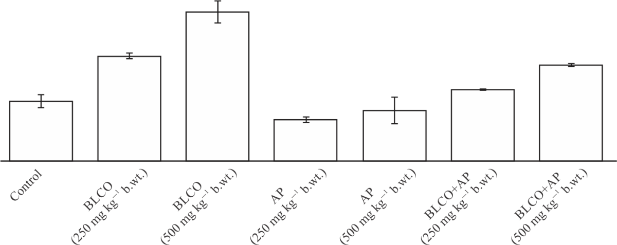 |
| Fig. 1: | Blood lead concentrations of Wistar albino rats orally exposed to Bonny Light Crude Oil and Andrographis paniculata leaf extract Values are reported as Mean±SEM of triplicate determination, values with different superscript alphabets are significantly different at p<0.05, the least significant difference (LSD) was used to test for the difference between individual treatments groups using Statistical Package for Social Sciences (SPSS), version 22.0, BLCO: Bonny Light Crude Oil, AP: Andrographis paniculata and mg kg–1 b.wt.: Milligram per kilogram body weight |
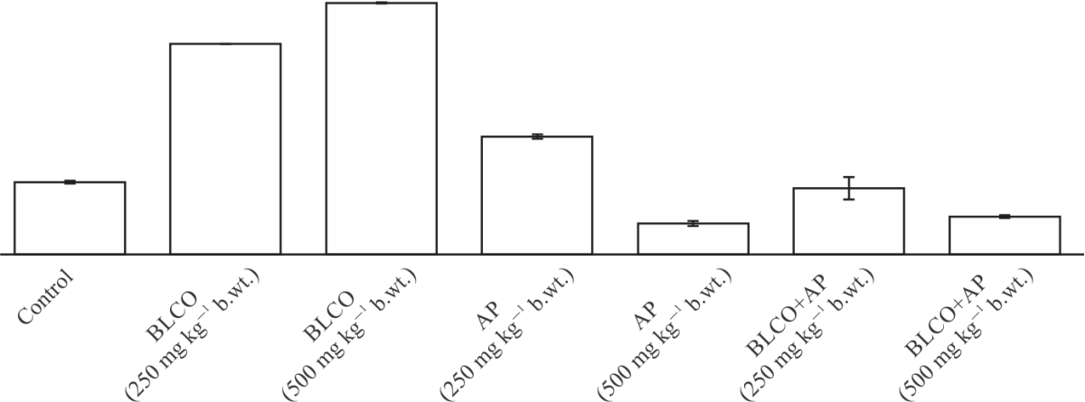 |
| Fig. 2: | Blood cadmium concentrations of Wistar albino rats orally exposed to Bonny Light Crude Oil and Andrographis paniculata leaf extract Values are reported as Mean±SEM of triplicate determination, values with different superscript alphabets are significantly different at p<0.05, the least significant difference (LSD) was used to test for the difference between individual treatments groups using Statistical Package for Social Sciences (SPSS), version 22.0, BLCO: Bonny Light Crude Oil, AP: Andrographis paniculata and mg kg–1 b.wt.: Milligram per kilogram body weight |
The blood heavy metals of the Wistar rats exposed orally to BLCO and AP are presented in Fig. 1-3. As revealed in Fig. 1, the Pb concentrations of the 250 mg kg–1 b.wt., BLCO and 500 mg kg–1 b.wt., BLCO treated groups were significantly (p<0.05) higher compared to both the control and the 250 mg kg–1 b.wt., AP and 500 mg kg–1 b.wt., AP. Likewise, for Cd and As shown in Fig. 2 and 3, respectively, both concentrations in the 250 mg kg–1 b.wt., BLCO and 500 mg kg–1 b.wt., BLCO treated groups were significantly (p<0.05) higher compared to both the control and the 250 AP and 500 mg kg–1 b.wt., AP. Notwithstanding, the 500 mg kg–1 b.wt., BLCO treated group showed a significantly (p<0.05) higher concentration compared to the 250 mg kg–1 b.wt., BLCO treated group. However, the 250 mg kg–1 b.wt., each of BLCO+AP and 500 mg kg–1 b.wt., each of BLCO+AP recorded significantly (p<0.05) lower concentration compared to the corresponding 250 mg kg–1 b.wt., BLCO and 500 mg kg–1 b.wt., BLCO treated groups.
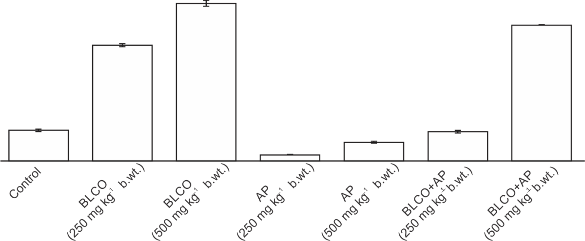 |
| Fig. 3: | Blood arsenic concentrations of Wistar albino rats orally exposed to Bonny Light Crude Oil and Andrographis paniculata leaf extract Values are reported as Mean±SEM of triplicate determination, values with different superscript alphabets are significantly different at p<0.05, the least significant difference (LSD) was used to test for the difference between individual treatments groups using Statistical Package for Social Sciences (SPSS), version 22.0, BLCO: Bonny Light Crude Oil, AP: Andrographis paniculata and mg kg–1 b.wt.: Milligram per kilogram body weight |
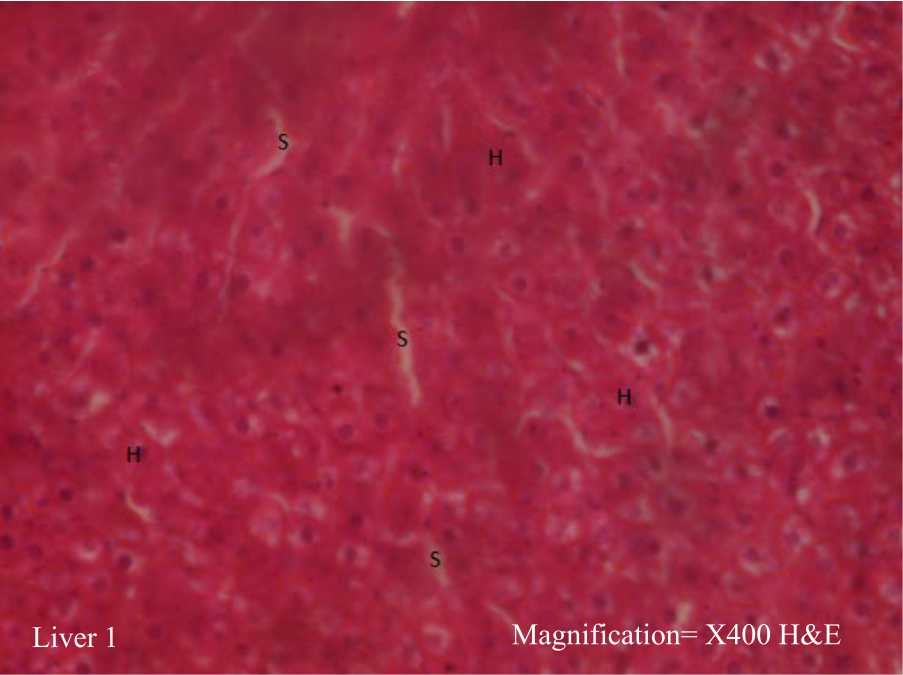 |
| Fig. 4: | Photomicrograph of liver from the control group Histologically normal liver showing, intact hepatocytes (H), sinusoids (S) contain kupffer cells |
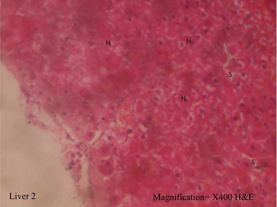
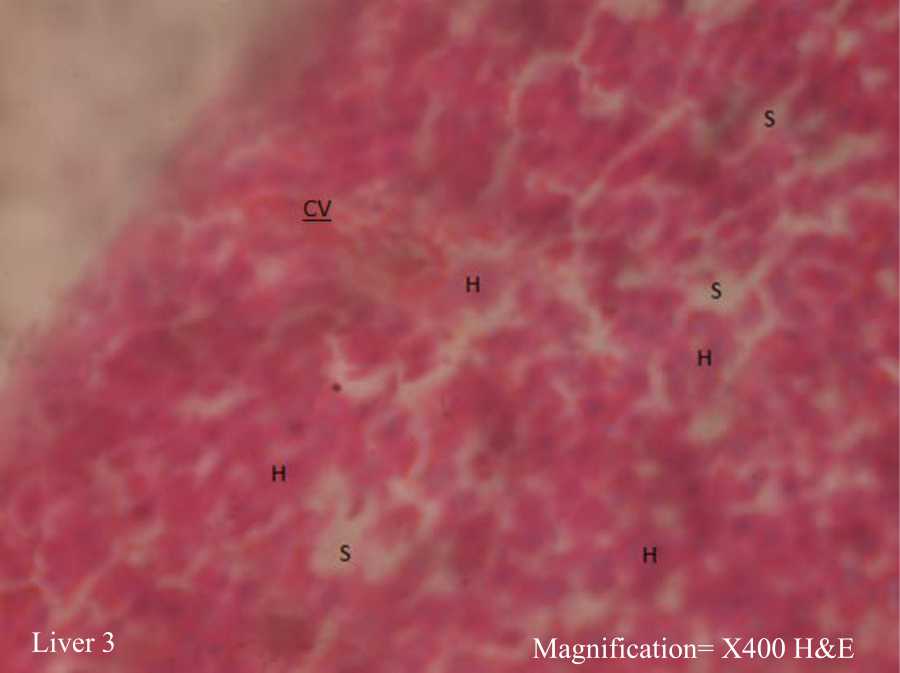
The results of the histopathological investigation of the Wistar rats orally exposed to BLCO and AP are presented in Fig. 4-17. Figure 4 showed a photomicrograph of the liver from the control group. It further revealed a histologically normal liver with intact hepatocytes (H) and sinusoids (S) containing kupffer cells. Figure 5 displayed a photomicrograph of the liver of Wistar rats administered 250 mg kg–1 b.wt., BLCO. Similar to the control group, there was evidence of histologically normal liver showing intact hepatocytes and sinusoids containing kupffer cells. Figure 6, on the other hand, illustrated a photomicrograph of the liver from the group-administered 500 mg kg–1 b.wt., BLCO, with evidence of histologically normal liver revealing intact hepatocytes, sinusoids containing kupffer cells but congested vein (CV). Additionally, the photomicrograph of the liver from the group-administered 250 mg kg–1 b.wt., AP as shown in Fig. 7, revealed histologically normal liver with intact hepatocytes, sinusoids containing kupffer cells and potent central vein (CV). Similar to the group administered 500 mg kg–1 b.wt., BLCO, Fig. 8 showed photomicrograph of liver from group-administered 500 mg kg–1 b.wt., AP with evidence of histologically normal liver with intact hepatocytes, sinusoids containing kupffer cells and congested central vein. Contrarily, Fig. 9 showed a photomicrograph of the liver from the group-administered 250 mg kg–1 b.wt., BLCO with evidence of intact hepatocytes, sinusoids containing kupffer cells and the portal triad of the hepatic artery (A), congested portal vein (V) and bile duct (D).
 |
| Fig. 5: | Photomicrograph of liver from the group-administered 250 mg kg–1 b.wt., of BLCO Histologically normal liver showing, intact hepatocytes (H), sinusoids (S) contain kupffer cells |
 |
| Fig. 6: | Photomicrograph of liver from the group-administered 500 mg kg–1 b.wt., of BLCO Histologically normal liver showing, intact hepatocytes (H), sinusoids (S) contain kupffer cells and congested central vein (CV) |
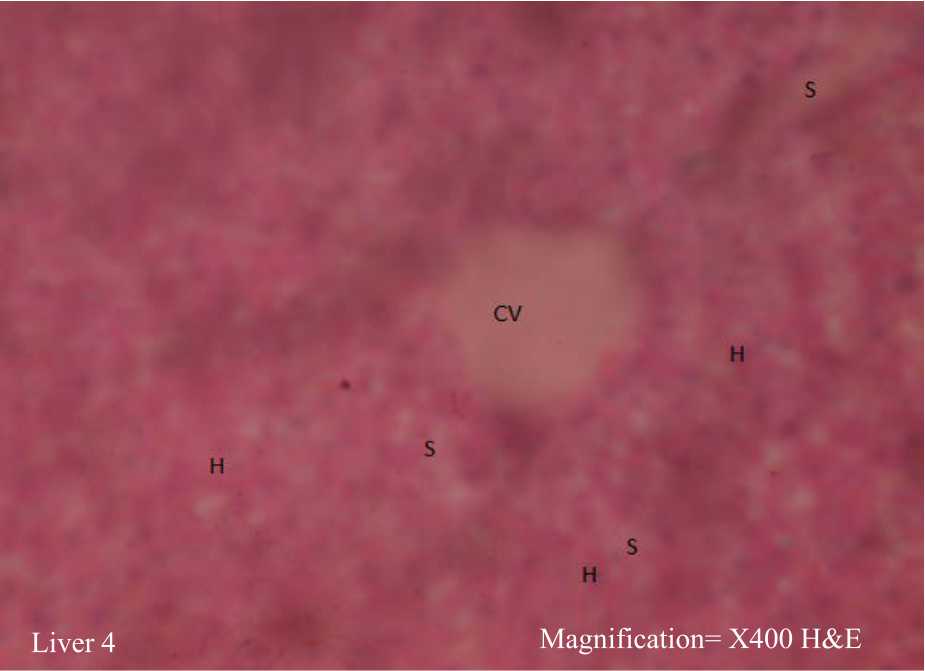 |
| Fig. 7: | Photomicrograph of liver from the group-administered 250 mg kg–1 b.wt., of AP Histologically normal liver showing, intact hepatocytes (H), sinusoids (S) contain kupffer cells and potent central vein (CV) |
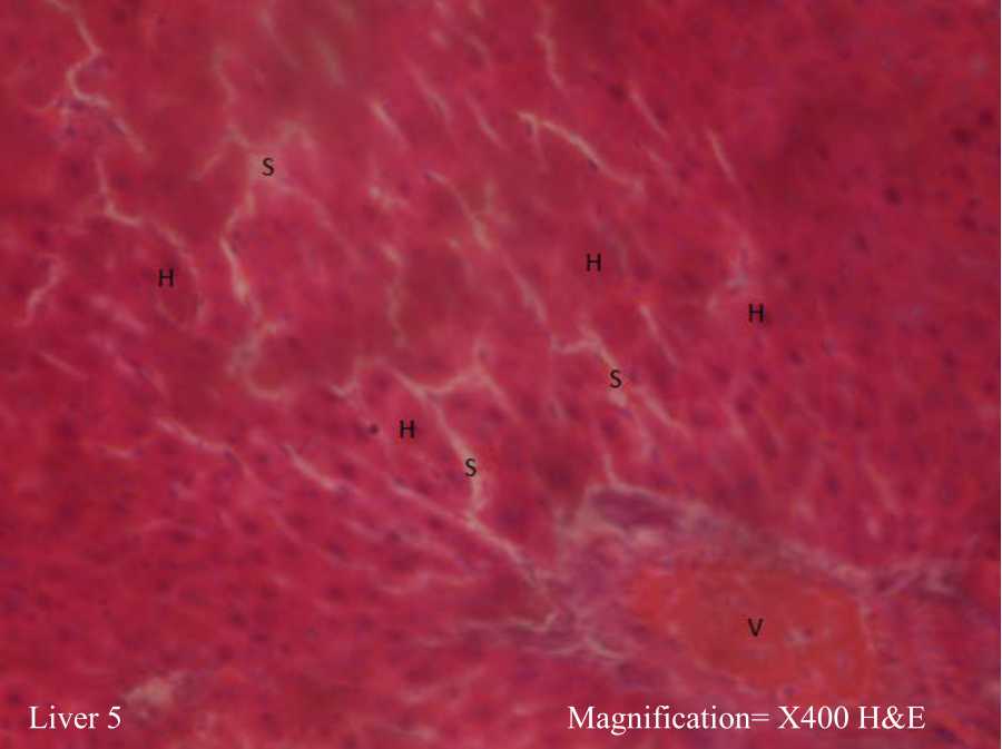 |
| Fig. 8: | Photomicrograph of liver from the group-administered 500 mg kg–1 b.wt., of AP Histologically normal liver showing, intact hepatocytes (H), sinusoids (S) contain kupffer cells and congested central vein (CV) |
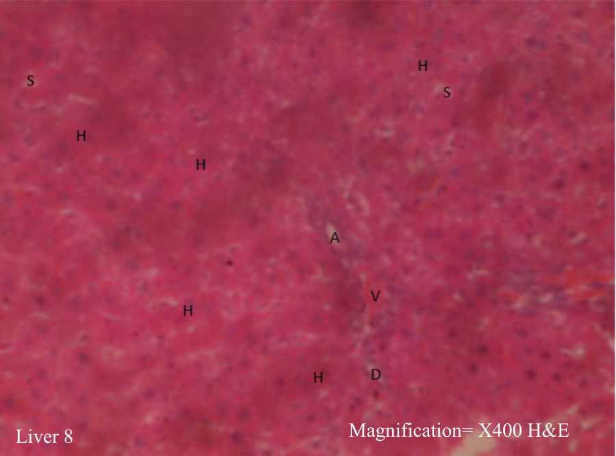 |
| Fig. 9: | Photomicrograph of liver from the group-administered 250 mg kg–1 b.wt., of AP+250 mg kg–1 b.wt., of BLCO Histologically normal liver showing, intact hepatocytes (H), sinusoids (S) contain kupffer cells, portal triad (Hepatic artery = A, Congested portal vein = V, Bile duct = D) |
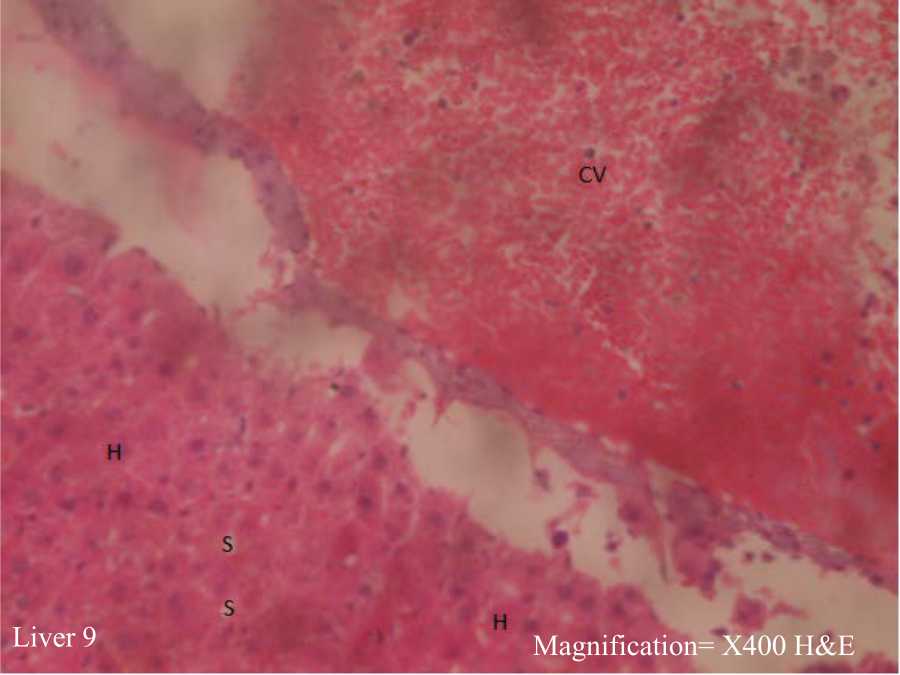 |
| Fig. 10: | Photomicrograph of liver from the group-administered 500 of AP+500 mg kg–1 b.wt., of BLCO Histologically distorted liver showing, intact hepatocytes (H), sinusoids (S) contain kupffer cells and enlarged and congested central vein (CV) |
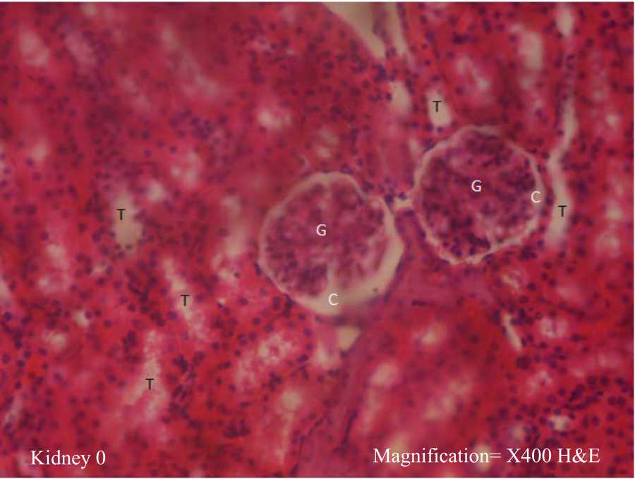 |
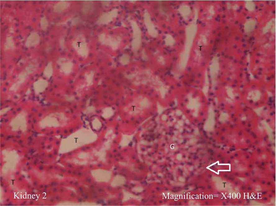
| Fig. 11: | Photomicrographs of kidney from the control group Histologically normal kidney showing, intact glomeruli (G) containing glomerular mesangial cells, glomerular matrix and capillaries, patent Bowman’s capsular spaces (C), renal tubules (T) and lined with simple columnar epithelial cells |
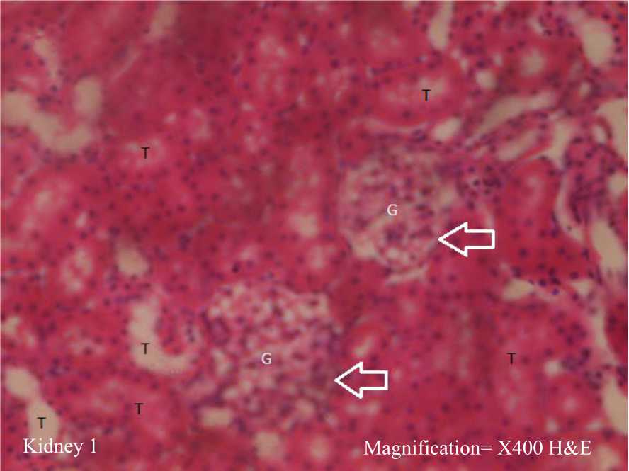 |
| Fig. 12: | Photomicrograph of kidney from the group administered 250 mg kg–1 b.wt., of BLCO Histologically distorted Kidney showing, enlargement of Glomerular tuft (G) with marked decrease/occlusion of Bowman’s capsular space (C) arrowed intact renal tubules (T) |
 |
| Fig. 13: | Photomicrographs of kidney from the group administered 500 mg kg–1 b.wt., of BLCO Histologically distorted kidney showing, enlargement of Glomerular tuft (G) with marked decrease/occlusion of Bowman’s capsular space (C) arrowed and intact renal tubules (T) |
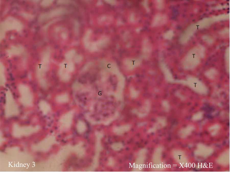 |
| Fig. 14: | Photomicrograph of kidney from the group administered 250 mg kg–1 b.wt., of AP Histologically normal kidney showing, intact Glomerular tuft (G) patent Bowman’s capsule (C) renal tubules (T) |
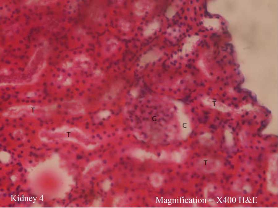 |
| Fig. 15: | Photomicrograph of kidney from the group administered 500 mg kg–1 b.wt., of AP Histologically distorted kidney showing, tubular necrosis (T), sloughing off of epithelial cells and necrotic debris in the tubular lumen intact Glomerular tuft (G) patent Bowman’s capsule (C) |
 |
| Fig. 16: | Photomicrographs of kidney the group from the group administered 250 of AP+250 mg kg–1 b.wt., of BLCO Histologically normal kidney showing, intact Glomerular tuft (G), patent Bowman’s capsule (C) and renal tubules (T) |
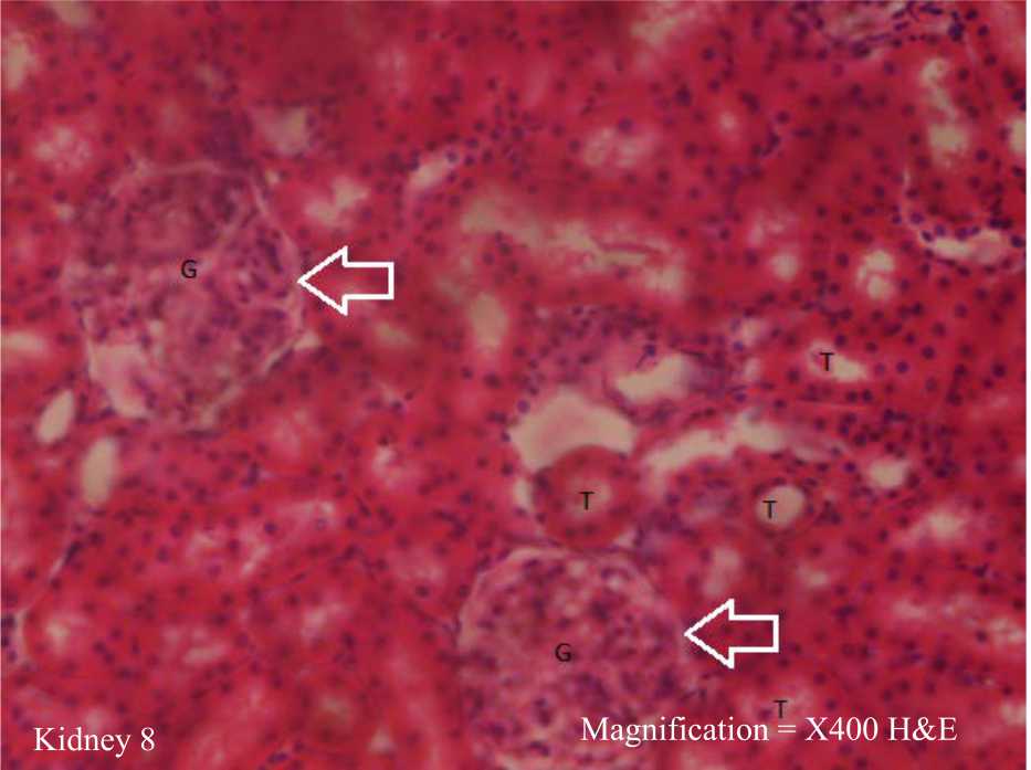 |
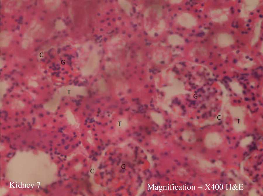
| Fig. 17: | Photomicrographs of kidney, from the group administered 500 of AP+500 mg kg–1 b.wt., of BLCO Histologically distorted kidney showing, enlarged Glomerular tuft (G) with occluded Bowman’s capsular space (C) arrowed intact renal tubules (T) |
Similarly, the photomicrograph of the liver from the group-administered 500 mg kg–1 b.wt., AP+500 mg kg–1 b.wt., BLCO as shown in Fig. 10, revealed a histologically distorted liver portraying intact hepatocytes, sinusoids containing kupffer cells and enlarged and congested central vein.
Figure 11 showed the photomicrograph of the kidney from the control group revealing histologically normal kidneys portraying intact glomeruli (G) containing glomerular mesangial cells, glomerular matrix and capillaries. It further portrayed patent Bowman’s capsular spaces (C) and renal tubules (T) lined with simple columnar epithelial cells. Figure 12 on the other hand, revealed a photomicrograph of the kidney from the group-administered 250 mg kg–1 b.wt., BLCO with evidence of histologically distorted kidney showing enlargement of glomerular tuft (G) with marked decrease/ occlusion of Bowman’s capsular space arrowed and intact renal tubules. Similarly, Fig. 13 showed a photomicrograph of the kidney from the group-administered 500 mg kg–1 b.wt., BLCO with evidence of histologically distorted kidney revealing enlargement of glomerular tuft with occluded Bowman’s capsule arrowed and intact renal tubules. However, the photomicrographs of kidneys from the groups administered 250 mg kg–1 b.wt., AP and 250 mg kg–1 b.wt., +250 mg kg–1 b.wt., BLCO as shown in Fig. 14 and 16, respectively revealed histologically normal kidney with evidence of intact glomerular tuft, patent Bowman’s capsule and renal tubules. Photomicrograph of the group administered 500 mg kg–1 b.wt., AP in Fig. 15 revealed histologically distorted kidney with tubular necrosis, sloughing off of epithelial cells, necrotic debris in the tubular lumen, intact glomerular tuft and patent Bowman’s capsule, while Fig. 17 revealed the photomicrograph of the kidney of the group administered 500 mg kg–1 b.wt., BLCO+500 mg kg–1 b.wt., AP, showing enlarged glomerular tuft with occluded Bowman’s capsular space arrowed and intact renal tubules.
DISCUSSION
The results obtained in this study revealed the biochemical implications of Wistar albino rats orally exposed to Bonny Light Crude Oil and Andrographis paniculata leaf extract. Chemical components of crude oil and dispersants can cause physiological damage in humans and wildlife, depending on the exposure dosage and susceptibility. On the other hand, traditional medicinal plants can be a source of biological and pharmacological products for the future, thereby offering protective effects on crude oil-orally exposed rats34. The present study demonstrated that at certain concentrations and periods of oral exposure, BLCO and AP leaf extract may not induce physiological damage. Could this be the justifiable reason why both BLCO and AP are used in folkloric medicine for the treatment of various kinds of diseases2,3,12,14,15? However, several studies have documented the use of such plants as AP and their products in managing petroleum-induced toxicity9,10.
The consumption of BLCO and AP leaf extract at doses of 250 and 500 mg kg–1 b.wt., for 21 days may not be injurious to animal health as indicated by the non-significance (p>0.05) difference in the serum liver function parameters (Table 1). However, the photomicrographs indicated that at 500 mg kg–1 b.wt., AP and 500 mg kg–1 b.wt., BLCO, there is congestion of the central vein. According to Hilscher and Sanchez35, the congestion of the central vein could lead to ischemia, atrophy of hepatocytes and distinction of sinusoids and this, in turn, could lead to hepatomegaly. This agrees with the histopathology results as shown in photomicrographs (Fig. 6 and 8). As revealed in the photomicrograph (Fig. 9), there was congestion of the portal triad. The portal triad is made up of three major tubes, the hepatic artery, portal vein and bile duct. Branches of the hepatic artery transport oxygenated blood to the hepatocytes, while the bile duct conveys bile products away from the hepatocytes to the larger ducts and gall bladder. The portal vein, on the other hand, carries blood with nutrients from the small intestine. Injury to the portal vein is usually due to penetrating trauma36. Given that the portal vein carries mostly oxygenated blood, the congested portal vein as shown in the photomicrograph suggests that blood in the liver is mostly poorly oxygenated, which over time could lead to cirrhosis of liver cells resulting from the insufficient oxygen supply.
However, the trend observed for AST, total bilirubin and total protein in the group treated with 500 mg kg–1 b.wt., BLCO+500 mg kg–1 b.wt., AP could be attributed to the elevated concentrations of both BLCO and AP used in combination. This finding is also reflected in the photomicrograph (Fig. 10) showing distorted liver and enlarged and congested central vein. Thus, a combination of BLCO and AP concentrations as adopted in this group may be injurious to animal health and could damage the liver cell membrane which could lead to cellular leakage of liver enzymes into general circulation. This finding is consistent with an earlier report10.
The kidney maintains a constant extracellular environment through its involvement in the excretion of metabolites including urea, creatinine and uric acid. It is also responsible for the regulation of water and electrolyte balance37. Abnormal levels of these catabolize and some electrolytes in the serum indicate impairment of renal function. Such impairment of the renal functions could result from exposure to different nephrotoxic substances38. The present study revealed no abnormality exists in the kidney markers analyzed. However, histological findings revealed that 250 mg kg–1 b.wt., BLCO (Fig. 12) and 500 mg kg–1 b.wt., BLCO (Fig. 13) showed evidence of histologically distorted kidney revealing enlargement of glomerular tuft with occluded Bowman’s capsule arrowed. Likewise, the group administered 500 mg kg–1 b.wt., AP (Fig. 15) showed tubular necrosis, sloughing off of epithelial cells and necrotic debris in tubular lines, while 500 mg kg–1 b.wt., BLCO+500 mg kg–1 b.wt., AP (Fig. 17) treated group showed enlarged glomerular tuft with occluded Bowman’s capsule space arrowed. This, thus, indicates that the doses of BLCO and AP leaf extract employed for the study may tend to cause renal damage. However, the trend observed in potassium and calcium concentrations in the group treated with 500 mg kg–1 b.wt., BLCO+500 mg kg–1 b.wt., AP are similar to the report on rats orally exposed to high concentrations of crude oil. Such alteration in electrolytes level is related to the development of hypertension39.
The severity of oxidative damage depends on the extent of disturbances in the normal redox state within the cells. Although a cell can regain its original functional state after overcoming small perturbations, more severe oxidative stress can cause cell death and necrosis. To counteract the damaging effect of reactive oxygen species (ROS), aerobic cells are provided with extensive antioxidant defence mechanisms40. Endogenous antioxidant enzymes such as SOD, CAT and GST as well as non-enzymatic antioxidants GSH can limit the effects of ROS but quickly become overwhelmed by large quantities of ROS41. The present study demonstrated that administration of BLCO for 21 consecutive days resulted in no dose-dependent difference in GSH and CAT. However, the increase in SOD may signify enzyme induction. An increase in SOD activity has been reported to be beneficial in the event of increased free radical generation42. Lipid peroxidation, on the other hand, is a degenerative pathway of membrane components mediated through free radicals produced in the cell43. This reaction leads to the formation of MDA which is cytotoxic and mutagenic. Because a large proportion of crude oil components is lipophilic, biological membranes may be the target sites where the adverse effect occurs. It is worthy of note that an increased MDA level indicates a state of stress in the liver possibly induced by BLCO or its metabolites41. Current findings, therefore, revealed that no stress occurred in the animals as a result of the ingested crude oil.
Although ingestion or exposure of crude oil fractions affects lipid metabolism causing the tissue to compromise its effectiveness in lipid metabolism, the present study revealed no significant difference in the lipid parameters of the exposed animals, thereby indicating no adverse effect posed by the quantity and duration of intake of the BLCO. However, it should be noted that constituents of petroleum are highly toxic to biological membranes and proteins. An example is a naphthalene which has been reported to cause haemoglobin denaturation and is one of the compounds responsible for the development of haemolytic anaemia in oiled wild life44. It has also been established that, the toxic constituents of petroleum such as benzene and lead are activated in the bone marrow, where these substances exert cytotoxic effects that could be mediated through disturbance in DNA function45. This leads to bone marrow depression which is characterized by inadequate production of red cells and other formed elements44. Nevertheless, findings revealed that the treatments did not affect the haematological parameters of the animals orally exposed to crude oil.
Heavy metals are taken into the body via inhalation, ingestion and skin absorption. If heavy metals enter and accumulate in body tissue faster than the body’s detoxification, pathways can dispose of them, a gradual buildup of these toxins will occur. The concentrations of the investigated heavy metals (Pb, Cd, As) revealed more accumulations in groups treated with 250 and 500 mg kg–1 b.wt., compared to the other groups. However, accumulation was higher in the 500 mg kg–1 b.wt., BLCO-treated group compared with 250 mg kg–1 b.wt., BLCO-treated group. This outcome is in line with Adedara et al.6, whose finding revealed differential heavy metals accumulation in the blood, liver and testes of rats. The decrease in metal concentrations experienced in the groups treated with similar concentrations of AP and BLCO could entail the detoxification property of the plant extract in comparison with the other groups.
CONCLUSION
In summary, the present study revealed that Andrographis paniculata leaf extract and BLCO-induction at concentrations of 250 and 500 mg kg–1 b.wt., for 21 consecutive days may be injurious to health affecting the liver and kidney. This is evident from the result of the histological analysis carried out on the tissue extracts. It is, therefore, recommended that the use of Andrographis paniculata and BLCO in folkloric medicine at concentrations similar to the ones employed in this study should be discontinued.
SIGNIFICANCE STATEMENT
Crude oil and Andrographis paniculata have been used in folkloric medicine for the treatment of various kinds of diseases. However, there has been a lot of concern bothering on the use of these materials and their implications in the physiological system. This study, therefore, provides an insight into the application of these materials in folkloric medicine and their possible safe doses and period. The investigation would help both native practitioners of folkloric medicine and researchers in academics to understand and further unravel the hidden treasures as well as drawbacks in the practice of folkloric medicine.
REFERENCES
- Brussaard, C.P.D., L. Peperzak, S. Beggah, L.Y. Wick and B. Wuerz et al., 2016. Immediate ecotoxicological effects of short-lived oil spills on marine biota. Nat. Commun., Vol. 7.
CrossRefDirect Link - Farombi, E.O., I.A. Adedara, A.P. Ebokaiwe, R. Teberen and T. Ehwerhemuepha, 2010. Nigerian Bonny Light Crude Oil disrupts antioxidant systems in testes and sperm of rats. Arch. Environ. Contam. Toxicol., 59: 166-174.
CrossRefDirect Link - Ebokaiwe, A.P. and E.O. Farombi, 2015. Influence of vitamin E and quercetin on Nigerian Bonny Light crude oil-induced neuronal and testicular toxicity in Wistar rats. J. Basic Clin. Physiol. Pharmacol., 26: 223-231.
CrossRefDirect Link - Achuba, F.I. and S.A. Osakwe, 2003. Petroleum-induced free radical toxicity in African catfish (Clarias gariepinus). Fish Physiol. Biochem., Vol. 29.
CrossRefDirect Link - Ovuru, S.S. and I.K.E. Ekweozor, 2004. Haematological changes associated with crude oil ingestion in experimental rabbits. Afr. J. Biotechnol., 3: 346-348.
CrossRefDirect Link - Adedara, I.A., A.P. Ebokaiwe and E.O. Farombi, 2013. Tissues distribution of heavy metals and erythrocytes antioxidant status in rats exposed to Nigerian Bonny Light Crude Oil. Toxicol. Ind. Health, 29: 162-168.
CrossRefDirect Link - Ita, S.O., E.O. Aluko, T.H. Olubobokun, U.A. Okon, A.B. Antai and E.E. Osim, 2014. The relationship between Nigerian Bonny Light Crude Oil-induced hypoglycaemia and endogenous serum insulin concentration in male Wistar rats: The role of antioxidant vitamins C and E. Am. J. Mol. Biol., 4: 81-88.
CrossRefDirect Link - Ogara, A.L., P.E. Joshua, K.O. Omeje and I.N.E. Onwurah, 2016. Effects of ingested crude oil contaminated diets on antioxidant enzyme and lipid profile in Wistar albino rat. J. Appl. Sci. Environ. Manage., 20: 927-932.
CrossRefDirect Link - Achuba, F.I. and C. Offor, 2020. Effects of Siam weed (Chromolaena odorata) leaf extract on crude oil-induced toxicity. Galician Med. J., Vol. 27.
CrossRefDirect Link - Hossain, M.S., Z. Urbi, A. Sule and K.M.H. Rahman, 2014. Andrographis paniculata (Burm. f.) Wall. ex Nees: A review of ethnobotany, phytochemistry, and pharmacology. Sci. World J., Vol. 2014.
CrossRefDirect Link - Salibay, C.C., T. Mahboob, A.K. Verma, J.S.S. Sebastian, H.A. Tabo, C.S. Raju and V. Nissapatorn, 2021. Natural Product-Derived Drugs for the Treatment of Inflammatory Bowel Diseases (IBD). In: Inflammation and Natural Products, Gopi, S., A. Kunnumakkara, A. Amalraj and S. Thomas (Eds.), Academic Press Elsevier Inc., London, UK, ISBN-13: 978-0-12-819218-4, Pages: 235-259.
CrossRefDirect Link - Panossian, A., T. Davtyan, N. Gukassyan, G. Gukasova, G. Mamikonyan, E. Gabrielian and G. Wikman, 2002. Effect of andrographolide and kan jang-fixed combination of extract SHA-10 and extract SHE-3-on proliferation of human lymphocytes, production of cytokines and immune activation markers in the whole blood cells culture. Phytomedicine, 9: 598-605.
CrossRefDirect Link - Okhuarobo, A., J.E. Falodun, O. Erharuyi, V. Imieje, A. Falodun and P. Langer, 2014. Harnessing the medicinal properties of Andrographis paniculata for diseases and beyond: A review of its phytochemistry and pharmacology. Asian Pac. J. Trop. Dis., 4: 213-222.
CrossRefDirect Link - Singha, P.K., S. Roy and S. Dey, 2007. Protective activity of andrographolide and arabinogalactan proteins from Andrographis paniculata Nees. against ethanol-induced toxicity in mice. J. Ethnopharmacol., 111: 13-21.
CrossRefPubMedDirect Link - Trivedi, N.P., U.M. Rawal and B.P. Patel, 2007. Hepatoprotective effect of andrographolide against hexachlorocyclohexane-induced oxidative injury. Integr. Cancer Ther., 6: 271-280.
CrossRefDirect Link - Pokala, N., N. Alasyam and K. Rasamal, 2019. Evaluation and comparison of antipyretic activity of aqueous leaf extracts of Vitex negundo and Andrographis paniculata in rabbits. Natl. J. Physiol. Pharm. Pharmacol., 9: 556-561.
CrossRefDirect Link - Verma, N. and M. Vinayak, 2008. Antioxidant action of Andrographis paniculata on lymphoma. Mol. Biol. Rep., 35: 535-540.
CrossRefPubMedDirect Link - Yu, B.C., C.R. Hung, W.C. Chen and J.T. Cheng, 2003. Antihyperglycaemic effect of andrographolide in streptozotocin-induced diabetic rats. Planta Med., 69: 1075-1079.
CrossRefDirect Link - Akhtar, M.T., M.S.B.M. Sarib, I.S. Ismail, F. Abas, A. Ismail, N.H. Lajis and K. Shaari, 2016. Anti-diabetic activity and metabolic changes induced by Andrographis paniculata plant extract in obese diabetic rats. Molecules, Vol. 21.
CrossRefDirect Link - Reyes, B.A.S., N.D. Bautista, N.C. Tanquilut, R.V. Anunciado and A.B. Leung et al., 2006. Anti-diabetic potentials of Momordica charantia and Andrographis paniculata and their effects on estrous cyclicity of alloxan-induced diabetic rats. J. Ethnopharmacol., 105: 196-200.
CrossRefDirect Link - Coon, J.T. and E. Ernst, 2004. Andrographis paniculata in the treatment of upper respiratory tract infections: A systematic review of safety and efficacy. Planta Med., 70: 293-298.
CrossRefDirect Link - Sheeja, K. and G. Kuttan, 2007. Modulation of natural killer cell activity, antibody-dependent cellular cytotoxicity, and antibody-dependent complement-mediated cytotoxicity by andrographolide in normal and Ehrlich ascites carcinoma-bearing mice. Integr. Cancer Ther., 6: 66-73.
CrossRefDirect Link - Chao, W.W., Y.H. Kuo, S.L. Hsieh and B.F. Lin, 2011. Inhibitory effects of ethyl acetate extract of Andrographis paniculata on NF-κB trans-activation activity and LPS-induced acute inflammation in mice. Evidence-Based Complementary Altern. Med., Vol. 2011.
CrossRefDirect Link - Isunu, L.E., F.O. Omoya, K.O. Ajayi, F. Akharaiyi, A.O. Ogundare and O.J. Babatunde, 2022. Evaluation of the antiplasmodial activities of methanol leaf extract of Andrographis paniculata (burm. f.): An in vitro and in vivo study. Microbes Infect. Dis., Vol. 3.
CrossRefDirect Link - Reddy, V.L.N., S.M. Reddy, V. Ravikanth, P. Krishnaiah and T.V. Goud et al., 2005. A new BIS-andrographolide ether from Andrographis paniculata Nees and evaluation of anti-HIV activity. Nat. Prod. Res., 19: 223-230.
CrossRefDirect Link - Zhou, J., S. Zhang, C.N. Ong and H.M. Shen, 2006. Critical role of pro-apoptotic Bcl-2 family members in andrographolide-induced apoptosis in human cancer cells. Biochem. Pharmacol., 72: 132-144.
CrossRefDirect Link - Mishra, K., A.P. Dash, B.K. Swain and N. Dey, 2009. Anti-malarial activities of Andrographis paniculata and Hedyotis corymbosa extracts and their combination with curcumin. Malar. J., Vol. 8.
CrossRefDirect Link - Uttekar, M.M., T. Das, R.S. Pawar, B. Bhandari and V. Menon et al., 2012. Anti-HIV activity of semisynthetic derivatives of andrographolide and computational study of HIV-1 gp120 protein binding. Eur. J. Med. Chem., 56: 368-374.
CrossRefDirect Link - Chukwuma, C.C., I.J. Aruorivwooghene, N.E. Oluchi and M.M. Okechukwu, 2019. Soil heavy metals remediation using Mariscus alternifolius, Fimbristylis ferruginea, Schwenkia americana and Spermacoce ocymoides. Singapore J. Sci. Res., 9: 77-85.
CrossRefDirect Link - Wu, C.C., L.Y. Sheen, H.W. Chen, S.J. Tsai and C.K. Lii, 2001. Effects of organosulfur compounds from garlic oil on the antioxidation system in rat liver and red blood cells. Food Chem. Toxicol., 39: 563-569.
CrossRefPubMedDirect Link - Han, Z.H., J.M. Ye and G.F. Wang, 2013. Evaluation of in vivo antioxidant activity of Hericium erinaceus polysaccharides. Int. J. Biol. Macromol., 52: 66-71.
CrossRefDirect Link - Nabavi, S.M., S.F. Nabavi, S. Eslami and A.H. Moghaddam, 2012. In vivo protective effects of quercetin against sodium fluoride-induced oxidative stress in the hepatic tissue. Food Chem., 132: 931-935.
CrossRefDirect Link - Wang, Y.S., S.P. Tian and Y. Xu, 2005. Effects of high oxygen concentration on pro- and anti-oxidant enzymes in peach fruits during postharvest periods. Food Chem., 91: 99-104.
CrossRefDirect Link - Asadbeigi, M., T. Mohammadi, M. Rafieian-Kopaei, K. Saki, M. Bahmani and M. Delfan, 2014. Traditional effects of medicinal plants in the treatment of respiratory diseases and disorders: An ethnobotanical study in the Urmia. Asian Pac. J. Trop. Med., 7: S364-S368.
CrossRefDirect Link - Hilscher, M. and W. Sanchez, 2016. Congestive hepatopathy. Clin. Liver Dis., 8: 68-71.
CrossRefDirect Link - Sabat, J., C.H. Hsu, Q. Chu and T.W. Tan, 2019. The mortality for surgical repair is similar to ligation in patients with traumatic portal vein injury. J. Vasc. Surg.: Venous Lymphatic Disord., 7: 399-404.
CrossRefDirect Link - Atherton, J.C., 2006. Regulation of fluid and electrolyte balance by the kidney. Anaesth. Intensive Care Med., 7: 227-233.
CrossRefDirect Link - Pazhayattil, G.S. and A. Shirali, 2014. Drug-induced impairment of renal function. Int. J. Nephrol. Renovascular Dis., 7: 457-468.
CrossRefDirect Link - Ojo, A.O., S.I. Jaja, B. Olubayode, D.B. Lawrence and F.F. Taiye, 2015. Effects of Nigeria Ekete Light Crude Oil on plasma electrolytes, packed cell volume (PCV) and lipids profile in Wistar (Rattus norvegicus) rats. Afr. J. Biotechnol., 14: 2047-2051.
CrossRefDirect Link - Alpsoy, L., A. Yildirim and G. Agar, 2009. The antioxidant effects of vitamin A, C, and E on aflatoxin B1-induced oxidative stress in human lymphocytes. Toxicol. Ind. Health, 25: 121-127.
CrossRefDirect Link - Adedara, I.A. and E.O. Farombi, 2012. Lack of recovery from hepatic oxidative damage in rats treated with Nigerian Bonny Light Crude Oil. Cell Biochem. Funct., 30: 480-486.
CrossRefDirect Link - Weydert, C.J. and J.J. Cullen, 2010. Measurement of superoxide dismutase, catalase and glutathione peroxidase in cultured cells and tissue. Nat. Protoc., 5: 51-66.
CrossRefDirect Link - Veena, C.K., A. Josephine, S.P. Preetha and P. Varalakshmi, 2007. Effect of sulphated polysaccharides on erythrocyte changes due to oxidative and nitrosative stress in experimental hyperoxaluria. Hum. Exp. Toxicol., 26: 923-932.
CrossRefDirect Link - Ita, S.O. and U.A. Udofia, 2011. Comparative study of some haematological parameters in rats following ingestion of crude oil (Nigerian Bonny Light), petrol, kerosene and diesel. Asian J. Biol. Sci., 4: 498-505.
CrossRefDirect Link - Okoro, A.M., E.J. Ani, J.O. Ibu and B.A. Akpogomeh, 2006. Effect of petroleum products inhalation on some haematological indices of fuel attendants in Calabar metropolis, Nigeria. Niger. J. Physiol. Sci., 21: 71-75.
CrossRefPubMedDirect Link