
Farnood Shokouhi Sabet Jalali
Department of Clinical Sciences
Siamak Saifzadeh
Department of Clinical Sciences
Hossein Tajik
Department of Food Hygiene, College of Veterinary Medicine, P.O. Box 57155/1177, Urmia University, Urmia, Iran
Solmaz Hobbi
Department of Food Hygiene, College of Veterinary Medicine, P.O. Box 57155/1177, Urmia University, Urmia, Iran
Asian Journal of Animal and Veterinary Advances
Year: 2008 | Volume: 3 | Issue: 3 | Page No.: 162-168


ABSTRACT
The aim of this investigation was a clinical and microbiological evaluation of the accelerating effect of Iranian garlic (1% aqueous extract) on the burn wound healing in dog. Ten apparently healthy mongrel dogs of both sexes, with mean weight of 20 ± 2.25 kg were studied. Based on Hoekstra standard model, two rectangular (2x3 cm2) wounds were created on the left (experiment) and right (control) caudodorsal areas of each dog. The experiment wounds were treated with 0.5 mL of 1% aqueous garlic extracts every 3 other day for 21 days. In the control group, the wounds were washed with normal saline at same frequency and time of day. The wounds were photographed and compared for rate of wound contraction with digital scanning software. The significant differences were seen between the experiment and control wounds for the rate of contraction. Moreover, a lesser total count of microorganisms was found (4 ± 0.5x104 cm-2) when garlic extract was applied on burn wounds compared to the control wounds (2 ± 0.5x105 cm-2) on the day 21 (p<0.05). According to the results of this study, topical usage of aqueous garlic extract could enhance burn wound healing process in the dog.
PDF Abstract XML References Citation
How to cite this article
Farnood Shokouhi Sabet Jalali, Siamak Saifzadeh, Hossein Tajik and Solmaz Hobbi, 2008. The Efficacy of Aqueous Extract of Iranian Garlic on the Healing of Burn Wound: A Clinical and Microbiological Study. Asian Journal of Animal and Veterinary Advances, 3: 162-168.
DOI: 10.3923/ajava.2008.162.168
URL: https://scialert.net/abstract/?doi=ajava.2008.162.168
DOI: 10.3923/ajava.2008.162.168
URL: https://scialert.net/abstract/?doi=ajava.2008.162.168
INTRODUCTION
Many of the spices and herbs used today were known to the people of the ancient cultures throughout the world and they were valued for their preservative and medicinal powers as well as their flavor and odor qualities (Zaika, 1988). Garlic is one of the edible plants, which has generated a lot of interest throughout human history as a medicinal panacea. Garlic (Allium sativum Linn.) a member of the Liliaceae family is used a public spice extensively in many parts of the world (Baghalian et al., 2005). Many species of genus Allium have been used for thousands years as vegetables, spices and as medicinal plant for the treatment of various diseases (Baruchin et al., 2001; Haciseferogullari et al., 2005; Sharma and Prasad, 2001).
Avecina, the famous ancient Persian physician, had suggested the usage of garlic extract in the curing of numerous infection diseases (Hosseini, 1988).
The various powerful flavors of these plants and their possible medical applications have attracted the attention of chemists and plant physiologists (Akgul, 1993; Graham and Graham, 1987). The most of its therapeutic effects are attributed to specific oil and water-soluble organosulfur compounds which are responsible for the typical odor and flavor of garlic (Sivam, 2001).
Garlic has an unusually high concentration of sulfur-containing compounds (1-3%) and its therapeutic properties are largely due to one particular class of sulfur-containing compounds, the thiosulfinates (Koch and Lawson, 1996; Lawson, 1996).
The thiosulfinate structure appears to be essential for the bactericidal, antifungal and antiprotozoal properties of garlic, likely reacting with SH-containing enzymes of these pathogens (Reuter et al., 1996).
One of the important of these thiosulfinates is allicin, which name taken from the Latin name of the garlic plant, Allium sativum (Ankri and Mirelman, 1999). Other studies were also carried out on the antimicrobial and antioxidant properties of garlic and its derivatives such as essential oil and oleoresin (Akgul, 1993; Zaika, 1988).
These bioactive components have been isolated from aqueous, ethanolic and fermented extracts of crushed garlic and have the potential to interact with a number of cellular targets, particularly those exhibiting reactive sulfhydryl moieties, whose functions range from control of cell cycle to expression of crucial antioxidant and detoxification enzymes. Interactions with these processes may underlie garlic`s putative therapeutic potential (Cooper and Pinto, 2005).
With attention to widespread usage of garlic in curing of various illnesses particularly infectious diseases, there is an absence of scientific investigation of application of this medicinal plant in wound healing. The present study is a clinical and microbiological evaluation of topical application of garlic extract in treatment of burn wound healing.
MATERIALS AND METHODS
The garlic extracts were freshly prepared everyday. The garlic was purchased from local vegetable market (Gilan, Iran). The garlic bulbs were peeled and ground to form a paste in 1 g quantity. The paste was then dissolved in 100 mL distilled water in a sterile tube. The solution was then centrifuged at 6000 rpm for 20 min at room temperature. The pellet was discarded and supernatant was diluted 10 times with distilled water to get 1% of garlic extract. The concentration of garlic extract used was based on Shukla and Taneja (Shukla and Taneja, 2002).
The extract was subjected to silica gel Column Chromatography (CC). On elution with hexane-ethyl acetate in a gradient in order to increase polarity (from 0 to 100% EtOAc and afterwards by washing the column with methanol) 4 fractions were obtained. These fractions were analyzed by GC-MS.
The prepared samples were irradiated with 25 KGy of gamma ray. Irradiation process was performed in the Atomic Energy Agency of the Islamic Republic of Iran.
This study was performed on ten apparently healthy adult mongrel dogs of both sexes, 4 to 5 years old with mean weight of 20 ± 2.25 kg. Animals were categorized randomly into control and garlic treated groups. The dogs were housed in individual cages and had access to water and food ad libitum. The investigators adhered to the Animal Welfare Act and the experimental protocol was also approved by the Animal Ethics Committee of the university. The model of the burn wound was produced according to Hoekstra standard (Brans et al., 1994). Food was withheld for 12 h before surgery. Dogs were premedicated with acetylpromazine (Hoogsrraten, Belgium) (0.1 mg kg-1, intravenously), anesthetized with sodium thiopental (Biochemie GmbH, Vienna, Austria) (10 mg kg-1, 2.5%, intravenously) and maintained with halothane (ICI Pharmaceuticals, Cheshire, England) in oxygen in a semiclosed circle system. Lactated Ringer`s solution (Shahid Ghazi Co., Tabriz, Iran) (10 mL kg-1 h-1, intravenously) was administered during the surgical procedure. Dogs were positioned in ventral recumbency and the area just behind the shoulders was shaved backwards and was prepared for aseptic surgery.
All animal were subjected to the rectangular burn wounds (2x3 cm2) using a hot (180°C) brass brick weighing 500 g which was pressed against the shaved skin for 10 sec on either side of the spine. Left side defect assigned to as experiment and the right one as control, so that each animal served as its own control. The experiment wounds received 0.5 mL of 1% aqueous garlic extract, in aseptic conditions, every three other days for 21 days. But in the control group, the wounds were washed with normal saline at same frequency and time of day. All the wounds were bandaged with routine sterile dressing, held in place with an elastic wrap. No antibiotic was used as a pre- or post-operative prophylaxis. To manage the pain and discomfort, Tramadol (KRKA, d. d., Novo mesto, Slovenia) (0.2 mg kg-1, IM) was administered every 3 h after surgery for 24 h and continued as needed.
Clinical and microbiological [quantitative (total plate count) and qualitative (using specialized microbial medias] examinations of the burn wounds were carried out on 0, 7, 14 and 21 days of the experiment. The clinical evaluation was including the general health conditions and the reaction to environment. The process of burn wound healing was carefully assessed as well as the granulation tissue formation and the progression of scar formation.
The wounds were photographed and all the photographs were scanned and wound areas were measured using digital scanning software (Sigma Scans Pro 5.0, SPSS Science, Chicago, IL). Time elapsed for wound healing was considered in both groups. The rates of wound contraction (percent decrease of wound area) (wound area on day 0 min wound area on day n, divided by the wound area on day 0 expressed as a percentage) were analyzed.
The results of total bacterial count, time taken for healing and rates of wound contraction were analyzed with a paired Student`s t-test. Differences were considered significant if p<0.05 (SigmaStat for Windows, version 2.03, Jandel Corporation, San Rafel, CA).
RESULTS
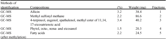
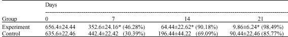
Results of biochemical analysis of the garlic extract were shown in Table 1. Also, from point of view of clinical evaluation, healing process of burn wounds was without any major complications during the study. Throughout the days after the experiment the animals showed a normal reaction to the environment and displayed no signs of suffering due to burn wounds. The rates of wound contraction (percent decrease of wound area) in experiment and control wounds were shown in Table 2.
The purpose of bacteriological examination was to evaluation of contamination of wound surface before and after topical application of garlic extract. The growth of Candida albicans, Staphylococcus aureus, Streptococcus pyogenes and Escherichia coli strains were observed during the microbiological examination of the skin before use of the preparations. The total number of the strains on the skin were 3 ± 0.5x104 cm-2 in control group and 5 ± 0.4x104 cm-2 in the experiment group.
In the control group, the number of microbes existing on 1 cm2 of the wound in the first day, were 3 ± 0.8x104 cm-2 and gradually increased during the following days to the value of 6 ± 0.6x105 cm-2 on day 7. In the 14th day the number of microbes imperceptibly to decrease until day 21, when reached to 2 ± 0.5x105 cm-2. In this group, Staphylococcus aureus, Streptococcus pyogenes, Escherichia coli and Candida albicans were also isolated on 7th day of the study. On the 14th day, the growth of Escherichia coli and Candida albicans was not observed. On 21st day, just Staph. aureus was isolated.
| Table 1: | The fraction composition of the garlic extract |
 | |
| Table 2: | Comparison of wound area (mm2) (mean ± SD) and percent decrease in the experiment and control wounds (n = 5 dogs) |
 | |
| *: Significant difference (p<0.05) Fig. 1: Results of quantitative microbiological examinations | |
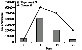 | |
| Fig. 1: | Results of quantitative microbiological examinations. |
In the garlic treated group, the numbers of microbes were 5 ± 0.6x104 cm-2 in the first 24 h of the experiment. On the 7th day of the experiment this count was 4 ± 0.8x105 cm-2 , however, on day 14 it decreased significantly to the value of 2 ± 0.6x105 cm-2. This value decreased significantly on the 21st day and reached to the 4 ± 0.5x104 cm-2. In this group, on the 7th day of the study, Streptococcus pyogenes, Staphylococcus aureus and Escherichia coli were isolated. Also on 14th day the growth of these microorganisms were observed, but just S. pyogenes was isolated on 21st day of the experiment.
Results of microbiological examinations was presented in Fig. 1.
DISCUSSION
Today usage of garlic as a natural and traditional therapy appears to become popular. There are many reports about therapeutic application of garlic in treatment of various diseases (Chowdhury et al., 1991; Dietz et al., 2004; Harris et al., 2001; Shadkchan et al., 2004; Sivam, 2001; Shukla and Taneja, 2002). But there are few researches on the accelerating effect of garlic on wound healing (Saifzadeh et al., 2006; Sardari et al., 2006a, b). The aim of this investigation is clinical and microbiological evaluation of efficacy of Iranian garlic extract on healing of burn wound.
The results of present study showed that the decrease in wound area was significantly greater (7, 14 and 21 days) in the experiment wounds compared to the control (p<0.05) (Table 2). These findings demonstrated that the rates of wound healing were affected significantly by 1% aqueous garlic extract treatment. These results are in agreement with other researches in this field (Saifzadeh et al., 2006; Sardari et al., 2006a, b). Although, these studies were accomplished on incisional wounds while our study was carried on burn wounds.
Most of the therapeutic effects of garlic are known to referable to its sulfur-containing compounds Biochemical analysis of garlic extracts in this study were revealed that concentration of two sulfur-containing compounds (Allicin and Methyl sulfonyl methane) which very important in healing accelerating efficacy and antimicrobial potency are more than 5% (Table 1).
These findings are in agreement with report about sulfur-containing compounds of Iranian garlic (Baghalian et al., 2005).
Methyl sulfonyl methane, also identified as dimethyl sulfone, is a naturally occurring sulfur compound found in a variety of foods, including onions and garlic. Methyl sulfonyl methane is an important nutrient and is essential for the maintenance of connective tissues, joint function, proper enzyme activity and hormone balance. Sulfur is very important for the formation of collagen and is a major component in the synthesis of cartilage and connective tissues. Sulfur is essential part of keratin which is necessary for the maintenance of healthy situation of the skin, hair and nails. Additionally, it gives strength, shape and hardness to their protein tissues. Methyl sulfonyl methane can also decrease scar tissue by changing the cross linking process in collagen to allow tissue repair and healing to take place (Naguib , 2002). Methyl sulfonyl methane may be responsible to the enhancing effect of aqueous garlic extract on wound healing.
From view point of microbiological evaluation, results of this experiment were showed on the day 21, the total number of microorganisms was lesser (4 ± 0.5x104 cm-2) when garlic extract were applied compared to the control wounds (2 ± 0.5x105 cm-2) (p<0.05).
Ankri and Mirelman (1999) attributed the antibacterial activity to the specific chemicals in garlic. The nature of these chemicals and the mechanisms of their action are not fully understood. Allicin is one of the sulfur components of garlic extract, this compound is known to be responsible for the most of the antibacterial property of garlic.
It is the most abundant thiosulfinate found in garlic and is generated when an enzyme alliinase reacts with its substrate alliin. Alliin was found to be a stable precursor that was converted to allicin by the action of an enzyme termed allinase which was also present in the cloves (Ellmore and Feldberg 1994). Allinase is present in extraordinarily large amounts in garlic cloves (10% of the total protein content) (Harris et al., 2001).
Enzyme and substrate are located in different compartments of the clove, the transformation of alliin into the biologically active allicin molecule upon crushing of a garlic clove is extremely rapid, being completed in seconds (Lawson, 1996).
The antibacterial effect of allicin is broad spectrum. In most cases the 50% lethal dose concentrations were somewhat higher than those required for some of the newer antibiotics. Interestingly, various bacterial strains resistant to antibiotics such as methicillin resistant Staphylococcus aureus as well as other multidrug-resistant enterotoxicogenic strains of Escherichia coli, Enterococcus, Shigella dysenteriae, S. flexneni and S. sonnei cells were all found to be sensitive to allicin (Chowdhury et al., 1991; Gonzalez-Fandos et al., 1994; Holzgartner et al., 1992; Shadkchan et al., 2004).
Garlic extracts are also effective against Helicobacter pylorus which is suspected of causing gastric ulcers (Celiini et al., 1996).
In view of the fact that the present experiment is the first study on the garlic as an accelerator of burn wound healing, our results are not comparable with those of previous works.
Considering these explanations, topical application of garlic causes significant enhancement in the rate of wound contraction and decreases the total bacterial count of burn wound surface.
Present results suggest that aqueous garlic extract may be used to accelerate the process of burn wound healing in the dog. However, it seems further studies are required to clarify other possible mechanisms involved in the would healing and to evaluate the effects of various doses of garlic extract during different periods of time.
REFERENCES
- Ankri, S. and D. Mirelman, 1999. Antimicrobial properties of allicin from garlic. Microbes Infect., 1: 125-129.
CrossRefDirect Link - Baghalian, K., S.A. Ziaib, M.R. Naghavic, H. Naghdi-Badib and A. Khalighia, 2005. Evaluation of allicin content and botanical traits in Iranian garlic (Allium sativum L.) ecotypes. Sci. Hortic., 103: 155-166.
Direct Link - Brans, T.A., R.P. Dutrieux, M.J. Hoekstra, R.W. Kreis and J.S. du Pont, 1994. Histopathological evaluation of scalds and contact burns in the pig model. Burns, 20: S48-S51.
Direct Link - Celiini, L., B. Di Campli, M. Masulli, S. Di Bartolomeo and N. Aliocati, 1996. Inhibition of Helicobacter pylori by Garlic extract. FEMS Immenol. Med. Micrbiol., 13: 273-277.
PubMedDirect Link - Chowdhury, A.K., M. Ahsan, S.N. Islam and Z.U. Ahed, 1991. Efficacy of aqueous extract of garlic and allicin in experimental shigellosis in rabbits. Indian J. Med. Res., 93: 33-36.
PubMed - Cooper, A.J.L. and J.T. Pinto, 2005. Aminotransferase, L-amino acid oxidase and b-lyase reactions involving L-cysteine S-conjugates found in allium extracts. Relevance to biological activity. Biochem. Pharmacol., 69: 209-220.
Direct Link - Dietz, D.M., J.R. Varcelotti and K.R. Stahlfeld, 2004. Garlic burns: A not-so-rare complication of a naturopathic remedy. Burns, 30: 612-613.
Direct Link - Gonzalez-Fandos, E., M.L. Garcia-Lopez, M.L. Sierra and A. Otero, 1994. Staphylococcal growth and enterotoxins (A-D) and thermonuclease synthesis in the presence of dehydrated garlic. J. Applied Bacteriol., 77: 549-552.
PubMed - Haciseferogullari, H., M. Ozcan, F. Demir and S. Calisir, 2005. Some nutritional and technological properties of garlic (Allium sativum L.). J. Food Eng., 68: 463-469.
CrossRef - Harris, L.C., S.L. Cottrel and S. Plummer, 2001. Antimicrobial properties of Allium sativum (garlic). Applied Microbiol. Biotechnol., 57: 282-286.
Direct Link - Saifzadeh, S., A. Tehrani, F. Shokouhi Sabet Jalali and R. Oroujzadeh, 2006. Enhancing effect of aqueous garlic extract on wound healing in the dog: Clinical and histopathological studies. J. Anim. Vet. Adv., 5: 1101-1104.
Direct Link - Sardari, K., A. Mirshahi, M. Maleki, M.R. Aslani and M.N. Barjasteh, 2006. Effects of topical allicin on second-intention wound healing in dogs (Histological aspects). Comp. Clin. Pathol., 15: 98-102.
Direct Link - Sardari, K., M.M. Dehgan, M. Mohri, M.R. Emami and A. Mirshahi et al., 2006. Macroscopic aspects of wound healing (contraction and epithelialisation) after topical administration of allicin in dogs. Comp. Clin. Pathol., 15: 231-235.
Direct Link - Shadkchan, Y., E. Shemesh, D. Mirelman, T. Miron, A. Rabinkov, M. Wilchek and N. Osherov, 2004. Efficacy of allicin, the reactive molecule of garlic, in inhibiting Aspergillus sp. in vitro and in a murine model of disseminated aspergillosis. J. Antimicrob. Chemother., 53: 832-836.
Direct Link - Sharma, G.P. and S. Prasad, 2001. Drying of garlic (Allium sativum) cloves by microwave-hot air combination. J. Food Eng., 50: 99-105.
CrossRefDirect Link - Shukla, Y. and P. Taneja, 2002. Antimutagenic effects of garlic extract on chromosomal aberrations. Cancer Lett., 31: 31-36.
Direct Link - Sivam, G.P., 2001. Protection against Helicobacter pylori and other bacterial infections by garlic. J. Nutr., 131: 1106S-1108S.
Direct Link - Zaika, L.L., 1988. Spices and herbs: Their antimicrobial activity and its determination. J. Food Saf., 9: 97-118.
CrossRefDirect Link