

Zhen-Yuan Lou
Department of Urology, Ninghai County First Hospital, Ningbo 315600, China
Wei-Jie Zhang
Department of Urology, Ninghai County First Hospital, Ningbo 315600, China
Jie Wang
Department of Urology, Ninghai County First Hospital, Ningbo 315600, China
Chong-Yi Yang
Department of Urology, Ninghai County First Hospital, Ningbo 315600, China
LiveDNA: 86.43231
International Journal of Pharmacology
Year: 2024 | Volume: 20 | Issue: 5 | Page No.: 883-891


ABSTRACT
Background and Objective: Benign Prostatic Hyperplasia (BPH) has been reported to be attributed to hyperproliferation, oxidative stress and chronic inflammation of epithelial cells in the prostate. Herein, the study aimed to explore the potential of diallyl disulfide (DADS), a garlic derivative with anti-inflammatory, antioxidant and anticancer activities, on BPH in vitro. Materials and Methods: The Human Prostate Epithelial cell line (RWPE-1) and immortalized Benign Prostatic Hyperplasia cell line (BPH-1) were utilized to observe the effect of DADS (0-100 μg/mL) on BPH in vitro. Cell proliferation, viability and apoptosis were analyzed by EdU staining, CCK-8 kit and Annexin V/PI staining. The commercial kits were applied to assess pro-inflammatory cytokines and oxidative stress-related markers. Western blot was carried out to investigate markers of AR signaling, apoptosis and Nrf2 signaling. Results: The DADS suppressed dihydrotestosterone-induced hyperproliferation in RWPE-1 cells and the cell viability, proliferation and AR, PSA and PCNA expressions in BPH-1 cells. However, there is no influence on BPH-1 cell apoptosis when exposing DADS with the concentration range of 10-100 μg/mL. Lipopolysaccharide (LPS) stimulated the increase of TNF-α, IL-8 and IL-6 secretion in RWPE-1 cells, which was significantly attenuated by DADS. Similarly, DADS could also reverse LPS-induced the decrease of GSH-Px and CAT and the increase of MDA. Moreover, LPS impaired the nucleus translocation of Nrf2 while this process was markedly suppressed by DADS. Conclusion: The DADS could suppress the hyperproliferation of dihydrotestosterone-treated RWPE-1 cells and BPH-1 cells and alleviate LPS-induced inflammation, oxidative stress and the Nrf2 signaling suppression in RWPE-1 cells.
PDF Abstract XML References Citation
Copyright: © 2024. This is an open access article distributed under the terms of the Creative Commons Attribution License , which permits unrestricted use, distribution and reproduction in any medium, provided the original author and source are credited.
How to cite this article
Zhen-Yuan Lou, Wei-Jie Zhang, Jie Wang and Chong-Yi Yang, 2024. In vitro Validation of the Therapeutic Potential of Diallyl Disulfide on Benign Prostatic Hyperplasia: Effect on Hyperproliferation, Oxidative Stress and Inflammation. International Journal of Pharmacology, 20: 883-891.
DOI: 10.3923/ijp.2024.883.891
URL: https://scialert.net/abstract/?doi=ijp.2024.883.891
DOI: 10.3923/ijp.2024.883.891
URL: https://scialert.net/abstract/?doi=ijp.2024.883.891
INTRODUCTION
Benign Prostate Hyperplasia (BPH), a noncancerous growth status, is histologically characterized by the prostatic epithelial and stromal cells with abnormal proliferation, of which prevalence reaches 80% among men over eighty years old1. During the aging process, the increase in the secretion of androgens to the prostate induces uncontrolled proliferation of epithelial cells in the transitional zone, which causes BPH, thereby resulting in lower urinary tract symptoms that seriously damage the quality of life2. Besides, BPH reportedly elevates the risk of bladder cancer and prostate cancer3. Hence, the exploration of treatment for patients with BPH should never be overlooked.
The pathogenesis and progression of BPH are thought to be related to several parameters, such as dietary factors4, sex hormones5, inflammation6 and oxidative stress7. Even though there is no consensus as to which is the primary one androgen receptor (AR) signaling is commonly assumed to exert a dominant role in the pathogenesis of BPH8. The 5-Alpha-Reductase (5AR) is responsible for regulating steroid metabolism, which can convert the testosterone that is generated in the testis to Dihydrotestosterone (DHT) in the prostate. Then, DHT binds to AR to trigger its transcription factor function, which in turn enhances the transcription of androgen-dependent genes, causing hyperproliferation and thereby contributing to BPH development. Thus, the 5AR-AR axis is used as the primary target for the treatment of BPH9. Finasteride, a 5AR inhibitor, was considered the first-line therapy when lower urinary tract symptoms first occur during BPH10. However, about 30% of patients with BPH have no response to the treatment with 5ARI, suffer worsened symptoms and even require surgery at last11,12. Therefore, novel treatment strategies with multiple targets are required.
Since the great accumulation of daily use experiences and appreciable treatment effects for multiple disorders, the use of compounds of natural origin for BPH has gained recent interest been an increasing interest worldwide13-15. Diallyl disulfide (DADS), an oil-soluble constituent that is abundant in garlic, exhibited various pharmacological activities, which include anticancer, antioxidant, anti-inflammatory, neuroprotective, as well as cardiovascular protective activities16. A previous study demonstrated that DADS is able to suppress the proliferation of prostate cancer cells via inducing apoptosis17. Moreover, the essential role of both oxidative stress and inflammation in BPH progression has been increasingly highlighted over the past decade, which suggested the therapeutic potential of DADS on BPH with multiple targets. However, no studies have investigated this. Accordingly, the present study conducted in vitro experiment behind the assumption that DADS, which has anti-inflammatory, antioxidant and anticancer activities, can be a potential therapeutic agent for BPH.
MATERIALS AND METHODS
Study area: This study was conducted at Ninghai County First Hospital from September 2022 to September 2023.
Cell culture and treatment: The Human Prostate Epithelial cell line (RWPE-1) was purchased from iCell Bioscience Inc. (Cat No. iCell-h286) while the human immortalized Benign Prostatic Hyperplasia cell line (BPH-1) was supplied by Shanghai Lianmai Biological Engineering Co., Ltd. (Cat No. LM8C1116). The RWPE-1 and BPH-1 cells were, respectively cultured in keratinocyte-serum-free and RPMI 1640 medium (Gibco) plus 10% fetal bovine serum (Gibco) and 1% penicillin-streptomycin (Gibco) in a 5% CO2 incubator at 37°C.
Depending on the experimental design, the RWPE-1 cells were exposed to DHT (Sigma–Aldrich) or lipopolysaccharide (LPS; Sigma–Aldrich) prior to the treatment of DADS (Sigma–Aldrich); while the BPH-1 cells only received DADS treatment.
Cell viability detection: To evaluate the optimal concentrations of DADS for the subsequent experiments, RWPE-1 cells were incubated with various concentrations of DADS (0-100 μg/mL) for 24 hrs. Cell viability was monitored using the Cell Counting Kit-8 (CCK-8) by the DOJINDO Laboratories as per the manufacturer’s protocol. After finishing the treatment, the media were removed and replaced with fresh media containing 10% CCK-8 reagent (v/v) and the cells were incubated for another 2 hrs. The absorbance was measured in a microplate reader (Bio-Rad, Hercules, California, USA) at 450 nm for cell viability calculation. Concentrations showing cytotoxicity to RWPE-1 cells were excluded from subsequent analyses.
The DHT (10 nM) was exploited to induce the hyperproliferation of RWPE-1 cells for the establishment of the in vitro BPH model. After exposing DHT for 24 hrs, RWPE-1 cells were incubated with various concentrations of DADS for another 24 hrs. The detection of cell viability was performed as above. The RWPE-1 cells without any treatment served as the control. Concentrations showing significant suppression of DHT-induced hyperproliferation were further chosen for subsequent analyses.
The BPH-1 cells were treated with various concentrations (designed according to the results of above analysis) of DADS for 24 hrs. After finishing the treatment, the media were removed and replaced with fresh media containing 10% CCK-8 reagent (v/v) and the cells were incubated for another 1.5 hrs. The absorbance was measured in a microplate reader at 450 nm.
EdU proliferation assay: The proliferative ability of BPH-1 cells was assessed using the EdU Cell Proliferation Kit (Beyotime Biotechnology). In brief, cells were labeled with EdU (10 μM) for 24 hrs, followed by fixation with 3.7% formaldehyde for 15 min and three time-washing with PBS containing 3% BSA. Cell nucleus staining was performed using DAPI. Finally, the images of the EdU-positive cells were captured using a fluorescent microscope. The cell proliferation was represented as the percentage of EdU-positive cells that was calculated by dividing the number of EdU-positive cells by the number of DAPI-stained cells.
Cell apoptosis analysis: Cell apoptosis was investigated by using the Annexin V-fluorescein isothiocyanate or Annexin V-propidium iodide (Annexin V-FITC/Annexin V-PI) apoptosis detection kit. After finishing the treatment with DADS (10, 50 and 100 μg/mL), BPH-1 cells were harvested, exposed to Annexin V-FITC and Annexin V-PI and then examined by flow cytometer. Annexin V positive cells were considered as apoptotic cells (of which PI negative means early stage while PI positive means lately).
Enzyme-Linked Immunosorbent Assay (ELISA): After inducing the inflammation with 10 μg/mL LPS for 24 hrs, RWPE-1 cells were administrated with 0, 10, 50 and 100 μg/mL of DADS for another 24 hrs. Then, the commercially available ELISA kits specific for tumor necrosis factor-alpha (TNF-α; Cat No. ABIN6574140), Interleukin (IL)-8 (Cat No. ABIN6574136) and IL-6 (Cat No. EH2IL6) supplied by antibodies-online Inc. and Thermo Fisher Scientific were used to analyze pro-inflammatory cytokine levels in cultured supernatant of RWPE-1 cells according to the manufacturer’s instructions.
Antioxidant defense and oxidative stress biomarkers measurement: After treating RWPE-1 cells with LPS alone or combined with 10, 50 and 100 μg/mL of DADS, glutathione (GSH; Cat No. KBH1462), catalase (CAT; Cat No. KBH1462) and malondialdehyde (MDA; Cat No. LS-F40103) in cultured supernatant of treated RWPE-1 cells were measured using commercial kits from Krishgen Biosystems and Lifespan Biosciences according to the manufacturer’s instructions.
Western blotting: Protein lysates of BPH-1 or RWPE-1 cells were prepared using RIPA buffer containing protease and phosphatase inhibitor cocktail. For the extraction of cytoplasmic and nuclear protein fractions, the nuclear and cytosol fractionation kit (Thermo Scientific) were applied according to the manufacturer’s instructions.
After quantification by BCA kit, 30 μg of protein from each sample was loaded onto an SDS-PAGE gel and then transferred onto a PVDF membrane via immunoblotting. The membranes were further incubated overnight at 4°C with primary antibodies to detect specific proteins in each sample, which included antibodies against as follows: AR (Cat No. A00542; Boster Bio), PSA (Cat No. MBS9403566; MyBioSource), PCNA (Cat No. FNab06217; Wuhan Fine Biotech Co., Ltd.), Bax (Cat No. FNab00810; Wuhan Fine Biotech Co., Ltd.), Bcl-2 (Cat No. FNab00839; Wuhan Fine Biotech Co., Ltd.), Nrf2 (Cat No. FNab05855; Wuhan Fine Biotech Co., Ltd.), Lamin B (Cat No. A01238-2; Boster Bio) and β-actin (Cat No. FNab00869; Wuhan Fine Biotech Co., Ltd.). Subsequently, membranes were rinsed three times and then incubated with a secondary antibody. Finally, protein bands were visualized and quantified by using the ECL Substrate Kit (Pierce, Rockford, Illinois, USA) and ImageJ software (version 1.41; National Institute of Health, Bethesda, Maryland, USA), respectively.
Statistical analysis: The values of quantitative analysis are quantified as Mean±Standard Error of the Mean (SEM).The one-way ANOVA test was utilized to analyze the difference among more than two groups. All statistical analyses in this study were conducted using GraphPad Prism software. Data were considered significant when p-values were less than 0.05.
RESULTS
DADS suppresses the hyperproliferation of BPH-1 and DHT-stimulated RWPE-1 cells: The chemical structure and formulation of DADS were displayed in Fig. 1a. Initially, RWPE-1 cells were treated with 0.1, 0.5, 1, 5, 10, 50 and 100 μg/mL of DADS. The CCK-8 assay revealed that DADS with a concentration range from 0.1 to 100 μg/mL has no cytotoxicity on RWPE-1 cells under normal physiological conditions (Fig. 1b). After exposure to DHT, the cell viability of RWPE-1 cells was significantly increased, which revealed that DHT could trigger the hyperproliferation of RWPE-1 cells, simulating the pathological characteristics of BPH in vitro (Fig. 1c). When the concentration reaches 1 μg/mL, DADS can significantly impair the DHT-stimulated hyperproliferation in RWPE-1 cells (Fig. 1c). Then, in order to confirm the anti-proliferative activity of DADS on BPH, BPH-1 cells were cultivated with 1, 5, 10, 50 and 100 μg/mL of DADS. THE DADS repressed the proliferation of BPH-1 cells in a concentration-dependent way (Fig. 1d), verifying the suppressive role of DADS in the hyperproliferation during BPH. Notably, this finding was further confirmed by the EdU staining (Fig. 1e).
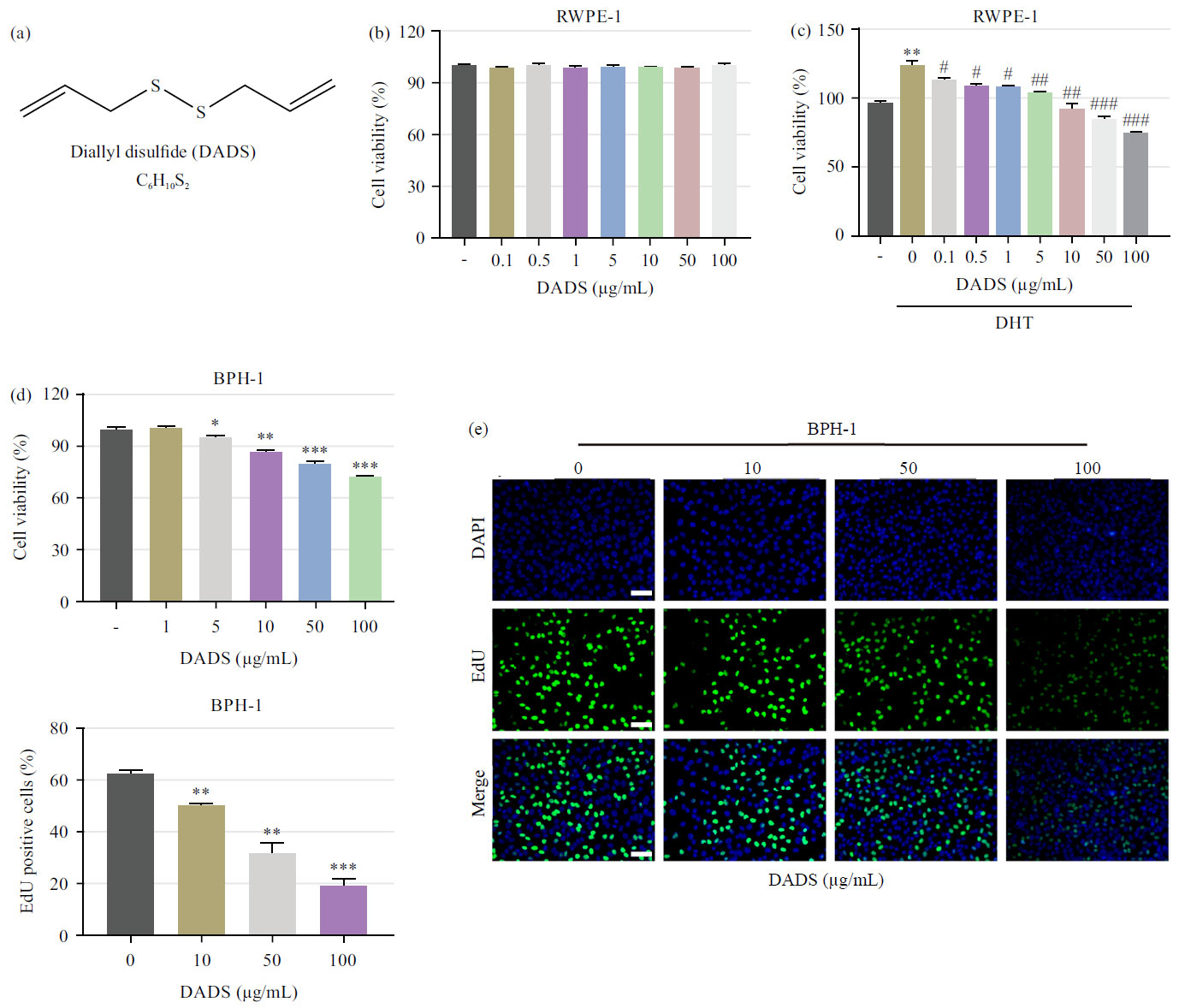 |
| Fig. 1(a-e): | Effect of DADS on the hyperproliferation of DHT-stimulated RWPE-1 and BPH-1 cells, (a) Chemical structure and formulation of DADS, (b) CCK-8 assay detects the cell viability of RWPE-1 cells after treatment with 0, 0.1, 0.5, 1, 5, 10, 50 and 100 μg/mL of DADS, (c) CCK-8 assay detects the cell viability of RWPE-1 cells with or without DHT stimulation after treatment with 0, 0.1, 0.5, 1, 5, 10, 50 and 100 μg/mL of DADS, (d) CCK-8 assay detects the cell viability of BPH-1 cells after treatment with 0, 1, 5, 10, 50 and 100 μg/mL of DADS and (e) EdU staining investigated the cell proliferation of BPH-1 cells after treatment with 0, 10, 50 and 100 μg/mL of DADS *p<0.05, **p<0.01 and ***p<0.001, vs. the 0 μg/mL DADS group and #p<0.05, ##p<0.01 and ###p<0.001, vs. the DHT group |
DADS inhibits the AR signaling pathway: Due to the pivotal role of the AR signaling pathway in BPH development, the effect of DADS on the expression of AR signaling-related proteins was investigated. As expected, the expression levels of AR, PSA, as well as PCNA in BPH-1 cells were significantly suppressed by the treatment of 10, 50 and 100 μg/mL of DADS (Fig. 2a-b).
DADS has little effect on the apoptosis of BPH-1 cells: A previous study indicated that DADS exerts a pro-apoptotic effect to suppress the proliferation of prostate cancer cells, we therefore explored whether DADS induce cell apoptosis and as a result, represses the hyperproliferation. Surprisingly, within the effective concentration that inhibits cell activity (10, 50 and 100 μg/mL), DADS has almost no effect on cell apoptosis. There was no significant change in the Annexin V positive cell ratio and the expression of Bax and Bcl-2 of BPH-1 cells following DADS treatment (Fig. 3a-d).
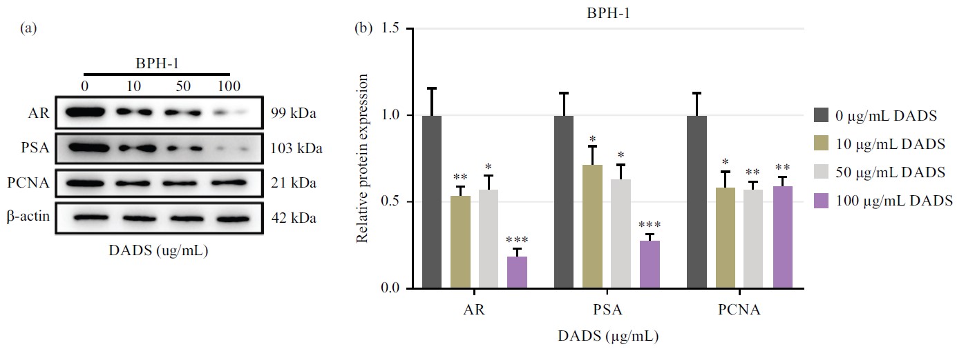 |
| Fig. 2(a-b): | Effect of DADS on the AR signaling pathway, (a) Bands of western blot showed the expression of AR, PSA and PCNA in BPH-1 cells after treatment with 0, 10, 50 and 100 μg/mL of DADS and (b) Quantification of the expression levels of AR, PSA and PCNA in BPH-1 cells after treatment with 0, 10, 50 and 100 μg/mL of DADS *p<0.05, **p<0.01 and ***p<0.001, vs. the 0 μg/mL DADS group |
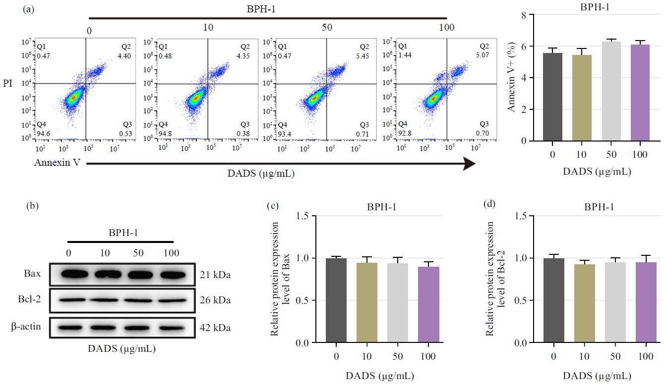 |
| Fig. 3(a-d): | Effect of DADS on cell apoptosis, (a) Flow cytometry combined with annexin V-FITC/PI double staining detected cell apoptosis of BPH-1 cells after treatment with 0, 10, 50 and 100 μg/mL of DADS and (b-d) Western blot detected apoptotic markers (Bax and Bcl-2) in BPH-1 cells after treatment with 0, 10, 50 and 100 μg/mL of DADS |
DADS alleviates the inflammation and oxidative stress of LPS-induced RWPE-1 cells: Next, to explore the potential of DADS on the inflammation and oxidative stress during BPH development, RWPE-1 cells were induced by LPS. It was observed that the secretion of pro-inflammatory cytokines including TNF-α, IL-6 and IL-8 was markedly strengthened following LPS stimulation (Fig. 4a-c).
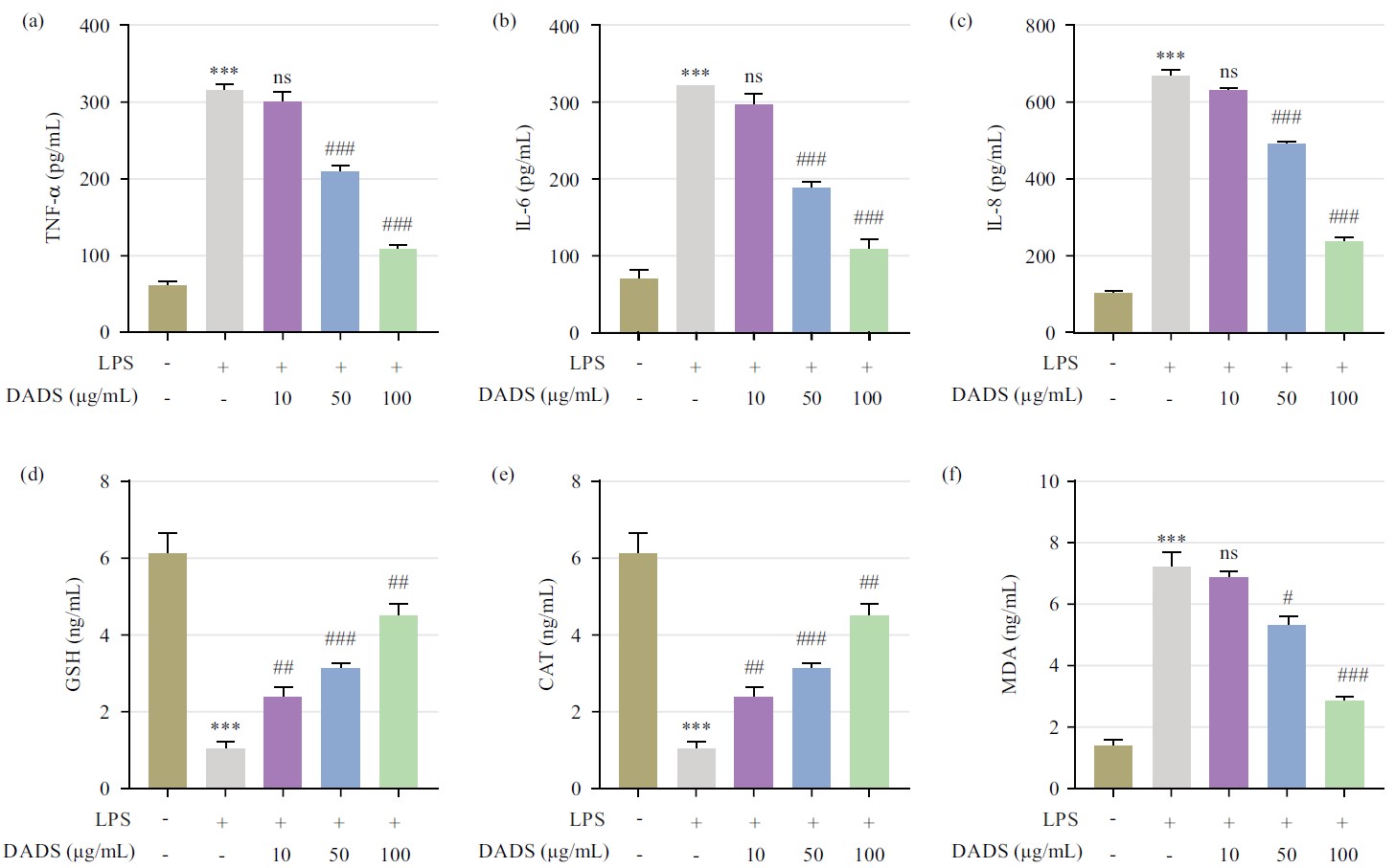 |
| Fig. 4(a-f): | Effect of DADS on LPS-induced inflammation and oxidative stress, (a-c) ELISA kits determined the secretion of TNF-α, IL-6 and IL-8 from RWPE-1 cells with or without LPS stimulation after treatment with 0, 10, 50 and 100 μg/mL of DADS and (d-f) Commercial kits detected the content of GSH, CAT and MDA from RWPE-1 cells with or without LPS stimulation after treatment with 0, 10, 50 and 100 μg/mL of DADS ***p<0.001, vs. the control group, nsp>0.05, #p<0.05, ##p<0.01 and ###p<0.001, vs. the LPS group |
In comparison with the LPS group, there was no significant difference in the secretion of pro-inflammatory cytokines after treatment with 10 μg/mL of DADS, while the secretion of pro-inflammatory cytokines was obviously reduced with the administration of 50 and 100 μg/mL of DADS (Fig. 4a-c). In the meantime, the trend of the secretion of antioxidant defense-related factors was contrary to that of pro-inflammatory cytokines, as revealed by that DADS significantly blocked LPS-induced the decrease of GSH and CAT (Fig. 4d-e). While the trend of the content of MDA was consistent with that of pro-inflammatory cytokines (Fig. 4f). These results suggested that DADS effectively attenuates LPS-induced inflammation and oxidative stress in RWPE-1 cells.
DADS regulates the activation of the Nrf2 signaling pathway: Finally, results found that compared with the control, the expression of Nrf2 in the nucleus was decreased while those in the cytoplasm were increased after LPS induction (Fig. 5a-c). The DADS at 10 μg/mL does not affect the nuclear translocation of Nrf2. After DADS was given at 50 and 100 μg/mL, DADS could significantly reverse the LPS-induced changes in the Nrf2 signaling pathway (Fig. 5a-c).
DISCUSSION
Accumulating evidence has supported multilateral and complementary theories behind the occurrence and progression of BPH. Herein, the study investigated the pharmacological effect of DADS against BPH because of its therapeutic potential with multiple targets for various diseases18. It was widely accepted that DHT functions as a critical regulator of the development of BPH due to its high affinity to AR activation19. Besides, an enlarged prostate caused by the aberrant proliferation of epithelial and stromal cells is the most obvious pathological feature of BPH20. Given these, the current research exploited two major prostate cell models, DHT-stimulated RWPE-1 and BPH-1 cell lines, to mimic the overgrowth of epithelial compartment in the development of BPH. As a result, our data indicated that DADS significantly repressed the hyperproliferation of both DHT-stimulated RWPE-1 and BPH-1 cells, as reflected in the reduced cell viability and EdU-positive cell ratio.
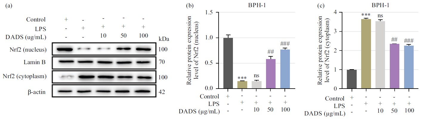 |
| Fig. 5(a-c): | Effect of DADS on the Nrf2 signaling pathway, (a) Bands of western blot showed the expression of Nrf2 in the cytoplasm and nuclear of RWPE-1 cells with or without LPS stimulation after treatment with 0, 10, 50 and 100 μg/mL of DADS and (b-c) Quantification of the expression levels of Nrf2 in the cytoplasm and nuclear of RWPE-1 cells with or without LPS stimulation after treatment with 0, 10, 50 and 100 μg/mL of DADS ***p<0.001, vs. the control group; nsp>0.05, #p<0.05, ##p<0.01 and ###p<0.001, vs. the LPS group |
As the basis of cell growth homeostasis, the balance of cell apoptosis and proliferation is thought to be disrupted during the progression of BPH21. So, the study further explore the pharmacological activity of DADS on the BPH-1 cell apoptosis. However, DADS has no effect on cell apoptosis in BPH-1 cells. Moreover, our research also found that DADS has the capability of modulating the AR signal in BPH-1 cells. These findings suggested that DADS may effectively suppress the overgrowth of epithelial compartments by modulating the AR signal but not inducing apoptosis.
The role of inflammation in the progression of BPH has drawn extensive attention since its degree seems to be related to the severity of urological lower urinary tract symptoms and a higher risk of acute urine retention22. The elevation of pro-inflammatory cytokines, such as TNF-α and ILs (IL-6 and IL-8), triggers tissue remodeling, resulting in the overgrowth of both epithelial and stromal cells23. Particularly, inflammation has recently been reported to influence the AR signaling pathway24. A previous analysis based on BPH specimens from 105 patients revealed a significant correlation between inflammation and AR, showing that BPH tissue samples having inflammatory focus display significantly higher expressions of PSA and AR25. Notably, current study data revealed that the increased pro-inflammatory cytokine contents from cultured supernatants were counteracted by DADS in LPS-induced RWPE-1 cells, a model mimicking BPH in vitro. This suggests that the suppressive role of DADS in inflammation may contribute to its effect on AR signaling in prostatic cells.
Oxidative stress and its resulting DNA damage usually occur in adult males, which might be another contributor to the BPH pathogenesis26. Accumulating evidence reported that oxidative stress in the BPH environment is related to the onset of inflammation27. Moreover, it has been assumed that oxidative stress might trigger compensatory cellular proliferation to aggravate the hyperplastic growth of the prostate 7. Antioxidant defenses such as superoxide dismutase enzyme, GSH and CAT play an essential role in neutralizing oxidative stress to protect cells against injury. However, in the case of BPH, their contents are relatively low. Herein, LPS induced not only the decrease of GSH and CAT but also the increase of MDA concentrations, which reflected an increase in cell damage by oxidative stress, while this condition was effectively reversed by the treatment with DADS.
The Nrf2 signaling pathway, a beneficial cytoprotective mechanism response to environmental deleterious stress activation, has been reported to be responsible for the effect of antioxidants, detoxification and metabolic control of multiple agents. The nuclear translocation of Nrf2 is a critical step in its activation28. Under BPH conditions, Nrf2, a nuclear transcription factor that functions as the primary regulator in redox homeostasis, was expressed low in prostate tissue from BPH patients29. Consistent with this report, current study finding found that the activation of Nrf2 was reduced in RWPE-1 cells after exposing LPS. Under homeostatic conditions, Nrf2 activation is blocked by the Nrf2-Keap1 complex that limits Nrf2 translocation to the nucleus. The nuclear translocation of Nrf2 enhances multiple vital functions, such as antioxidant activity, maintaining redox homeostasis, as well as detoxification, by binding to antioxidant-responsive elements in cells30. A previous study demonstrated that DADS acts as an anti-inflammation and antioxidative agent for emphysema by activating the Nrf2 pathway31. In this investigation, DADS promotes the nuclear accumulation of Nrf2 in LPS-treated RWPE-1 cells, suggesting the notion that Nrf2 activation mediates the protection of DADS in BPH.
CONCLUSION
Overall, current study first emphasized the role of DADS in orchestrating cell proliferation, inflammation and oxidative during the progression of BPH. However, prostatic inflammation is attributed to the multitude of pathogenic factors, LPS alone may not entirely mimic the complexity of the in vivo environment. Hence, it is required to perform in vivo experiments based on the BPH animal model to validate the therapeutic potential of DADS on BPH in future exploration. This study, for the first time, provides evidence of the beneficial effects of DADS on BPH in vitro by alleviating cell proliferation, inflammation and oxidative stress. Mechanistically, the effect of DADS on the development of BPH might be mediated by the activation of Nrf2.
SIGNIFICANCE STATEMENT
The pathogenesis of BPH has been reported to be attributed to multiple complex steps and mechanisms. Hence, novel treatment strategies with multiple targets are required. This study provides evidence of the beneficial effects of DADS on BPH in vitro by attenuating cell proliferation, inflammatory response and oxidative stress. Mechanistically, the effect of DADS on the development of BPH might be mediated by the activation of Nrf2. Our findings suggest DADS may serve as a promising treatment for BPH.
REFERENCES
- GBD 2019 Benign Prostatic Hyperplasia Collaborators, 2022. The global, regional, and national burden of benign prostatic hyperplasia in 204 countries and territories from 2000 to 2019: A systematic analysis for the global burden of disease study 2019. Lancet Healthy Longevity, 3: E754-E776.
CrossRefDirect Link - Chughtai, B., J.C. Forde, D.D.M. Thomas, L. Laor and T. Hossack et al., 2016. Benign prostatic hyperplasia. Nat. Rev. Dis. Primers, Vol. 2.
CrossRefDirect Link - Dai, X., X. Fang, Y. Ma and J. Xianyu, 2016. Benign prostatic hyperplasia and the risk of prostate cancer and bladder cancer: A meta-analysis of observational studies. Medicine, Vol. 95.
CrossRefDirect Link - Russo, G.I., G. Broggi, A. Cocci, P. Capogrosso and M. Falcone et al., 2021. Relationship between dietary patterns with benign prostatic hyperplasia and erectile dysfunction: A collaborative review. Nutrients, Vol. 13.
CrossRefDirect Link - Nicholson, T.M. and W.A. Ricke, 2011. Androgens and estrogens in benign prostatic hyperplasia: Past, present and future. Differentiation, 82: 184-199.
CrossRefDirect Link - Gandaglia, G., A. Briganti, P. Gontero, N. Mondaini and G. Novara et al., 2013. The role of chronic prostatic inflammation in the pathogenesis and progression of benign prostatic hyperplasia (BPH). BJU Int., 112: 432-441.
CrossRefDirect Link - Minciullo, P.L., A. Inferrera, M. Navarra, G. Calapai, C. Magno and S. Gangemi, 2015. Oxidative stress in benign prostatic hyperplasia: A systematic review. Urol. Int., 94: 249-254.
CrossRefDirect Link - Izumi, K., A. Mizokami, W.J. Lin, K.P. Lai and C. Chang, 2013. Androgen receptor roles in the development of benign prostate hyperplasia. Am. J. Pathol., 182: 1942-1949.
CrossRefDirect Link - Aggarwal, S., S. Thareja, A. Verma, T.R. Bhardwaj and M. Kumar, 2010. An overview on 5α-reductase inhibitors. Steroids, 75: 109-153.
CrossRefDirect Link - Salisbury, B.H. and P. Tadi, 2024. 5-Alpha-Reductase Inhibitors. StatPearls Publishing, Treasure Island.
Direct Link - Kang, P.M., Y.J. Kim, W.T. Seo, S.H. Kang and T.S. Kim et al., 2019. Correlation between 5-α reductase type 2 protein expression and methylation of 5-α reductase type 2 promotor gene of benign prostatic hyperplasia. World J. Urol., 37: 709-718.
CrossRefDirect Link - Pérez, P.R., 2022. Post-finasteride syndrome. Literature review. Archivos Españoles Urología, 75: 382-399.
CrossRefDirect Link - Kim, H.J., B.R. Jin and H.J. An, 2023. Hesperidin ameliorates benign prostatic hyperplasia by attenuating cell proliferation, inflammatory response, and epithelial-mesenchymal transition via the TGF-β1/Smad signaling pathway. Biomed. Pharmacother., Vol. 160.
CrossRefDirect Link - Youn, D.H., J. Park, H.L. Kim, Y. Jung and J. Kang et al., 2018. Berberine improves benign prostatic hyperplasia via suppression of 5 alpha reductase and extracellular signal-regulated kinase in vivo and in vitro. Front. Pharmacol., Vol. 9.
CrossRefDirect Link - Jin, B.R. and H.J. An, 2021. Oral administration of berberine represses macrophage activation-associated benign prostatic hyperplasia: A pivotal involvement of the NF-κB. Aging, 13: 20016-20028.
CrossRefDirect Link - Song, X., Z. Yue, L. Nie, P. Zhao, K. Zhu and Q. Wang, 2021. Biological functions of diallyl disulfide, a garlic-derived natural organic sulfur compound. Evidence-Based Complementary Altern. Med., Vol. 2021.
CrossRefDirect Link - Arunkumar, A., M.R. Vijayababu, P. Kanagaraj, K. Balasubramanian, M.M. Aruldhas and J. Arunakaran, 2005. Growth suppressing effect of garlic compound diallyl disulfide on prostate cancer cell line (PC-3) in vitro. Biol. Pharm. Bull., 28: 740-743.
CrossRefDirect Link - He, H., Y. Ma, H. Huang, C. Huang and Z. Chen et al., 2021. A comprehensive understanding about the pharmacological effect of diallyl disulfide other than its anti-carcinogenic activities. Eur. J. Pharmacol., Vol. 893.
CrossRefDirect Link - Carson, C. and R. Rittmaster, 2003. The role of dihydrotestosterone in benign prostatic hyperplasia. Urology, 61: 2-7.
CrossRefDirect Link - Prajapati, A., S. Gupta, B. Mistry and S. Gupta, 2013. Prostate stem cells in the development of benign prostate hyperplasia and prostate cancer: Emerging role and concepts. BioMed Res. Int., Vol. 2013.
CrossRefDirect Link - Liu, J., D. Liu, X. Zhang, Y. Li and X. Fu et al., 2021. NELL2 modulates cell proliferation and apoptosis via ERK pathway in the development of benign prostatic hyperplasia. Clin. Sci., 135: 1591-1608.
CrossRefDirect Link - Liu, Y., Z. Wang, Y. Gan, X. Chen and B. Zhang et al., 2021. Curcumin attenuates prostatic hyperplasia caused by inflammation via up-regulation of bone morphogenetic protein and activin membrane-bound inhibitor. Pharm. Biol., 59: 1024-1033.
CrossRefDirect Link - Raafat, M., A.A. Kamel, A.H. Shehata, A.S.F. Ahmed and A.M.A. Bayoumi et al., 2022. Aescin protects against experimental benign prostatic hyperplasia and preserves prostate histomorphology in rats via suppression of inflammatory cytokines and COX-2. Pharmaceuticals, Vol. 15.
CrossRefDirect Link - Debelec-Butuner, B., C. Alapinar, L. Varisli, B. Erbaykent-Tepedelen, S.M. Hamid, C. Gonen-Korkmaz and K.S. Korkmaz, 2014. Inflammation-mediated abrogation of androgen signaling: An in vitro model of prostate cell inflammation. Mol. Carcinogen., 53: 85-97.
CrossRefDirect Link - Wu, Z.L., Y. Yuan, H. Geng and S.J. Xia, 2012. Influence of immune inflammation on androgen receptor expression in benign prostatic hyperplasia tissue. Asian J. Androl., 14: 316-319.
CrossRefDirect Link - Ahmad, M., N. Suhail, T. Mansoor, N. Banu and S. Ahmad, 2012. Evaluation of oxidative stress and DNA damage in benign prostatic hyperplasia patients and comparison with controls. Indian J. Clin. Biochem., 27: 385-388.
CrossRefDirect Link - Kaya, E., Y. Ozgok, M. Zor, A. Eken and S. Bedir et al., 2017. Oxidative stress parameters in patients with prostate cancer, benign prostatic hyperplasia and asymptomatic inflammatory prostatitis: A prospective controlled study. Adv. Clin. Exp. Med., 26: 1095-1099.
CrossRefDirect Link - Brigelius-Flohé, R. and L. Flohé, 2011. Basic principles and emerging concepts in the redox control of transcription factors. Antioxid. Redox Signaling, 15: 2335-2381.
CrossRefDirect Link - Hao, B., Z. Miao and Yuan, 2016. Clinical significance of Nrf2 expression in benign prostatic hyperplasia and prostate cancer tissues. Int. J. Clin. Exp. Pathol., 9: 118-123.
Direct Link - Dinkova-Kostova, A.T. and A.Y. Abramov, 2015. The emerging role of Nrf2 in mitochondrial function. Free Radical Biol. Med., 88: 179-188.
CrossRefDirect Link - Liu, Y., A. Li, X. Feng, X. Sun, X. Zhu and Z. Zhao, 2018. Pharmacological investigation of the anti-inflammation and anti-oxidation activities of diallyl disulfide in a rat emphysema model induced by cigarette smoke extract. Nutrients, Vol. 10.
CrossRefDirect Link