
E. P. Setyowati
Faculty of Pharmacy, Gadjah Mada University, Sekip Utara, Yogyakarta, Indonesia
U. A. Jenie
Faculty of Pharmacy, Gadjah Mada University, Sekip Utara, Yogyakarta, Indonesia
Sudarsono
Faculty of Pharmacy, Gadjah Mada University, Sekip Utara, Yogyakarta, Indonesia
L.B. S. Kardono
Indonesian Institute of Science (LIPI), Jl. Gatot Subroto 10, Jakarta, Indonesia
R. Rahmat
Indonesian Institute of Science (LIPI), Jl. Gatot Subroto 10, Jakarta, Indonesia
Pakistan Journal of Biological Sciences
Year: 2008 | Volume: 11 | Issue: 22 | Page No.: 2560-2566


ABSTRACT
Identification of cytotoxic constituent of Indonesian sponge Kaliapsis sp. has been conducted. The structure identification was judged based on the spectroscopic data, namely, ultraviolet, MS, one and two-dimensional 1H-NMR and 13C-NMR methods. The cytotoxic constituent was identified as 1-(tetrahydro-4-hydroxy-5-(hydroxymethyl)furan-2-yl)-5-methyl pyrimidine-2,4(1H,3H)-dione. This constituent hasn’t been isolated from sponges as natural product.
PDF Abstract XML References Citation
How to cite this article
E. P. Setyowati, U. A. Jenie, Sudarsono, L.B. S. Kardono and R. Rahmat, 2008. Identification of Cytotoxic Constituent of Indonesian Sponge Kaliapsis sp. (Bowerbank). Pakistan Journal of Biological Sciences, 11: 2560-2566.
DOI: 10.3923/pjbs.2008.2560.2566
URL: https://scialert.net/abstract/?doi=pjbs.2008.2560.2566
DOI: 10.3923/pjbs.2008.2560.2566
URL: https://scialert.net/abstract/?doi=pjbs.2008.2560.2566
INTRODUCTION
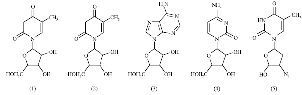
Sponge is a multicellular organism, spineless and porous (Castro and Huber, 2003). Bergmann and Feeney (1951) isolated strong tumor inhibitor nucleoside compounds spongotimidine (1) and spongouridine (2) isolated from Caribean sponge Cryptotethia crypta (Tethylida). Following this finding, sponge become potential source for marine bioactive secondary metabolites. This compounds led the researchers to synthesized the analogs, namely, Ara-A (3, Vidarabine®, Vidarabin Thilo®) and Ara-C (4, Cytarabine, Alexan®, Udicil®), which were found enhancing the antiviral activity (Kijjoa and Sawangwong, 2004; Proksch et al., 2003). Further development of the analogs, the 3’-azido-3’ deoxythymidine (5, AZT, Zidovudin) is currently being used to treat cancer and AIDS (Acquired Immune Deficiency Syndrome) (Müller et al., 2004; García et al., 2007; Newman and Cragg, 2004).
Kaliapsis sp. sponge is Lithistide family (Hooper and Soest, 2002). It is a unique family containing various functional groups of natural products. The sponge genera from this family are famous for their spineless characteristics and for their ability to produce various bioactive metabolites, such as, poliketide, cyclic peptide, alkaloid, pigments and sterols. Sponges in this family are also known for their various cytotoxic constituents. Sponge in the family of Theonellidae the genus of Discodermia contained an anticancer Discodermolide (6). This compound was also an immunosuppressant which has been tested in phase I clinical evaluation I (Newman and Cragg, 2004; Bewley and Faulkner, 1998).
We collected Kaliapsis sp. sponge in the sea around Menjangan island, West Bali, Indonesia. Earlier research showed that the ethanolic extract of this sponge and the isolate were cytotoxic active on Myeloma cell having IC50 0.18 μg mL-1 (Setyowati et al., 2007). Now we are reporting the identity of the isolate (isolate 1).
 |
 |
MATERIALS AND METHODS
Materials: Kaliapsis sp. sponge was collected on 15 October 2004 in the sea around Menjangan island, West Bali National Park, 20 m under sea level.
General instrument: Infrared spectrophotometer (IR) (FTIR 8201 PC Shimadzu, Ultraviolet Spectrometer (UV) (Milton Roy 3000), mass spectrometer EIMS (Electron Impact Mass Spectroscopy) of INCOS 50 (Finigan MT). Spektrometer Resonance Magnetic Nuclear (NMR) 500 MHz (Jeol), operating at radiofrequency of 500 MHz.
Isolation procedure: The bioactivity guided extraction, fractionation and isolation of the active isolate were conducted based on standard procedure as reported previously (Setyowati et al., 2007; Houssen and Jaspars, 2005).
Cytotoxicity evaluation: The cytotoxic evaluation was performed following an established standard procedure (Doyle and Griffiths, 1998).
RESULTS AND DISCUSSION
Isolate 1, transparent colorless needle crystals, MP (uncorrected) 132-134°C. The isolate was soluble in methanol and DMSO but water insoluble. TLC detection on Cerium (IV) sulfate showed brown color after 110°C heating for 10 min.
Isolate 1 showed infra red absorption at v 3500-3200 cm-1 indicating the hydroxide (OH stretching) or amide (NH stretching) functional groups (Silverstein and Webster, 1998; Jenie et al., 2006). The isolate contained aliphatic alcohol as shown on the infra red absorption of aliphatic alcohol v 1200 and 1000 cm-1. The absorption at v 2931.6 and 2839 cm-1 showed the methyl and methylene functional groups. It was supported also by the infra red absorption at v 1477.4, 1434.9 and 1400.2 cm-1. The absorption at v 1662.5 cm-1 was the characteristics of carbonyl (-C = O) functional group of an amide and the absorption of v 1272.9 cm-1 was due to the occurrence of ether linkage –C-O-C-. Therefore, the isolate 1 contained -NH-CO-H, -CH3, CH2, R-OH and –C-O-C functional groups.
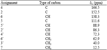
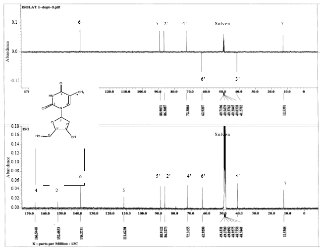
The 1H-NMR spectra showed that the isolate 1 (Fig. 1) showed the chemical shift of methyl (s), at δH 1.8 ppm (3H), two metilen protons at δH 3.8 and 2.4 ppm and one methin proton of secondary alcohol at δH 3.9 ppm.
From the spectra, it was clear the occurrence of proton at δH 11 ppm for amide proton (-CONH).
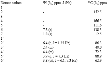
The 1H-1H Cosy spectrum (Fig. 2), showed the proton-proton correlations of isolate 1. This spectrum confirmed the proton chemical shifts (δH) ppm of 7.8 (s), 1.8 (s), 6.4 (t, J = 1.35 Hz), 2.4 (m), 4.4 (m), 3.9 (q, J = 7.3 Hz) and 3.8 (dd, J = 6.1, 7.3 Hz), assigned for protons at C-6, 7, 2’, 3’, 4’, 5’ and 6’, respectively.
 | |
| Fig. 1: | The 1H-NMR spectra of isolate 1 in CD3OD |
 | |
| Fig. 2: | 1H-1H COSY spectra of isolate 1 |
Figure 3, showed the 13C-NMR spectra indicating that isolate 1 had 10 carbon atoms. Two carbons was in a very downfield position, δC 166.5 and 152.5 ppm were assigned as the carbonyl carbons. The methyl peak was showed at δC 12.5 ppm. The APT (Attached Proton Test) showed the number of -CH3, -CH2, -CH and -C (quaternary carbon) of isolate 1 and this isolate consisted of one methyl (-CH3), two methylenes (-CH2) and five methines (-CH) and two quaternary carbons. Table 1 showed the chemical shifts assignment of the 13C signals.
Correlation of protons and carbon signals as showed at Table 2, were confirmed by the HMQC (Heteronuclear Multiple Quantum Coherence) spectrum. Long-range proton-carbon correlation was judged by the HMBC (Heteronuclear Multiple Bond Coherence) spectrum.
| Table 1: | The 13C-NMR assignment of isolate 1 in CD3OD |
 | |
This spectrum showed the correlation of CH3 correlated to CO (C-4), H-6 to (C-2’), OH to C-4’. Figure 4 and Table 2, showed long-range correlation of 1H-13C correlation of isolate 1 resulted from the HMBC spectrum.
 | |
| Fig. 3: | 13C-NMR and APT of isolate 1 in CD3OD |
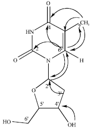 | |
| Fig. 4: | HMBC long-range correlation of isolate 1 |
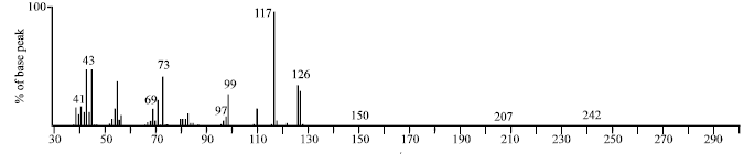
The mass spectrum (EI-MS) (Fig. 5), showed that the isolate 1 had molecular weight of 242 for chemical formula of C10H14O5N2 based on its molecular ion peak of m/z 242.
| Table 2: | HMBC long-range correlation of isolate 1 |
 | |
The EI-MS spectrum of isolate 1 showed the ion fragments at m/z 242 (3%), 207 (3%), 150 (3%), 126 (41%), 117 (100%, base peak), 99 (32%), 97 (11%), 73 (47%), 69 (18%), 43 (49%), 41 (15%). Figure 6 showed the structure of isolate 1 and Fig. 7 showed fragmentation pattern of isolate 1.
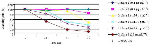
Cytotoxicity evaluation showed that isolate 1 inhibit the growth of HeLa, Raji, Myeloma and T47D cells in vitro. The isolate 1 decreased the percentage of living cells of HeLa (Fig. 8a), Raji (Fig. 8b), Myeloma (Fig. 8c) and T47D (Fig. 8d). It was likely a dose dependent since the more extracts in the cells, the more decreasing of the percentage of living cells.
 | |
| Fig. 5: | EI-MS spectrum of isolate 1 |
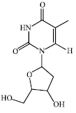 | |
| Fig. 6: | Structure of isolate 1, 1-(tetrahydro-4-hydroxy-5-(hydroxymethyl)furan-2-yl)-5-methylpyrimidine-2,4(1H,3H)-dione |
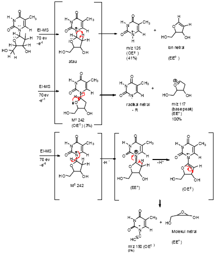 | |
| Fig. 7: | Splitting of fragmentation pattern of isolate 1 isolated from Kaliapsis sp. sponge |
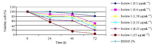 | |
| Fig. 8a: | Percentage of living HeLa cells vs. isolate 1 concentration |
 | |
| Fig. 8b: | Percentage of living Raji cells vs. isolate 1 concentration |
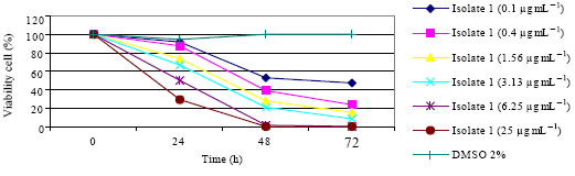 | |
| Fig. 8c: | Percentage of living Myeloma cells vs. isolate 1 concentration |
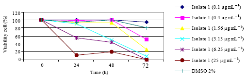 | |
| Fig. 8d: | Percentage of living T47D cells vs. isolate 1 concentration |
It was showed that the cell growth for HeLa, Myeloma and Raji cells, in media control being increased up to 72 h however, for T47D cells being increased up to 48 h, then showed the cell death starting fro 72 h. It was likely that the nutrition in the media was not sufficient for cell growth. The IC50 in vitro cytotoxicity of isolate 1 was 0.18, 7.9, 6.9 and 5.8 μg mL-1 on Myeloma, T47D, HeLa and Raji cells, respectively.
ACKNOWLEDGMENTS
The author (EPS) would like to acknowledge the funding support from Indonesian Ministry of National Education (Hibah Bersaing Grant No. XV/1/2007). Mass spectra and NMR were provided by LIPI Research Center for Chemistry Serpong Indonesia. The staff is acknowledged.
REFERENCES
- Bergmann, W. and R.J. Feeney, 1951. Contributions to the study of marine products. XXXII. The nucleosides of sponges. I. Org. Chem., 16: 981-987.
CrossRefDirect Link - Bewley, C.A. and D.J. Faulkner, 1998. Lithistid sponges: Star performers or hosts to the stars. Angew. Chem. Int., 37: 2162-2178.
Direct Link - Martinez-Garcia, M., M. Diaz-Valdes, A. Ramos-Espla, N. Salvador, P. Lopez, E. Larriba and J. Anton, 2007. Cytotoxicity of the ascidian cystodytes dellechiajei against tumor cells and study of the involvement of associated microbiota in the production of cytotoxic compounds. Mar. Drugs, 5: 52-70.
Direct Link - Kijjoa, A. and P. Sawangwong, 2004. Drugs and cosmetics from the sea. Mar. Drug, 2: 73-82.
CrossRefDirect Link - Muller, W.E.G., R. Batel, H.C. Schroder and I.M. Muller, 2004. Traditional and modern biomedical prospecting: Part I-the history: Sustainable exploitation of biodiversity (sponges and invertebrates) in the Adriatic Sea in Rovinj (Croatia). Evidence-Based Complement. Altern. Med., 1: 71-82.
CrossRefDirect Link - Newman, D.J. and G.M. Cragg, 2004. Marine natural products and related compounds in clinical and advanced preclinical trials. J. Natl. Prod., 67: 1216-1238.
PubMed - Proksch, P., R.A. Edrada and R. Ebel, 2003. Drugs from the sea-opportunities and obstacles. Rev. Mar. Drugs, 1: 5-17.
Direct Link - Setyowati, E.P., U.A. Jenie, Sudarsono, B. Kardono, R. Rahmat and E. Meiyanto, 2007. Isolation of cytotoxic substance from kaliapsis sponge. Indonesian J. Pharm., 18: 183-189.
Direct Link