
F.E. Uboh
Department of Biochemistry, Faculty of Basic Medical Sciences, University of Calabar, Calabar, Nigeria
M.I. Akpanabiatu
Department of Biochemistry, Faculty of Basic Medical Sciences, University of Uyo, Uyo, Nigeria
A.N. Aquaisua
Department of Anatomy, Faculty of Basic Medical Sciences, University of Uyo, Uyo, Nigeria
Eno-obong I. Bassey
Department of Anatomy, Faculty of Basic Medical Sciences, University of Uyo, Uyo, Nigeria
Journal of Pharmacology and Toxicology
Year: 2012 | Volume: 7 | Issue: 2 | Page No.: 78-86


ABSTRACT
Measurement of the levels of total protein, creatinine, uric acid, urea, Blood Urea Nitrogen (BUN), potassium (K+), chloride (Cl-), bicarbonate (HCO3-), sodium (Na+) and calcium (Ca2+) in the blood is useful in assessing the functional integrity of the renal tissues. In this study, the effect of oral exposure to 10, 20 and 30 mg kg-1 b.wt. of nitrocellulose thinner on these serum renal function indices and histopathology of the renal tissues was assessed. Twenty four adult male albino rats (120-150 g), divided into one control and three test groups of six rats each, were used in this study. The rats in the control group were administered with normal saline, while the graded concentrations of the nitrocellulose solvent were each administered orally, as a single daily dosage, for 30 days. The results showed that exposure to nitrocellulose thinner caused a significant (p<0.05) dose-dependent increase in the levels of serum creatinine, uric acid, urea, BUN and K+, as well as decrease in the levels of total serum protein, Cl-, HCO3- and Na+ in rat model. However, there was no significant (p>0.05) difference in serum Ca2+ levels obtained for the rats between and within the test groups, compared to the control. Also, the result of microscopic examinations showed a dose dependent histopathological damage to the renal tissues of rats exposed to nitrocellulose thinner. The observations made from the tissue microscopic analysis, in correlation with that of biochemical assay, indicated the existence of disturbances in the filtration function of the kidneys in rats exposed to higher dosages of the solvent. Hence, the results obtained from our biochemical and histopathogical findings suggested that nitrocellulose thinner possesses the potential (s) of inducing nephrotoxicity in rats.
PDF Abstract XML References Citation
Received: November 05, 2011;
Accepted: January 31, 2012;
Published: March 14, 2012
How to cite this article
F.E. Uboh, M.I. Akpanabiatu, A.N. Aquaisua and Eno-obong I. Bassey, 2012. Oral Exposure to Nitrocellulose Thinner Solvent Induces Nephrotoxicity in Male Albino Wistar Rats. Journal of Pharmacology and Toxicology, 7: 78-86.
DOI: 10.3923/jpt.2012.78.86
URL: https://scialert.net/abstract/?doi=jpt.2012.78.86
DOI: 10.3923/jpt.2012.78.86
URL: https://scialert.net/abstract/?doi=jpt.2012.78.86
INTRODUCTION
Environmental pollution and health hazards are known to be associated with the use of various industrial solvents including hexane, toluene, benzene, among others. Generally, different types of chemical solvents are used by different industries for different purposes. Some of these solvents may have very toxic effects in humans and other species of organisms. Nitocellulose thinner is one of the known solvents with various use in different industries. This solvent is generally used in furniture, paint and automobile spray painting industries. It may also be used in mixtures with other solvents in domestic or industrial products. Nitrocellulose thinner is an important industrial solvent containing different organic chemical substances, including ethylbenzene or toluene and butyl acetate. These chemical substances are known to constitute chemical pollutants in the environments where they are used. Typically, it has been reported that these chemical agents, among others, have been detected in household and workplace air (WHO, 1996, 2005).
The kidney is known to be responsible for the maintenance of the constant extracellular environment through its involvement in the excretion of such catabolites as urea, creatinine, uric acid and regulation of water and electrolyte balance. Abnormal concentration of these catabolites and some electrolytes in the plasma or serum therefore serve as a clear indication of renal function impairment (Gidado et al., 2001; Nwankwo et al., 2006; Haddy et al., 2006; Crook, 2007; Zanna et al., 2008). Impairment of the renal functions may be caused by several diseased conditions and exposure to certain reactive or toxic metabolites, i.e., nephrotoxic substances (Chatterjea and Shinde, 2002; Jimoh and Odutuga, 2004; Crook, 2007). For instance, certain mixtures of hydrocarbon, gasoline vapours, lead, insecticides, pesticides and other chemical solvents have been reported to pose some degrees of adverse effects on the functional integrity of the renal tissues in humans and experimental animals (Wang et al., 2002; Dioka et al., 2004; Boogaard et al., 2005; Adeniran et al., 2006; Hernandez-Serrato et al., 2006; Patil et al., 2007; Uboh et al., 2008, 2009, 2011; Salawu et al., 2009). Also, exposure to lead from automobile exhaust is reported to be a risk factor for nephrotoxicity among traffic policemen (Mortada et al., 2001). Renal function impairment manifests in a variety of different clinical presentations, some of which may be asymptomatic. The renal function impairment with asymptomatic presentations can only be detected by routine laboratory examinations. Azotaemia, a clinical condition associated with renal function impairment, is one of such presentations that can rightly be detected by laboratory findings. The condition is characterized by elevated levels of serum creatinine, urea and BUN (Cotran et al., 1999). A persistently increased serum creatinine is reported to be one of the risk factors for chronic kidney disease, which may results in renal failure (Mortada et al., 2001).
The kidney functions may therefore be assessed from the level of some electrolytes (such as K+ Na+, Cl- and HCO3-), metabolites (such as creatinine, uric acid, urea and blood urea nitrogen) in the plasma and the histological analysis of the ultrastructural status of the renal tissues (Nwankwo et al., 2006; Atangwho et al., 2007; Crook, 2007; Uboh et al., 2008, 2009, 2011). Nephrotoxic response, resulting in renal tissue dysfunction, has been reported to be characterized by the distortions in the architectural integrity of the glomerular tubules. This may be evidenced by lesions on different segments of the nephron, including exacerbation of hyaline droplet formation as the primary change, development of granular casts as a sequelae and finally chronic nephrosis as a consequence of granular cast formation obstructing the nephron. Renal dysfunction of any kind may affect all or parts of the nephron to some extent, impairing either the glomerular or tubular functions. Generally, the net effect of renal dysfunction on the plasma and urine depends on the proportion of the glomeruli to tubules that are affected and on the number of nephrons involved. In this present study, the changes in some serum renal function indices and histopathology of the renal tissue, hence, nephrotoxicity, associated with oral exposure to nitrocellulose thinner were assessed in male albino Wistar rats.
MATERIALS AND METHODS
Chemicals and reagents: Nitrocellulose thinner (POLYGARD®, MISWA CHEMICALS LTD, England), Randox, Dialab diagnostic, CRESCENT diagnostic and AXIOM Gestllschaft for Dignostica and Biochemica mBH reagent kits were used for the biochemical and electrolyte assays. All the other chemicals used in this study were of high grade of purity commercially available.
Animal handling and experimental design: Twenty four mature male albino Wistar rats, weighing between 120 to 150 g were obtained from Biochemistry Department Experimental Research Animal House of the University of Calabar, Calabar, Nigeria. They were fed with a standard laboratory diet and tap water. Illumination was 12 h light/dark cycle and room temperature was 25±2°C. The animals were divided into four groups, [i.e., one control (I) and three experimental groups (II, III and IV)], which consisted of six apparently normal albino Wistar rats per group. The experimental groups II, III and IV were exposed daily to 10, 20 and 30 mg kg-1 body weight, respectively of nitrocellulose thinner by oral administration for 30 days, while the control group was given normal saline. In this study, all the animal experimentations were carried out following the guidelines for the care and use of laboratory animals obtained from the Institutional Animal Ethics Committee. This research work carried out between September and October, 2011.
Collection and handling of blood serum for analyses: Twenty-four hours after last exposure, the animals were anaesthetized with chloroform vapour and dissected. Whole blood from each animal was collected by cardiac puncture into well labelled non-heparinized sample tubes and allowed to clot for 3 h in iced water. The serum was separated from the clots after centrifuging at 10,000 rpm for 5 min into well-labelled plain sample bottles and used for assays. The kidneys were also collected for histopathological examinations.
Biochemical assays
Serum protein: The concentration of total serum protein was assayed spectrophotometrically by Biuret method using Randox reagent kits (Randox Laboratory Ltd, United Kingdom, BT29 4QY).
Serum urea and blood urea nitrogen: The concentration of urea in serum was estimated by the endpoint colorimetric method using Dialab reagent kits (Searcy et al., 1967). In this method, urease enzyme hydrolyses urea to ammonia and carbon dioxide. The ammonia so formed reacts with alkaline hypochloride and sodium salicylate in the presence of sodium nitroprusside to form a coloured chromophore which was measured with DREL 3000 HACH (England) model spectrophotometer.
Serum uric acid level: The concentration of uric acid in the serum of the experimental animal was estimated by enzymatic colorimetric test using CRESCENT diagnostic kit (Saudi Arabia) according to the procedure given in the kit protocol.
Serum creatinine: The concentration of serum creatinine was assayed based on the reaction of creatinine with an alkaline solution of sodium pirate to form a red complex. The red coloured complex which is proportional to the concentration of creatinine in the sample was measured spectrophotometrically.
Serum calcium level: The level of calcium in the serum samples of both the test and control animals was measured using AXIOM Gestllschaft for Dignostica and Biochemica mBH kit (Deutschland).
Serum sodium: Serum sodium concentration was estimated using Mg-Uranylacetate reaction method described in Dialab diagnostic kits (Trinder, 1957). Sodium in serum is precipitated with Mg-Uranylacetate, the remaining uranyl ions form a yellow-brown complex with thioglycolic acid. The difference between reagent blank analyses is proportional to the sodium chloride.
Serum chloride: The level of chloride in serum was determined using mercuric thiocyanate reaction method described in Dialab diagnostic kits (Tietz, 1976). Chloride ions in the sample react with mercuric thiocyanate displacing the thiocyanate ions. The displaced thiocyanate ions react with ferric ions producing a coloured complex.
Serum bicarbonae ion: Serm bicarbonate ion (HCO3-) concentrations were determined as described by Tietz et al. (1999), using Randox reagent kit.
Histopatholoigical examination: For light microscopic examination, liver and kidney tissues from each groups were fixed with 10% buffered formalin, embedded with paraffin. After routine processing, paraffin sections of each tissue were cut into 4 μm thickness and stained with haematoxylin and eosin.
Statistical analysis: All the data obtained for the biochemical assay results were presented as Mean±SEM. These results were analyzed using the Statistical Package for Social Sciences (SPSS for windows, version 17.0). Comparison were made between and within the experimental groups using one-way Analysis of Variance (ANOVA), followed by pair wise comparison between each test and control groups using Student’s t-test. Values of less than 0.05 (i.e., p≤0.05) were regarded as statistically significant.
RESULTS
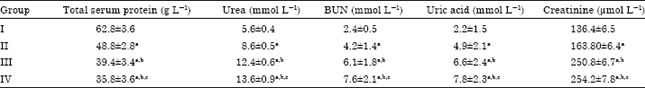
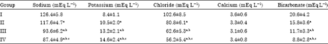
The results of this study on the effect of oral exposure to nitrocellulose thinner on the concentrations of serum indices of renal function test (total serum protein, urea, BUN (Blood Urea Nitrogen), uric acid, creatinine, Na+, Cl-, HCO3-, Ca2+ and K+) in albino Wistar rats are presented in Table 1 and 2. The results showed that exposure to nitrocellulose thinner caused a significant (p<0.05) increase in the concentrations of creatinine, uric acid, urea and BUN and K+, in a dose-dependent pattern, among and within the test groups, compared to the control group (Table 1, 2).
The results of the study also recorded a significant (p<0.05) dose-dependent decrease in the levels of total serum protein, chloride (Cl-), hydrogen carbonate (HCO3-) and sodium (Na+), among and within the test groups, compared to the control group (Table 1, 2). However, it was observed from the results of this study that serum calcium (Ca2+) levels obtained for the rats exposed to nitrocellulose thinner were not significant (p>0.05) different, between and within the test groups, compared to the control (Table 2).
| Table 1: | Effect of oral and inhalation exposure to endosulfan on some renal function test serum indices in rats |
 | |
| Data are presented as Means±SD, n = 6, ap≤0.05 compared with group I, bp≤0.05 compared with group II, cp≤0.05 compared with group III, NcT = Nitrocellulose thinner | |
| Table 2: | Effect of oral and inhalation exposure to endosulfan on some renal function test serum indices in rats |
 | |
| Data are presented as Means±SD, n = 6, ap≤0.05 compared with group I, bp≤0.05 compared with group II, cp≤0.05 compared with group III, NcT = Nitrocellulose thinner | |
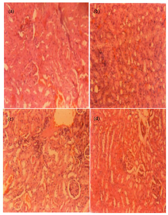 | |
| Fig. 1(a-d): | The photomicrographs of kidney (x160) from rats administered with normal saline, 10, 20 and 30 mg kg-1 nitrocellulose thinner (i.e., a, b, c and d), respectively. (a): Normal architectural structure of the glomerular tubules, (b): Swollen edematous glomerular tubules and (c and d): Swollen edematous, with atrophic and degenerated, glomerular tubules |
The result of microscopic examinations showed a dose dependent histopathological damage to the renal tissues of rats exposed to nitrocellulose thinner, compared to the tissues from the control rats (Fig. 1a-d). The glomerular tubules of rats in the test groups were observed to have developed swollen edematous with atrophies and degeneration, in dose-dependent pattern (Fig. 1b-d). This gave an indication that exposure to nitrocellulose thinner may induce glomerular tubules damage, distorting the functional integrity of the renal tissues. The observations made from the tissue microscopic analysis, in correlation with that of biochemical assay, indicated the existence of disturbances in the filtration function of the kidneys in rats exposed to graded concentrations of the solvent.
DISCUSSION
This study investigated the effect of nitrocellulose thinner on the kidney functions in rat model. In assessing the renal effects of nitrocellulose thinner, the concentration of creatinine, uric acid, urea, blood urea nitrogen, protein, hydrogen carbonate (HCO3-), chloride (Cl-), potassium (K+), sodium (Na+) and calcium (Ca2+) in the serum, as well as the histopathology of the renal tissues were examined. In this study, elevated levels of serum creatinine, urea, BUN, uric acid and K+, as well as decreased levels of serum protein, Na+, Ca2+, HCO3- and Cl- are reported to be associated with oral exposure to nitrocellulose thinner in rats. Also, significant distortions in the architectural integrity of the ultrastructural status of the renal tissues were observed for rats exposed to nitrocellulose thinner. Specifically, the ultrastructure of the cells of renal proximal tubules and vacuoles with damaged external membrane were observed, as well as swollen and pleomorphic mitochondria. The observations made from this study indicated a condition of nephrotoxicity and correlated the previous reports on the nephrotoxicity effects of certain mixtures of hydrocarbon, gasoline vapours, lead, insecticides and pesticides in humans and experimental animals (Mortada et al., 2001; Wang et al., 2002; Tos-Luty et al., 2003; Dioka et al., 2004; Gidlow, 2004; Boogaard et al., 2005; Adeniran et al., 2006; Hernandez-Serrato et al., 2006; Farrag and Shalby, 2007; Patil et al., 2007; Afshar, 2008; Saadi et al., 2008; Uboh et al., 2008, 2009, 2011; Attia and Nasr, 2009; Salawu et al., 2009). The results of this study therefore give a clear indication that nitrocellulose thinner contain some chemical substances with nephrotoxic potentials. The specific chemical constituent (s) and mechanism (s) responsible for the nephrotoxic effect reported in this study to be associated with oral exposure to nitrocellulose thinner is (are) not very clear. However, it may be assumed that the reactive metabolites of the nitrocellulose thinner’s constituents could have interacted with the renal tissues to cause derangements in glomerular functions.
It is hereby suggested that the chemical constituents in nitrocellulose thinner, like other known xenobiotics, might have been metabolically transformed into various metabolites in the body (Hu and Wells, 1994). According to Page and Mehlman (1989) and Nygren et al. (1994), some of these metabolites may be very reactive, interacting in various ways with the metabolizing and excreting tissues (mainly the liver and kidneys) to express their toxicity effects. The interaction of these metabolites with the renal tissues may cause cellular injury, hence, damage to the tissues. Once the renal tissues are damaged, the overall functionality of the kidneys may be compromised. It is recorded in this study that exposure to nitrocellulose thinner caused a significant increase in serum creatinine, uric acid, urea and blood urea nitrogen, decrease in serum protein, Na+, Ca2+, HCO3- and Cl- levels and adverse alterations in the architectural integrity of the renal tissues in rats. These results agree with the research reports for malathion and other pesticides, indicating that exposure to these pesticides induced severe physiological and biochemical disturbances in the renal tissues of the experimental animals (Yousef et al., 2003; Kerem et al., 2007; Attia and Nasr, 2009). Studies from other authors also showed that some insecticides, pesticides and other solvents induced liver and kidney histopathological alterations in experimental animals (Farrag and Shalby, 2007; Afshar, 2008; Saadi et al., 2008; Uboh et al., 2008, 2009, 2011; Attia and Nasr, 2009). Particularly, Tos-Luty et al. (2003) reported that malathion intoxication led to severe effects on the structures of the kidney, covering parenchymatous degeneration of the cells of renal tubules and hyperemia of the cortical part of the kidney, especially of renal glomeruli, as well as infiltrations. An elevation in circulating serum creatinine, uric acid, urea and blood urea nitrogen has been reported to be strongly associated with the development of hypertension and renal disease (Mazzali et al., 2001; Hernandez-Serrato et al., 2006; Patil et al., 2007). All these literature reports correlate the results of our findings in this present study, thereby documenting an hypothetical evidence that exposure to nitrocellulose thinner may be a risk factor for the development of hypertension and renal disorders.
The significant increase in the concentration of creatinine in the serum reported in this study might have resulted from its decreased excretion which, in turn, is related to renal insufficiency. The concentration of creatinine in the blood is known to correlate inversely with the volume of glomerular filtration. Hence, creatinine is considered to be among the useful markers of the filtration function of kidneys, particularly that creatinine is excreted only via the kidneys (Appelton, 1995; Birkner et al., 2000; Grucka-Mamczar et al., 2005). Also, increased serum urea and uric acid concentrations also explain the impaired renal function implicated in rats treated with nitrocellulose thinner. The high serum urea and uric acid levels may result from a decrease in the rate of urea and uric acid secretion into urine, which may likely results from renal insufficiency (Wang et al., 2002; Dioka et al., 2004; Hernandez-Serrato et al., 2006; Patil et al., 2007). According to Appelton (1995), increase in serum urea, uric acid and creatinine concentration is a reflection of impaired renal function. Moreover, decrease in the concentration of serum calcium and protein has been reported to implicated in renal insufficiency, resulting from the tissue’s function impairment (Appelton, 1995; Birkner et al., 2000; Grucka-Mamczar et al., 2005). The hyperkalaemia, hyponatraemia, hypochloridaemia, hyperuricaemia and azotaemia, as reported in our findings with rats treated with nitrocellulose thinner, are indicators of impairment of renal functions.
CONCLUSION
From the results recorded in this present study, it may be concluded that exposure to nitrocellulose thinner induced adverse effects on the renal function in rat model. These observations therefore indicated that exposure to nitrocellulose thinner is a risk factor for renal function impairment and the associated disorders. Hence, a clear indication and recommendation that the use of nitrocellulose thinner in our environments be well regulated by the various environmental protection agencies is documented by the results of this study.
REFERENCES
- Farrag, A.R.H. and S.E.M. Shalby, 2007. Comparative histopathological and histochemical studies on IGR, lufenuron and profenofos insecticide albino rats. J. Applied Sci. Res., 3: 377-386.
Direct Link - Adeniran, O.Y., M.A. Fafunso, O. Adeyemi, A.O. Lawal, A. Ologundudu and A.A. Omonkhua, 2006. Biochemical effects of pesticides on serum and urinological system of rats. J. Applied Sci., 6: 668-672.
CrossRefDirect Link - Afshar, S., A.A. Farshid, R. Heidari and M. Ilkhanipour, 2008. Histopathological changes in the liver and kidney tissues of Wistar albino rat exposed to fenitrothion. Toxicol. Ind. Health, 24: 581-586.
CrossRefDirect Link - Appelton, J., 1995. Changes in the plasma electrolytes and metabolites of the rat following acute exposure to sodium fluoride and strontium chloride. Arch. Oral. Biol., 40: 265-268.
PubMed - Atangwho, J.J., P.E. Ebong, M.U. Eteng, E.U. Eyong and A.U. Obi, 2007. Effect of Vernonia amygdalina del leaf on kidney function of diabetic rats. Int. J. Pharmacol., 3: 143-148.
CrossRefDirect Link - Attia, A.A. and H.M. Nasr, 2009. Dimethoate-induced changes in biochemical parameters of experimental rat serum and its neutralization by black seed (Nigella sativa L.) oil. Slovak J. Anim. Sci., 42: 87-94.
Direct Link - Birkner, E., E.G. Mamczar, Z. Machoy, R. Tarnawski and R. Polaniak, 2003. Disturbance of protein metabolism in rats after acute poisoning with sodium fluoride. Fluoride, 33: 182-186.
Direct Link - Dioka, C.E., O.E. Orisakwe, F.A.A. Adeniyi and S.C. Meludu, 2004. Liver and renal function tests in artisans occupationally exposed to lead in mechanic village in nnewi, nigeria. Int. J. Environ. Res. Public Health, 1: 21-25.
CrossRefDirect Link - Gidado, A., J.Y. Bashirat, G.M. Gana, A.A. Ambi, M.A. Milala and H. Zanna, 2001. Effects of aqueous extract of the seeds of Datura stramonium on some indices of liver and kidney function in rats. Nig. J. Exp. Applied Biol., 2: 123-127.
Direct Link - Grucka-Mamczar, E., E. Birkner, J. Zalejska-Fiolka and Z. Machoy, 2005. Disturbances of kidney function in rats with fluoride-induced hyperglycemia after acute poisoning by sodium fluoride. Fluoride, 38: 48-51.
Direct Link - Haddy, F.J., P.M. Vanhoutte and M. Feletou, 2006. Role of potassium in regulating blood flow and blood pressure. Am. J. Physiol. Regul. Integr. Comp. Physiol., 290: R546-R552.
CrossRef - Hernandez-Serrato, M.I., T.I. Fortoul, R. Rojas-Martinez, L.R. Mendoza-Alvarado and L. Canales-Trevino et al., 2006. Lead blood concentrations and renal function evaluation: Study in an exposed Mexican population. Environ. Res., 100: 227-231.
CrossRefPubMedDirect Link - Hu, Z. and P.G. Wells, 1994. Modulation of benzo-(a)-pyrene bioactivation by glucuronidation in lymphocytes and hepatic microsomes from rats with hereditary deficiency of UDP-glucuronosyl transferase. Toxicol. Applied Pharmacol., 127: 306-313.
PubMed - Jimoh, F.O. and A.A. Odutuga, 2004. Histological changes of selected rat tissues following ingestion of thermally oxidized groundnut oil. Biokemistri, 16: 1-10.
Direct Link - Kerem, M., N. Bedirli, N. Gurbuz, O. Ekinci and A. Bedirli et al., 2007. Effects of acute fenthion toxicity on liver and kidney function and histology in rats. Turk. J. Med. Sci., 37: 281-288.
Direct Link - Mazzali, M., J. Hughes, Y. Kim, A. Jefferson and D. Kang et al., 2001. Elevated uric acid increases blood pressure in the rat by a novel crystal-independent mechanism. Hypertension, 38: 1101-1106.
Direct Link - Mortada, W.I., M.A. Sobh, M.M. El-Defrawy and S.E. Farahat, 2001. Study of lead exposure from automobile exhaust as a risk for nephrotoxicity among traffic policemen. Am. J. Nephrol., 21: 274-279.
Direct Link - Nygren, J., B. Cedewal, S. Erickson, M. Dusinska, and A. Kolman, 1994. Induction of DNA strand breaks by ethylene oxide in human diploid fibroblasts. Environ. Mol. Mutagen., 24: 161-167.
CrossRefDirect Link - Page, N.P. and M. Mehlman, 1989. Health effects of gasoline refueling vapours and measured exposures at service stations. Toxicol. Ind. Health, 5: 869-890.
PubMed - Patil, A.J., V.R. Bhagwat, J.A. Patil, N.N. Dongre, J.G. Ambekar and K.K. Das, 2007. Occupational lead exposure in battery manufacturing workers, silver jewelry workers, and spray painters in Western Maharashtra (India): Effect on liver and kidney function. J. Basic Clin. Physiol. Pharmacol., 18: 87-100.
PubMed - Saadi, L., N. Lebaili and M. Benyoussi, 2008. Exploration of cytotoxic effect of malathion on some rat organs structure. Commun. Agric. Applied Biol. Sci., 73: 875-881.
PubMedDirect Link - Salawu, E.O., A.A. Adeleke, O.O. Oyewo, E.A. Ashamu, O.O. Ishola, A.O. Afolabi and T.A. Adesanya, 2009. Prevention of renal toxicity from lead exposure by oral administration of Lycopersicon esculentum. J. Toxicol. Environ. Health Sci., 1: 22-27.
Direct Link - Tos-Luty, S., D. Obuchowska-Przebirowska, J. Latuszynska, M. Tokarska-Rodak and A. Haratym-Maj, 2003. Dermal and oral toxicity of Malathion in rats. Ann. Agric. Environ. Med., 10: 101-106.
PubMedDirect Link - Uboh, F.E., M.I. Akpanabiatu and Y. Alozie, 2008. Comparative effect of gasoline vapours on renal functions in male and female albino wistar rats. J. Pharmacol. Toxicol., 3: 478-484.
CrossRefDirect Link - Uboh, F.E., M.I. Akpanabiatu, J.I. Ndem, Y. Alozie and P.E. Ebong, 2009. Comparative nephrotoxic effect associated with exposure to diesel and gasoline vapours in rats. J. Toxicol. Environ. Health Sci., 1: 68-74.
Direct Link - Uboh, F.E., E.N. Asuquo, M.U. Eteng and E.O. Akpanyung, 2011. Endosulfan-induces renal toxicity independent of the route of exposure in rats. Am. J. Biochem. Mol. Biol., 1: 359-367.
CrossRefDirect Link - Yousef, M.I., F.M. El-Demerdash, K.I. Kamel and K.S. Al-Salhen, 2003. Changes in some hematological and biochemical indices of rabbits induced by isoflavones and cypermethrin. Toxicology, 189: 223-234.
CrossRefPubMedDirect Link - Zanna, H., S. Adeniji, B.B. Shehu, S. Modu and G.M. Ishaq, 2008. Effects of aqueous suspension of the root of Hyphaene thebaica (L.) mart on some indicators of liver and kidney function in rats. J. Pharmacol. Toxicol., 3: 330-334.
CrossRefDirect Link