
R. Aliyu
Department of Biochemistry, Faculty of Medical Sciences, University of Jos, PMB 2084 Jos, Nigeria
A. Adebayo
Department of Natural Sciences, College of Science and Technology, Covenant University, PMB 1 023, Canaanland OTA, OGl_x state, Nigeria
D. Gatsing
Department of Biochemistry, Faculty of Science, University of Dschang,
P.O. Box 67 Dschang, Cameroon
H. Garba
Industrial Chemistry Programme, School of Science,
Abubakar Tafawa Balewa University, PN �B 0248 Bauchi, Nigeria
Journal of Pharmacology and Toxicology
Year: 2007 | Volume: 2 | Issue: 4 | Page No.: 373-379


ABSTRACT
The effects of Commiphora africana ethanolic leaf extract on some biochemical markers of liver and kidney functions were investigated in rats. The results showed a significant (p<0.05) decrease in serum albumin after 10 days of treatment for the group administered 100 mg kg-1 body weight of the extract. There was no significant (p>0.05) change in alkaline phosphatase activity, while groups administered 100 and 150 mg kg-1 showed significant (p<0.05) increases in serum total bilirubin, after 10 days of treatment. Also, aspartate aminotransferase activity was significantly increased in groups administered 100 mg kg-1 (p<0.01) and 150 mg kg-1 (p<0.05), while alanine aminotransferase showed no significant (p>0.05) increase. The group administered 25 mg kg-1 showed significant (p<0.05) increase in serum creatinine after 24 h of treatment. The results of the liver and kidney histology showed that there was no noticeable damage to the liver tissues of rats administered the extract. However, hydropic degeneration of the cortical-tubular epithelium and glomerulus was seen with the group administered 100 mg kg-1. Similarly, the group treated with 150 mg kg-1 showed acute glomerulonephritis and proliferation of the mesangial cells. These results suggest that C. africana extract may enhance liver function at low doses and may cause adverse effects at high doses.
PDF Abstract XML References Citation
How to cite this article
R. Aliyu, A. Adebayo, D. Gatsing and H. Garba, 2007. The Effects of Ethanolic Leaf Extract of Commiphora africana (Burseraceae) on Rat Liver and Kidney Functions. Journal of Pharmacology and Toxicology, 2: 373-379.
DOI: 10.3923/jpt.2007.373.379
URL: https://scialert.net/abstract/?doi=jpt.2007.373.379
DOI: 10.3923/jpt.2007.373.379
URL: https://scialert.net/abstract/?doi=jpt.2007.373.379
INTRODUCTION
The world consumption of medicinal plants is growing rapidly. The need to verify claims of medicinal properties as well as determine their safety limits cannot be overemphasized. Commiphora africana belongs to the family of Burseraceae (Evans, 1989) and it is found on dry sites and savannah forest of Africa (Irvine, 1961). It is traditionally used for the treatment of a number of ailments including the treatment of typhoid and wound healing (Lewis and Elvin-Lewis, 1977).
The phytochemical constituents as well as the antimicrobial properties of this plant has been reported by Aliyu et al. (2002). Medicinal potentials of other members of this plant’s family have been reported extensively by Tariq et al. (1985). The ethanolic fraction of the leaf extract of this plant has been reported to contain phenolic compounds and tannins, whereas the hexane fraction contained alkaloids, triterpenes and sterols (Aliyu et al., 2002). This study was undertaken to assess the toxicological risk of the consumption of the plant extract as an antimicrobial agent.
MATERIALS AND METHODS
Source of Plant Materials
Commiphora africana was collected from Kwa-Kwa village of Suleija in Niger State (Northern Nigeria) in the month of October and was identified by Mallam Ibrahim Muazam of the Herbarium Department of the National Institute for Pharmaceutical and Research Development (NIPRD), Idu-Abuja, Nigeria, where a voucher specimen (No. 3592) was deposited.
Source of Experimental Animal
Female albino Wistar rats weighing 125-180 g and aged 8-10 weeks were used in the study. They were purchased from the animal house of the Department of Pharmacology, University of Jos. The animals were maintained at room temperature of 25±2°C and fed with standard animal feed, grower’s mash (Grand Cereals LTD) and they had free access to regular tap water.
Treatment of Plant Material
The leaves of C. africana were collected and air dried in the laboratory for a week after which they were pounded in a mortar. One hundred gram of the pounded C. africana leaves were extracted in ethanol solvent using the soxhlet extraction apparatus. The extraction processes were allowed to continue for about 18 h. Thereafter, the extract was concentrated using a rotary evaporator and finally dried in open beaker 6 h duration. Percentage yield obtained was 6.73. This was kept in clean, dried bottle and placed in a dessicator until ready for use.
Treatment of Animals
Acute Toxicity Study
A total of 50 female rats were used for the acute toxicity study. Five Groups of 10 rats each were administered 0, 25, 50, 100 and 150 mg kg-1 body weight of the extract by gastric intubation. After 24 h of treatment, 5 rats per group were anaesthesized under ether and blood collected by cardiac puncture into Serum Separator Tubes (SST). The remaining 5 rats in each group were administered the extracts for another 24 h after which they were anaesthesized under ether and blood samples collected by cardiac puncture into SST bottles for biochemical analysis. Sera were prepared in both cases by centrifuging at 10,000 rpm for 10 min.
Sub-Chronic Toxicity Study
A total of 25 female rats were used for the study. Five groups of 5 rats each were treated with 0, 25, 50, 100 and 150 mg kg-1 body weight of the ethanolic leaf extract for 10 days. The animals were observed for all external symptoms of toxicity and mortality. After this duration of treatment, the rats were anaesthesized under ether and blood collected by cardiac puncture into SST while the liver and kidney were extracted for biochemical and histological examinations, respectively.
Biochemical Studies
The sera were used to estimate Aspartate Aminotransferase (AST), Alanine Aminotransferase (ALT), Alkaline Phosphatase (ALP), total bilirubin, total protein, albumin, urea and creatinine. These parameters were analysed on reflotron (Roche) and spectrophotometer (spectro Sc) by using test combination reagents/strips (Roche Diagnostic and Human Biochemica and Diagnostica GmbH).
Histological Studies
The extracted liver and kidney tissues were fixed in 10% normal saline for 72 h after which the tissues were sliced to a thickness of 2.1 mm each. These were dehydrated using alcohol of graded concentrations. They were further treated with paraffin wax and cast into blocks; sections of the tissues were cut on a microtome to 5 μm. These were later attached to a slide and dried. The samples slides were then stained in haematoxylin and eosin. The slides were then viewed on a photographic microscope (Nikon, Model 240) to detect any damage.
Statistical Analysis
Statistical analyses were performed with the aid of SPSS for Windows software programme (Release 10.0). Group comparisons were done using the analysis of variance (ANOVA) and the Student’s t-test. A p-value of <0.05 was considered statistically significant.
Ethics
This study was conducted with respect for the welfare of animals, as recommended by WHO (1992).
RESULTS
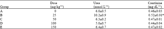
The results of the study as presented in Table 1-6 showed that there was a significant (p<0.05) decrease in ALT activity after 24 h of treatment, in the group administered 150 mg kg-1 of the ethanolic leaf extract of Commiphora Africana. Also, AST activity showed a decrease in the group treated with 150 mg kg-1, but this was not statistically significant (p>0.05) (Table 1). The group treated with 25 mg kg-1 had significantly (p<0.05) increased creatinine after 24 h of treatment. Also, an insignificant (p>0.05) increase was observed in serum urea level at 25 mg kg-1 (Table 2). Similarly, after 48 h of treatment with C. africana, the group treated with 25 mg kg-1 showed significant (p<0.05) decrease in AST activity (Table 3). No significant (p>0.05) changes were observed in urea and creatinine activities after 48 h of treatment (Table 4).
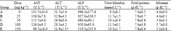
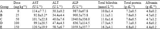
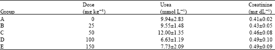
Also, after 10 days of treatment with C. africana ethanolic leaf extract, there was significant increase in AST activity in groups treated with 100 mg kg-1 (p<0.01) and 150 mg kg-1 (p<0.05) (Table 5). ALT activity was not significantly (p>0.05) increased for these two groups after 10 days duration. The results also show a significant (p<0.05) increase in total bilirubin level with groups administered 100 and 150 mg kg-1 and a significant (p<0.05) decrease in albumin level with the group administered 100 mg kg-1 (Table 5). The result showed no significant (p>0.05) changes in ALP activity during the entire duration of treatment. No significant (p>0.05) changes were observed in urea and creatinine activities after 10 days of treatment (Table 6).
The results of the liver and kidney histology showed that there was no noticeable damage to the liver tissues of rats administered the extract. However, hydropic degeneration of the cortical-tubular epithelium and glomerulus was seen with the group administered 100 mg kg-1. Similarly, the group treated with 150 mg kg-1 showed acute glomerulonephritis and proliferation of the mesangial cells.
| Table 1: | Effects of Commiphora africana ethanolic leaf extract some liver functions parameters after 24 h of treatment |
 | |
| Tabulated values are the Mean±SEM of 5 determinations, *p<0.05 vs control | |
| Table 2: | Effects of Commiphora africana leaf extract on some kidney function test after 24 h of treatment |
 | |
| Tabulated values are Mean±SEM of 5 determinations, *p<0.05 vs control | |
| Table 3: | Effects of Commiphora africana leaf extract on some liver function parameters after 48 h of treatment |
 | |
| Tabulated values are Mean±SEM of 5 determinations, *p<0.05 vs control | |
| Table 4: | Effects of Commiphora africana leaf extract on some kidney function parameters after 48 h of treatment |
 | |
| Tabulated values are Mean±SEM of 5 determinations | |
| Table 5: | Effects of Commiphora africana leaf extract on some liver function parameters after 10 days of treatment |
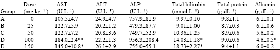 | |
| Tabulated values are Mean±SEM of 5 determinations, *p<0.05 vs control and **p<0.01 vs control | |
| Table 6: | Effects of Commiphora africana leaf extract on some kidney function parameters after 10 days of treatment |
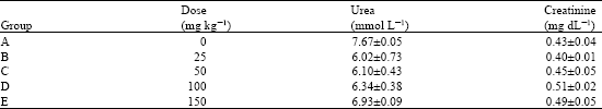 | |
| Tabulated values are Mean±SEM of 5 determinations | |
DISCUSSION
The result showed no significant changes in ALP activity during the entire duration of treatment. Tissue damage is usually associated with the release of enzymes specific to the affected organ or tissue into circulation. The consequence is an increase in activity of such enzymes in body fluids (Garba et al., 2006). ALP is used as the marker of obstructive jaundice and intrahepatic cholestasis (Davern and Scharschmidt, 2002) and it is also a marker of the kidney (Wright et al., 1992), placenta and bones (Mayne, 1999). This may seem to suggest that there were no significant liver and kidney damages.
The transaminases (AST and ALT) are often used as specific markers of active hepatic injury and represent markers of hepatocellular necrosis (Davern and Scharschmidt, 2002). These liver enzymes catalyse the transfer of α-amino groups of aspartate and alanine to the α-keto group of α-ketoglutaric acid (Davern and Scharschmidt, 2002). Whereas ALT activity is primarily localised in the liver and largely specific for parenchymal diseases (Deneke and Riiffersdorf, 1984; Nduka, 1999; Gatsing et al., 2005), AST activity is present in a wide variety of tissues including heart, skeletal muscle, kidney, brain and the liver (Nduka, 1999; Gatsing et al., 2005). The decreased in ALT (significant at 150 mg kg-1) and AST (not significant) activities in serum observed after 24 h of treatment seem to suggest that the leaf extract may not exert adverse effect on the liver within this duration of treatment. The results of this study correlate with the work of Rao et al. (2001) where no significant effect was observed in AST and ALT activity after treating rats with Commiphora molmol for 24, 48 and 72 h. Although, this plant is not the same species of Commiphora, it belongs to the same family.
However, after 10 days of administration, groups administered 100 and 150 mg kg-1 of the ethanolic leaf extract showed significant rise in AST activity as compared to the control group. This may seem to suggest a possible damage to the liver, heart or kidney. AST is a non-specific marker that is present in a number of tissues (Mayne, 1999). The significant increase in the activity of AST of groups treated with 100 and 150 mg kg-1 body weight seems to corroborate the results from kidney histology. Whereas the group treated with 100 mg kg-1 showed hydropic derangement in the cortical tubular epithelium and glomerulus and suggesting glomeruli necrosis, the group treated with 150 mg kg-1 showed among other derangement, glomerulonephrititis. ALT activity was not significantly increased for these two groups after 10 days duration; this may suggest that the extract may not induce hepatocellular necrosis. The significant decrease in serum albumin of rats in group treated with 100 mg kg-1 body weight further explains the injury done to the kidney. Albumin levels are usually reduced in chronic liver disease, congestive heart failure and nephritis (Sclavo, 1987).
Enhancement in serum total protein is an indication of tissue injury while a significant decrease in total protein contents of the liver is a reflection of hepatic toxicity (Gatsing et al., 2005). After 24 and 48 h of treatments, there was no significant change in serum protein as compared with the control group, suggesting that within these doses and duration of treatment, C. africana ethanolic leaf extract may not exert tissue damage, particularly the liver. Similarly, after 10 days of treatment, there was no significant decrease in the serum protein, suggesting that there may be no significant tissue damage.
Bilirubin is derived mainly from the haem moiety of haemoglobin molecules and synthesized in the liver. High level of bilirubin (hyperbilirubinaemia) is commonly found in bilirubin abnormal metabolism, which results in chemical condition called jaundice (Mayne, 1999; Nduka, 1999). Increase in serum bilirubin may arise from excessive haemolysis, cytotoxicity to the liver or from obstruction into the bile ducts. Rats treated with 100 and 150 mg kg-1 showed significant rise in serum bilirubin as compared with the control, which suggests that the extract may induce haemolysis, since the other markers did not show any hepatotoxic effect of this extract.
Creatinine is a waste product derived from creatine and it is excreted by the kidneys. Creatinine values are used as indicators of renal functions; usually increased creatinine levels do not appear unless significant renal impairment exist (Tietz, 1982). Thus, the increased serum creatinine level observed for the treated groups, though statistically significant only at 25 mg kg-1 after 24 h of treatment and not statistically significant at high doses and durations, as compared to the control group, seems to corroborate the noticeable damage to the cortex and glomerulus for the groups treated with high doses after 10 days of treatment. The insignificant increase in creatinine at high doses may possibly be due to some regenerative mechanisms by the kidney in response to the effect of C. africana and studies have equally shown that before any of the kidney markers (creatinine or urea) becomes significantly increased in the blood, about 75% of the nephrons must have been damaged (Boyd, 1983).
CONCLUSION
In the light of the foregoing, it is evident that Commiphora africana enhances the liver and kidney functions at low doses, but at high doses of 100 and 150 mg kg-1 body weight may be potentially toxic to the kidney as seen from histological studies.
Aliyu et al. (2002) recommended the use of the ethanolic leaf extract of Commiphora africana in the treatment of microbial infections. This extract was found to inhibit Salmonella typhi, E. coli, Staphylococcus aureus, Streptococcus, Candida albicans, Bacillus subtilis and P. aeruginosa. The minimum inhibitory concentration was generally 3.125 mg mL-1 (Aliyu et al., 2002), corresponding to a therapeutic dose of 223.214 mg kg-1 body weight, which is greater than the doses already showing nephrotoxicity in the present study (i.e., 100 and 150 mg kg-1). Since the kidney tissues seem to be damaged at doses lower than the therapeutic dose, it therefore suggests that the ethanolic leaf extract of C. africana may not be used to treat those infections with an acceptable margin of safety.
ACKNOWLEDGMENT
The authors are grateful to Miss. H. Umar and Mallam Ibrahim Muazu of the Department of Medicinal Plant Research and Traditional Medicine, National Institute for Pharmaceutical Research and Development (NIPRD) Abuja, for introducing them to this plant. They equally appreciate the kind gesture of the management and staff of Faith Alive Foundation, Jos, for allowing them use their special laboratory.
REFERENCES
- Garba, I.H., D. Gatsing and G. Ubom, 2006. Elevated total and isoenzyme forms of acid phosphatase in falciparum malaria. Comptes Rendus Biol., 329: 75-78.
Direct Link - Wright, P.J., P.D. Leathwood and D.T. Plummer, 1972. Enzymes in rat urine: alkaline phosphatase. Enzymology, 42: 317-327.
PubMed