

Razak Wahab
Universiti Malaysia Sabah, Locked Bag 2073, 88999 Kota Kinabalu, Sabah
Mahmud Sudin
Universiti Malaysia Sabah, Locked Bag 2073, 88999 Kota Kinabalu, Sabah
Hashim W. Samsi
Forest Research Institute Malaysia, 52109 Kepong, Kuala Lumpur
Journal of Applied Sciences
Year: 2005 | Volume: 5 | Issue: 5 | Page No.: 897-902





ABSTRACT
The development of fungal colonization and decay in the culms of a tropical bamboo Gigantochloa scortechinii, through ground contact tests in a tropical soil were described. Observations were made using Scanning Electron Microscopy (SEM). Both untreated and samples treated with either Borax-Boric Acid mixture (BBA) Ammoniacal Copper Quaternary (ACQ) ammonium compound or Copper-Chrome-arsenic (CCA) were investigated. Over 24-months of exposure, untreated and ineffectively treated culms exhibited extensive degradation and colonisation of all tissues (ground tissue parenchyma, fibers and vascular elements) by fungi. Fungal colonists were observed in the cell lumina, the degraded cell walls and in the intercellular spaces. The morphology of decay was a characteristic of degradation by white and soft rot fungi and occasionally, was accompanied by bacterial attack. In contrast, the tissues of culms that had received effective preservative treatments had restricted hyphal colonisation with infrequent hypal invasion into cell via pits; or without cell wall degradation.
PDF Abstract XML References Citation
How to cite this article
Razak Wahab, Mahmud Sudin and Hashim W. Samsi, 2005. Fungal Colonisation and Decay in Tropical Bamboo Species. Journal of Applied Sciences, 5: 897-902.
DOI: 10.3923/jas.2005.897.902
URL: https://scialert.net/abstract/?doi=jas.2005.897.902
DOI: 10.3923/jas.2005.897.902
URL: https://scialert.net/abstract/?doi=jas.2005.897.902
INTRODUCTION
Bamboos have been used in various forms throughout mankind’s history. They are used intensively for a wide range of purposes such as handicrafts, paper, joss-sticks, barbecue sticks, chopsticks, light construction, flooring, paneling and also in traditional social functions such as weddings and religious ceremonies. For a number of years, research work has been carried out in a number of countries in order to enhance the utilisation and range of products that can be manufactured from bamboos[1]. One of the limitations to wider application of bamboo materials in certain environments is its non-durability against insect pests and fungal decay. Whilst a number of traditional methods and treatment approaches derived from wood preservation technology have been applied to bamboos[2-5] there have been only relatively few detailed reports of the fundamental aspects of colonization and decay of bamboo by fungi[6-9].
The main objective of this study was to investigate the breakdown of the fiber and parenchyma cells of the bamboo, G. scortechinii, receiving ACQ, BBA or CCA treatments by dacaying fungi. Scanning electron microscopy (SEM) was used to investigate the growth of bamboo colonising and decay fungi into the tissue structure and the modes of attack on bamboo cell walls in a natural tropical field soil. SEM offers advantage over most other types of microscopy for such studies in that a wide range of magnifications is available, great depth of field and a fully three dimensional image is obtained and specimen preparation is relatively straightforward.
MATERIALS AND METHODS
Specimens: Samples of G. scortechinii of young (2 year-old) and matured (4 years-old) were prepared as small stakes (dimensions 20 mm wide x thickness of culm wall (approximately 10 mm) x 500 mm in length) from within the portion of nodes 6 and 7. Several specimens were then treated using vacuum-pressure, sap-displacement or soaking with a 2 and 4% solution strength of copper-chrome arsenate (CCA), ammoniacal copper quaternary ammonium compound (ACQ) or a borax-boric acid (BBA at 1.54:1 ratio) preservative. The preservative chemicals were allowed to fix for 1 week and then air-dried for a further 3-4 weeks. Untreated and treated stakes were buried to a depth of 400 mm in soil in site cleared of rubberwood plantation some 3 years previously. The site is located in a lowland area. The site is in an agriculture land with good drainage having hot and humid climate throughout the year with an average daily temperature vary from 21 to 32°C and average rainfall of about 2540 mm. The stakes were exposed in soil for 24 months and on removal from soil were placed in Formalin-Acetic acid-Alcohol (FAA) and stored for up to 2 weeks in the laboratory. Some samples were taken from the ground after 4 months exposure for observations and testings. Prior to preparation for SEM the samples were first rinsed with running tap water for 2-3 h before final rinsing with distilled water for 30 min.
Surface preparation: Two methods of surface preparation were used; fresh razor cut and freeze fracture following the method used by Exley et al.[10], Kucera[11] and Sulaiman[7]. The parts of the bamboo stakes for preparation were selected ranging from 10 to 30 cm beneath the ground level.
Fresh razor blade cut: The method was described by Exley et al.[10] and Kucera[11] was used. After hand-cut the bamboo using was razor blade, the sample was firmly held and cut to 0.1 mm transverse surfaces using a sledge microtome. All the micrographs presented use this method of surface preparation except one prepared by the freeze fracture technique.
Freeze fracture: Rinsed samples were placed in liquid nitrogen (approx. –195°C) for about 30 min before placed in between two supports. The back of a scalpel was placed centrally on the sample between the supports and shock load was applied to the scalpel to make the sample breaks. The breakage plane was then used for examination in the SEM. This technique was engaged in an attempt to develop clean transverse surfaces free from knife smear for examination. Specimen size after surface preparation for the SEM observation was approximately 4x4x4 mm
Dehydration: The samples were dehydrated by freeze drying. Samples were quenched in liquid nitrogen at –195°C and then were freeze-dried in Edward Moduyo freeze dryer at –60°C for 16 to 24 h. Samples were then kept dry in a desiccator until mounting. Freeze drying gave useful results producing less collapse of the fungal hyphae than CPD and preserving extracellular mucilaginous material (ECM) surrounding decay microorganisms.
Mounting: The samples were mounted on aluminum SEM stubs with double sided adhesive tape.
Sputter coating: The samples were coated with gold in argon using a Polaron Sputter Coater to give an ultra-fine grain deposit of gold (grain size of 15-10 Angstrom). Satisfactory coating for these samples were normally about 700 Angstrom. The samples were then viewed at appropriate kV (12 to 25 kV) to avoid beam damage of the specimen using a Philips SEM 500 Scanning Electron Microscope.
A wide range of specimens were prepared and examined. Photomicrographs of selected portions were taken and a selection illustrating the characteristic features were observed.
RESULTS AND DISCUSSION
The microscopic features of the G. scortechinii culm in relation to the decay patterns and treatment processes showed various characteristics (Fig. 1 to 12). It was initially anticipated that the freeze fracture technique would allow a clear transverse surfaces of fibers and parenchyma to be examined and reveal unambiguous representations of the decay morphology[8-12]. However, after repeated attempts it was found that careful interpretation is necessary as the technique could produce ‘false’ cavities due to the tensile forces generating from the pull-out of some layers of the cell wall.
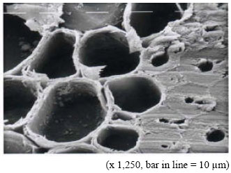 | |
| Fig. 1: | Transverse surface of fibers and ground tissues parenchyma cells in an unexposed 4 year old culm (fresh razor cut, freeze dried) |
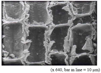 | |
| Fig. 2: | Longitudinal surface of ground tissue parenchyma cells of an unexposed 4 year old culm.. Numerous simple pits are evident (fresh razor cut, freeze dried) |
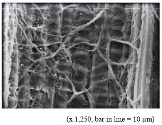 | |
| Fig. 3: | Protoxylem vessel extensively colonized by fungal hyphae and bacteria in a 2 year old culm treated with 2% BBA solution by high pressure sap-displacement and exposed for 24 month in soil, (fresh razor cut, freeze dried) |
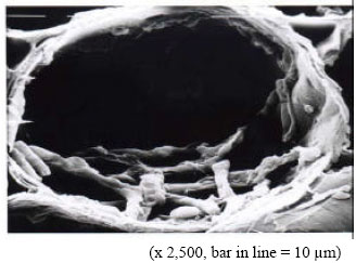 | |
| Fig. 4: | The lumen of ground tissue parenchyma heavily colonized by hypae in a 2 year-old culm treated with 2% ACQ by pressure sap-displacement |
Furthermore, the location of the fracture was highly variable.
Numerous pitting system (Fig. 2 and 5) such as bordered and simple pits which play an important role in absorbing and diffusing of preservatives solution from the vessels to the surrounding fibers and ground tissues parenchyma were clearly seen in the vessel and parenchyma cell walls of unexposed materials. These pitting systems also provide a pathway for the passage of hyphae from cell to cell (Fig. 5 and 12). Hyphal development was extensive in the protoxylem vessels after the prolonged exposure to soil, the hyphae appearing to be appressed to the sides of these vessels intercellular spaces in the ground tissue parenchyma were also seen (Fig. 6).
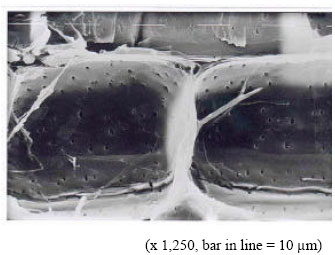 | |
| Fig. 5: | Fungal hypha colonising intercellular space in ground tissue parenchyma of a 4 year old culm treated with 4% BBA solution by high pressure sap-displacement and exposed in soil for 24 months, (fresh razor cut, freeze dried, bar = 10 μm) |
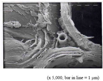 | |
| Fig. 6: | Fungal hyphae growing in intercellular spaces/middle lamella between adjacent parenchyma cells of a 4 year old culm treated with 4% BBA solution by high pressure sap-displacement-exposed in soil for 24 months. The adjacent cell walls of the two cells seem to separated and degraded to some extent (fresh razor blade cut, freeze drying, bar = 10 μm) |
Observation of the untreated and inadequately treated samples (mostly samples treated with BBA) showed extensive colonisation by bacteria and fungi and cases of extensive cell wall degradation by fungi (Fig. 3).
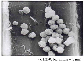 | |
| Fig. 7: | Granules (crystals) of boron preservative on a fibre cell wall surface in A 2 year old culm treated with a 4% BBA solution by vacuum pressure–exposed in soil for 24 months. The boron granules or deposits were left crystalized after the treatment process. Bore holes (arrow) indicate attack by the hyphae of white rot fungi. Possible ECM also observed on the cell surfaces. (fresh razor cut, freeze dried) |
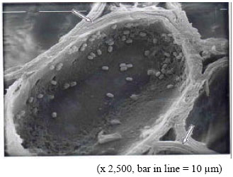 | |
| Fig. 8: | Transverse view of the ground tissue parenchyma of a 4 year old culm treated with 2% BBA solution by vacuum-pressure-exposed for 4 month in soil. Extensively colonized by bacteria, extracellular mucilaginous material (ECM) evident on lumen surface. Note also the multi-layered parenchyma cell wall (see arrow), (fresh razor cut, freeze dried) |
A characteristic feature of both groups of microorganisms was the presence of ECM in appreciable quantities which was well preserved in the freeze drying preparation (Fig. 8, 9 and 11).
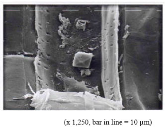 | |
| Fig. 9: | Rhomboid crystal, probably calcium oxalate, in lumen of parenchyma cell in longitudinal section form a 4 year old culm treated with 4% ACQ solution by soaking and exposed for 24 months in soil. ECM and possible hyphal debris also evident in the same cell (fresh razor cut, freeze dried) |
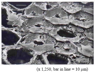 | |
| Fig. 10: | Transverse view of fibers and parenchyma cells of a 2 year old culm treated with 2% CCA solution by vacuum-pressure and exposed in soil for 24 months. The cell walls of some of the fibers show indications of soft rot decay cavities (arrows) (freeze fractured, freeze dried) |
Most of the tissues particularly the parenchyma cells in the untreated control samples, were attacked by these organisms. In all cases, the fungi grew principally on the cell lumen inner surface, progressed into the cell wall secondary layers leading to extensive cell wall breakdown and the separation of the fibre and parenchyma cells (Fig. 6). In Fig. 7 the heavy degradation of the parenchyma cells appears to result from both fungal and bacterial activities.
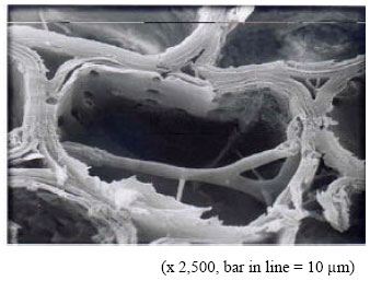 | |
| Fig. 11: | A longitudinal view at a boundary between two parenchyma cell of a 2 year old culm treated with 2% CCA solution by soaking and exposed in soil for 24 months. Several fungi hypae are shown (fresh razor blade cut, freeze drying). |
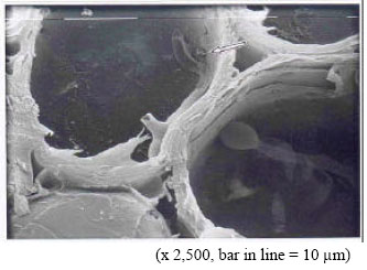 | |
| Fig. 12: | Hyphae in adjacent parenchyma cells of 2 year old culm treated with 2% CCA solution by sap-displacement and exposed in soil for 24 months. Some ECM evident in the lumen surfaces by a pit (arrow), (fresh razor cut, freeze dried) |
Features of the cell wall degradation and hyphae/hyphal remains were suggestive of white rot attack (Fig. 7 and 9) where bore holes and rhomboidal calcium oxalate crystals were observed[13]. White rot appeared to be associated with ineffective preservative treatments (either the wrong preservative chemical were used such as the case of BBA which were known to be leachable when exposed to the environment) or the application method (soaking in the case of ACQ) for soil exposure. In the case of the presence of oxalate crystals and ECM it should be noted that no decay was observed and it is possible that there was limited hyphal colonisation without decay occurring. Indications of soft rot attack were obtained in several samples, although the freeze fracture technique did not generally yield surfaces with completely unambiguous characteristics and the fresh razor cut specimens exhibited smear on transverse surfaces, especially where decay was heavy. The example given in Fig. 10 is typical of the appearance of freeze fracture surfaces. Treatments which gave a good decay resistance (such as those bamboo samples treated with ACQ and CCA at 2 and 4% concentrations through either soaking and vacuum treatments) over the 24 month period of soil exposure did show some fungal colonisation (Fig. 4 and 12) but this was commonly restricted to sparse growth in cell lumena and occasional passage from cell to cell via pits as opposed to cell wall degradation.
In general, the SEM observations made on the colonisation of G. scortechinii in the tropical soil exposure has indicated an extensive hyphal and bacterial growth in all tissues of the bamboo in untreated or ineffectively treated material. Colonising hyphae were able to develop in parenchyma, fibre and vascular cell lumena and to grow through pits and within intercellular spaces in the parenchyma. Parenchyma and fibre cells were clearly subject to widespread degradation. Conversely, distinctly limited growth of hyphae and very few bacteria were seen in G. scortechinii which had received effective treatments such as in the case of samples treated with ACQ and CCA. The decay morphologies recognised in the susceptible material were characteristic of white and soft rot fungi with evidence also for bacterial degradation. In view of the difficulties noted in the preparation of transverse surfaces for observation of soft rot decay morphologies via SEM it is clearly desirable that further studies are conducted and additional microscopic techniques employed.
REFERENCES
- Ashaari, Z. and N. Mamat, 2000. Traditional treatment of malaysian bamboos: Resistance towards white rot fungus and durability in service. Pak. J. Biol. Sci., 3: 1453-1458.
CrossRefDirect Link