
Md. Mesbah Uddin Talukder
Department of Pharmacy, The University of Asia Pacific, Dhaka-1209, Bangladesh
Tasnuva Haque
Department of Pharmacy, Stamford University Bangladesh, 51 Siddeswari Road, Dhaka-1217, Bangladesh
Deepannita Barua
Department of Pharmacy, The University of Asia Pacific, Dhaka-1209, Bangladesh
Singapore Journal of Chemical Biology
Year: 2012 | Volume: 1 | Issue: 1 | Page No.: 1-12


ABSTRACT
A new emulsion-gelation method was used to prepare oil (soyabean and mustard oil) entrapped diclofenac sodium beads using sodium alginate as the polymer. The prepared beads were evaluated by their morphological, physical and release parameters. From the result, it was seen that in 30% soyabean oil containing beads oil droplets were distributed in uniform size in the beads than the mustard oil. Most of the beads of F-1, F-3 and F-7 were broken down in pH 7.4 phosphate buffer rather than in 0.1 N HCl. Among the seven formulations, 30% soyabean oil and mustard oil containing beads were present in floating condition up to 24 h in pH 7.4 phosphate buffer, 0.1 N HCl and 0.9% NaCl media. Beads were found to be fragmented in buffer from the beginning but not in 0.9% NaCl medium. The mean diameters of the beads were increased with the increased concentration of oils. With the incorporation of oil, rate retarding actions of the beads were increased. The release pattern of all formulation were fitted to zero order model and the release exponent showed the release mechanism was non-fickian or anomalous. Finally, it can be concluded that highest oil containing beads showed most rate retarding activities and mustard oil was most sustaining than soyabean oil.
PDF Abstract XML References Citation
How to cite this article
Md. Mesbah Uddin Talukder, Tasnuva Haque and Deepannita Barua, 2012. Development and Evaluation of Ionotropically Emulsion Gelled Sodium Alginate Beads and its Morphological Characterization by Optical Micrographs. Singapore Journal of Chemical Biology, 1: 1-12.
URL: https://scialert.net/abstract/?doi=sjchbio.2012.1.12
URL: https://scialert.net/abstract/?doi=sjchbio.2012.1.12
INTRODUCTION
An emulsification/internal gelation method is proposed for producing small diameter alginate beads in large quantity. The difficulty in using dispersion/external gelation techniques with ionic polysaccharide is that the calcium source (CaCl2) is insoluble in the oil phase. As an alternative, internal gelation of the dispersed alginate droplets may be initiated by releasing Ca2+ from an insoluble complex (calcium salt) through pH reduction (Lencki et al., 1989). The use of hydrogel systems for controlling the release of drugs has increasingly become important in the formulation of pharmaceuticals. It is well known that hydrogel can respond to surrounding conditions such as pH, ionic strength, temperature and electric current. The pH-sensitivity of hydrogels an important factor in designing polymers for controlled drug release in the gastrointestinal tract, which has a variation of pH from the stomach to the intestine. Hydrogels from natural polymers, especially polysaccharides have been widely used because of their advantageous properties such as non-toxicity, biocompatibility and biodegradability (Peppas, 1997).
Sodium diclofenac (DS, C14H10Cl2NO2Na) is a widely used Non-Steroidal Anti-Inflammatory Drug (NSAID) that exhibits antirheumatic, analgesic, osteoarthritis and antipyretic activities. It has a short half-life in plasma (1-2 h). The daily dose varies between 75 and 200 mg person-1, given in 3 or 4 divided portions depending on the route of administration. The most common adverse effects of the drug are gastritis, peptic ulceration and depression of renal functions (Tapia et al., 2004; Gillman et al., 1990). Because of the short biological half-life and associated adverse effects, it is considered an ideal model drug for controlled drug delivery.
Alginate is a naturally occurring biopolymer that is finding increasing applications in the biotechnology industry. Alginate has been used successfully for many years in the food and beverage industry as a thickening agent, a gelling agent and a colloidal stabilizer. Alginate also has several unique properties that have enabled it to be used as a matrix for the entrapment and/or delivery of a variety of proteins and cells. These properties include: (1) a relatively inert aqueous environment within the matrix; (2) a mild room temperature encapsulation process free of organic solvents; (3) a high gel porosity which allows for high diffusion rates of macromolecules; (4) the ability to control this porosity with simple coating procedures and (5) dissolution and biodegradation of the system under normal physiological conditions. They are widely used in food and pharmaceutical industries, such as disintegrate and tablet binder, thickening stabilizing agents in mixtures and as gelling agents inconfectionary. Recently, they have been employed as a matrix for entrapment of drugs (Bodmeier and Paeratakul, 1989), macromolecules and biological cells (Lanza et al., 1995).
The purpose of the present study was to prepare beads by emulsion-gelation technique using two different oils (soyabean and mustard oil) with a fixed amount of sodium alginate and to evaluate the beads in respect of morphology, physical and release parameters.
MATERIALS AND METHODS
Materials: DS was a gift sample from Eskayed Bangladesh Ltd. Sodium alginate and CaCl2 were purchased from Loba Chemie, India and Merck Spacialities Private Ltd., India. Soyabean oil was from Teer brand of Deepa Food Products Ltd. And mustard oil was from Radhuni brand of Square Consumers Ltd. Both of them were purchased from local market of Bangladesh. Among the reagents, sodium dihydrogen orthophosphate, disodium hydrogen orthophosphate anhydrous were from Qualikems Fine Chemicals Pvt. Ltd Qualikems Fine Chemicals Pvt. Ltd., India. Hydrochloric acid and sodium chloride (NaCl) were obtained Merck, Germany. The project was conducted from 01.06.2009 to 31.03.2010 in the pharmacy laboratory of The University of Asia Pacific and Stamford University Bangladesh.
Preparation of calcium chloride (CaCl2) solution: At first, 5 g of CaCl2 salt was weighted with the help of an electronic balance (Shimadzu, Japan) and it was taken in a 100 mL volumetric flask. Water was added to dissolve it and the volume was made up to 100 mL. Thus, 5% CaCl2 solution was prepared.
Preparation of gel beads (Emulsion-gelation method): Firstly, according to Table 1, the desired amount of sodium alginate was weighted in an electronic balance and was taken in a beaker. Then, sodium alginate solution was prepared by adding distilled water with constant heating. Sodium alginate was mixed properly with water by continuous stirring (Hwasin Technology Co., Korea).
| Table 1: | Formulation of Sodium alginate based Diclofenac sodium beads by using different concentrations of oil where the ratio of drug: polymer is 1:2 |
 | |
 | |
| Fig. 1: | (a) Beads of F-1(containing no oil), (b) beads of F-4 (Containing 30% soybean oil) and (c) beads of F-7 (Containing 30% mustard oil) |
When the sodium alginate solution was completely prepared, then DS and soybean oil (for F-2 to F-4) or mustard oil (for F-5 to F-7) was added immediately. Then these were dissolved and mixed properly by using glass rod or stirrer. When all the ingredients were mixed completely a gel was formed. Then the gel was taken in a syringe with nozzle of 2.5 mm outer diameter and extruded into a beaker filled with chilled CaCl2 solution in a drop wise fashion with gentle agitation at room temperature. The distance from the nozzle to the CaCl2 solution was 5 cm. When gel was poured into the CaCl2 solution from syringe as droplet, droplets become spherical as soon as it reaches to CaCl2 solution; it became hard since the temperature was below the gelling point of sodium alginate. Thus sodium alginate beads were prepared, where DS was incorporated with soybean oil and mustard oil of different concentrations. The beads were allowed to stand in the solution not more than 10 min. Then beads were collected removing the CaCl2 solution by filtration followed by rinsing with the chilled water. The washed beads were then placed in a Petri-dish and placed in the open space for air-dry for over-night (using fan, if required). Finally, the oil entrapped sodium alginate beads were prepared by emulsion gelation method (Fig. 1a-c) (Choudhury and Kar, 2005).
Evaluation of physical parameters of oil entrapped sodium alginate based DS beads
Swelling study: The swelling behavior of the oil entrapped sodium alginate beads were studied in two dissolution media, that is, 0.1 N HCl (pH 1.06) and phosphate buffer (pH 7.4). Beads of different formula were immersed in both 0.1 N HCl (pH 1.06) and phosphate buffer (pH 7.4) media. Before immersing, the initial diameter of the beads were measured using digital Vernier Caliper (SDK, China). At the end of one hour, the diameter of the swollen beads was determined. Similarly, the diameters of the swollen beads were determined at 2, 3, 4, 6, 7 and 8 h, respectively. The swelling behavior of the beads of the different formula was determined by measuring the difference between the initial diameter of the beads and final diameter of the beads at time t. The swelling index for each formula determined at time t was calculated using the following equation:
| (1) |
where, Dt is the diameter of the beads at time t and D0 is the initial diameter of the dried beads (Piyakulawat et al., 2007).
The buoyancy test of the beads: The buoyancy test of the oil entrapped sodium alginate-DS beads was determined in three different media, such as; 0.1 N HCl (pH 1.06), phosphate buffer (pH 7.4) and 0.9% NaCl.
Ten beads of each of the seven formulations were immersed in the 25 mL beaker containing four different media. The floating ability or buoyancy behavior of the beads was measured by visual observation for 24 h: 0 min, every 10 min for the first 30 min, every 15 min for the next 1 h, 1 h 30 min and then every 30 min till 10 h and kept it stand for overnight (24 h). The preparation was considered to have buoyancy in the test solution only when all of the beads floated (Elmowafy et al., 2009).
Determination of fragmentation index: The fragment index of the oil entrapped sodium alginate-DS beads was determined in two different media (0.9% NaCl and pH 7.4 phosphate buffer). Seven beads of each of the seven formulations were immersed in the 25 mL beaker containing the media. The fragmentation of the beads was measured by visual observation for 24 h: 0 min, every 10 min for the first 30 min, every 15 min for the next 1 h, 1 h 30 min and then every 30 min till 10 h and stands it for overnight (24 h) (Choudhury and Kar, 2005). The beads were considered to have fragmentation when all the beads were fragmented or divided into two parts.
Mean diameter: The mean diameter of the oil entrapped DS loaded sodium alginate beads were measured by digital vernier caliper. The diameter of fourteen beads was determined and then calculates their mean diameter by using the following equation:
| (2) |
where, n is the total number of beads which was used for determining the mean diameter and n=14 (Piyakulawat et al., 2007).
In vitro dissolution study: The in vitro dissolution study was conducted in two different stages: acid stage and buffer stage. In acid stage, the medium was 0.1 N HCl (pH 1.06). It should be noted that the release of the drug was very low in the acidic medium. Because the drug (DS) itself is an acidic drug. That’s why, the buffer stage was considered during the dissolution test for the treating the data in various pharmacokinetic models.
In the buffer stage, the dissolution profile of the beads was determined using USP (XXIII) dissolution apparatus-I (rotating basket method) (Veego, India) taking 900 mL of phosphate buffer pH 7.4 for 8 h. The dissolution media was maintained at a temperature of 37±1°C. The speed of rotation of basket maintained was 50 rpm. To prepare pH 7.4 phosphate buffer, 2.96 g of sodium dihydrogen orthophosphate (NaH2PO4.2H2O) and 11.5 g of di-sodium hydrogen-o-phosphate anhydrous (Na2HPO4) were dissolved in distilled water to obtain 1000 mL of solution. Then the pH of the solution was checked using pH meter (Hanna Instrument, Portugal. In the seven baskets, 100 mg DS containing equivalent beads of each formula was taken. The samples were withdrawn from dissolution vessel after each hour interval. The samples were filtered and if necessary diluted. The absorbance of the samples was determined by UV-visible spectrophotometer (Shimadzu, Japan) at the wavelength of maximum absorbance at 276 nm. Corresponding dissolution medium was replaced into the vessel after each interval (Manjunatha et al., 2007).
Release kinetics: The suitability of several equations that are reported in the literature to identify the mechanisms for the release of DS from beads with respect to the release data. The data were evaluated according to the following equations:
Zero-order model (Donbrow and Samuelov, 1980):
| (3) |
Higuchi model (Higuchi, 1961, 1963):
| (4) |
Korsmeyer-Peppas model (Korsmeyer et al., 1983; Peppas, 1985):
| (5) |
where, Mt is the amount of drug dissolved in time t, M0 is the initial amount of drug, K0 is the zero-order release constant, KH is the Higuchi rate constant, K is a release constant and n is the release exponent that characterizes the mechanism of drug release.
First order model (Merchant et al., 2006):
| (6) |
where, C is the cumulative percent of drug release, Co is the initial concentration of drug and k is the first order rate constant.
The magnitude of the exponent n indicates that the release mechanism is Fickian diffusion, case II transport or anomalous transport. In the present study (spherical shape) the limits considered were n </= 0.43 (indicates a classical Fickian diffusion-controlled drug release) and n = 0.85 (indicates a case II relaxational release transport: polymer relaxation controls drug delivery). Values of n between >0.45 and <0.89 can be regarded as indicators of both phenomena (transport corresponding to coupled drug diffusion in the hydrated matrix and polymer relaxation), commonly called anomalous non-Fickian transport. Values of n greater than 0.85 indicates super case II transport, in which a pronounced acceleration in solute release by a film occurs toward the latter stages of release experiments, resulting in a more rapid relaxation-controlled transport (Korsmeyer et al., 1983; Peppas, 1985).
Due to the differences in drug release kinetics, the constant k, though one of the measures of release rate, should not be used for comparison. Therefore, to characterize the drug release rates in different experimental conditions, Mean Dissolution Time (MDT) was calculated from dissolution according to Mockel and Lippold (1993) using the following equation:
| (7) |
where, n is the release exponent and K is the kinetic constant calculated from Eq. 5.
The similarity factor was used to compare the difference of dissolution profiles of the test matrix tablets is given below:
| (8) |
where, Rt and Tt are the percentage of drug dissolved at each time point for the test and reference products, respectively and n is the number of dissolution samples taken The US Food and Drug Administration and the European Agency for the Evaluation of Medicinal Products have suggested that 2 dissolution profiles can be considered similar if f2 is between 50 and 100 (CDER, 1997; EMEA, 2006).
Statistics: To compare the means of all release data and to assess statistical significance between them, one way repeated measures Analysis of Variance (ANOVA) performed at the 5% significance level, using SPSS software Version 16.00.
RESULTS AND DISCUSSION
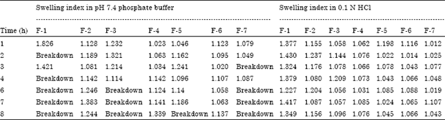
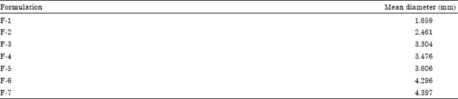
The potical micrograph of 30% soyabean oil and mustard oil containing beads were shown in Fig. 2a and b. From Table 2, it was seen that beads of F-1, F-3 and F-7 showed highest break down whereas F-2, F-4 and F-6 showed no breakdown in pH 7.4 phosphate buffer. The beads of F-4 were highest swelled (1.339) than F-2 and F-6. No breakdown observed in case of swelling study of beads in 0.1 N HCl. From Table 3, it was observed that no fragmentation index was found for the beads in 0.9%NaCl but it was very high in case of pH 7.4 Phosphate buffer. In buffer media, F-7 showed highest fragmentation index of 100, which was followed by F-1 and F-5 (90). Table 4 indicates mean diameter of 14 beads. F-1 had the diameter of 1.659 mm. In case of soyabean oil and mustard oil containing formulations, F-4 had the highest diameter of 3.394 mm and F-7 had that of 4.397 mm. The beads of F-1 released highest amount of drug (87.19%) at 8 h. Highest oil containing formulations showed less release of drug. That is, F-4 released 51.11% and F-7 released 41.12% at 8 h.
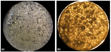 | |
| Fig. 2: | Optical microscopic photographs of oil droplet of emulsion gelled beads prepared using (a) 30% soybean oil and (b) 30% mustard oil |
| Table 2: | Swelling index of beads in pH 7.4 phosphate buffer and 0.1 N HCl media |
 | |
Optical microscopic photographs of oil entrapped emulsion gelled beads: The Optical microscopic photographs using optical microscope (Zeiss, USA) of oil droplets of emulsion gelled beads with different concentrations of oil are shown in Fig. 2a and b. In the above figures, it has been shown that oils were perfectly and carefully incorporated into emulsion gelled beads. That’s why, with the help of optical microscopic photographs, the dispersion of oil droplets of three different formulations were clearly visible. The process was done by cross section of oils so that the droplets were shown clearly. Thirty percent soybean oil was incorporated into F-4 and the oils were uniformly distributed into the formulation and the droplets were apparently found to be almost uniform in size as shown in Fig. 2a. On the other hand, 30% mustard oil was incorporated into F-7 and the oils were uniformly distributed in to the formulation but the droplets were not uniform in size (Fig. 1b) (Zhou et al., 2007).
Swelling study of oil entrapped DS beads: The swelling behavior study of the oil entrapped DS beads was performed in pH 7.4 phosphate buffer and 0.1 N HCl media. The results were shown in Table 2. In the Phosphate buffer (pH 7.4); among the seven beads of F-1, most of the beads (five beads) were breakdown after 2, 4, 6, 7 and 8 h. The rest two beads were swelled in 1st and 3rd h with diameters of 1.826 and 1.421 mm, respectively. The beads of F-2 (10% soybean oil) were swelled up to 8 h with different diameters. After 1st and 8th h, the diameters of the beads were 1.128 and 1.244 mm, respectively. Four beads of F-3 (20% soybean oil) were swelled with various diameters that were 1.232, 1.321, 1.214 and 1.114 mm, respectively at 1st to 4th h. After 6th to 8th h, the beads of F-3 were breakdown completely. Without any breakdown, the beads of F-4 (30% soybean oil) were swelled with various diameters. The beads were swelled with the diameter from 1.023 to 1.339 mm at 1st and 8th h, respectively. Similarly, the beads of F-5 (10% mustard oil) swelled with 1.046 and 1.186 mm diameter at 1st and 8th h and F-6 (20% mustard oil) were swelled with the diameter of 1.123 and 1.137 mm after 1st and 8th h time interval. Three beads of F-7 (20% soybean oil) were swelled with the diameters of 1.079, 1.049 and 1.087 mm at an interval of 1, 2 and 4 h, respectively. Then, they broke down.
In 0.1 N HCl media, no breakdown of beads was observed. F-1 had the diameter of 1.377 after 1st h of swelling, it was highest at the 7th h (1.417 mm). There was a slight rise in diameter from 1st to 8th h in case of F-2 to F-4 and F-7. But in case of F-5 and F-6, a gradual drop in diameter was observed in Table 2.
The buoyancy test for beads: From the buoyancy tests (conducted in 0.1N HCl, in pH 7.4 phosphate buffer and in 0.9% NaCl), it was observed that the results of F-1 to F-7 were almost same indifferent of media. A wide range of change in buoyancy was seen depending on the type and concentration of oils. It was observed that in 0.1 N HCl beads of F-1 were present in sink condition from first time to 40 min then they were floated and remained in floating condition till 600 min. Finally, they sinked. But in pH 7.4 phosphate buffer and in 0.9% NaCl media the beads of F-1 remained in sink condition throughout 24 h. The beads of F-2, F-3, F-5 and F-6 both in three media were present in sink condition till 24 h. But beads of F-4 and F-7 were floated from starting to 24th h in the three media. 30% soyabean oil and 30% mustard oil containing formulation showed floating properties which is necessary for sustaining the release of drug. F-1 contained no oil that’s why it showed varied buoyancy results.
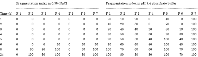
Fragmentation index of the gelled beads: The fragmentation index of seven formulations of beads in 0.9% NaCl solution and in pH 7.4 phosphate buffer media upto 24 h were shown in Table 3. From the table it can be observed that beads of F-1 (with no oil) were not fragmented in NaCl media but in buffer they were fragmented 20% at 1st h and 100% fragmented at 24th h. Again F-2 and F-3 (containing 10% and 20% soyabean oil respectively) fragmented 80 and 40% at 8th h in NaCl solution, whereas they started to be fragmented from the 1st h in buffer. Beads of F-4 (having 30% soyabean oil) did not fragment in NaCl but started to fragment from 3rd h in buffer. Again fragmentation index of F-5 and F-6 (containing 10 and 20% mustard oil) was high in buffer media than in NaCl media. F-7 (with 30% mustard oil) were completely fragmented in buffer media from the 1st h.
| Table 3: | Fragmentation index of DS beads in 0.9% NaCl and pH 7.4 phosphate buffer media |
 | |
| Table 4: | Mean diameter of beads (n = 14) |
 | |
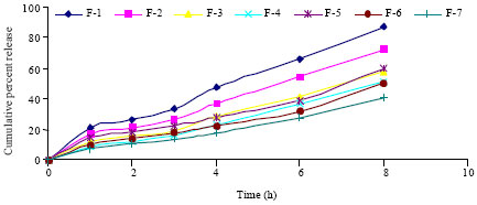 | |
| Fig. 3: | Cumulative percent release vs. time curve of beads of F-1 to F-7 |
Mean diameter of the beads of different formulations: To determine the mean diameter of beads, at first diameter of 14 beads were measured by digital vernier caliper and it was then divided by n (number of beads =14). The mean diameter was presented in table 4. Among the seven formulations, the diameter of F-7 was highest (4.397 mm) and F-1 was the least (1.659 mm). It was also observed that beads with no oil were the smallest one and increasing the oil concentration from 10 to 30%, the diameter of the beads were increased. Also, mustard oil containing beads were larger than soyabean oil containing beads.
In vitro dissolution studies: The dissolution of beads of F-1 to F-7 was carried out in 0.1N HCl followed by pH 7.4 phosphate buffer. As the release rate of DS in acid media was very little, then it was not considered for constructing cumulative percent release vs. time curve (Fig. 3). From the curve it can be said that DS release rate from F-1 was highest (87.19%) at 8 h. The cumulative release of DS from F-2 to F-4 were 72.03, 57.95 and 51.11% at 8 h. As the soyabean oil concentration of F-2 to F-4 increased from 10 to 30%, the release rate were found to be decreased respectively. Similarly, in case of F-5 to F-7, concentrations of mustard oil were increased from 10 to 30% and the release rate of DS from them were decreased (59.69, 50.36 and 41.12% at 8 h, respectively). From the curve it was also found that the rate retarding action of soyabean oil was less than mustard oil.
The release data of DS were fitted according to Zero order, Higuchi and Korsmeyer model. The regression coefficient (R2), rate constant, release exponent (n), Mean Dissolution Time (MDT) and similarity factor (f2) were shown in Table 5. From the table it can be observed that release kinetics of all of the seven formulations was most fitted to zero order model. So release of DS from the beads was nearly independent on the concentration of drug in depot. The release exponent (n) value found to be within 0.43 to 0.85 indicates that the probable release mechanism from the beads were diffusion and erosion controlled.
| Table 5: | Regression coefficient (R2), release rate constant, release exponent (n), mean dissolution time (MDT) and similarity factor (f2)* of zero order, Higuchi and Korsmeyer model of F-1 to F-7 |
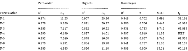 | |
| *Similarity factor (f2) was constructed considering F-4 as reference standard. | |
MDT is the indicator of sustaining action of formulation (Reza et al., 2003). The MDT values of F-1 were the least (8.694 h) as no rate retaining oil was incorporated in it. MDT of F-2 to F-4 was found to be increased (9.447, 0.742 and 11.330 h) with the incorporation of increased amount of soyabean oil. Again, MDT of F-5 to F-7 was again increased (9.742, 11.330 and 13.210 h) with the increased incorporation of mustard oil. From the MDT value it was also found that mustard oil had more sustaining capability than soyabean oil.
f2 is the indicator of similarity of release pattern of a formulation with the reference one. Here F-4 was taken as reference standard. Except F-1 all the formulations showed f2 value greater than 50, indicating the release profile of those formulations were very similar to F-4.
From the output of one way repeated measure ANOVA and post hoc test was conducted for finding out the differences in release pattern of the seven formulations. The multiple comparisons (Bonferroni and Dunnett) were also carried out. Dunett t tests treat one group as a control and compare all other groups against it. The paired comparison of the six groups with the control group give p-value = 0.000 to 0.005, that is, less than 0.05. So, it can be stated that there was significant difference between the release patterns of DS from the beads.
CONCLUSION
Beads were prepared successfully by emulsion-gelation technique. Here the rate retarding action of two oils was investigated. From the result, it was found that soyabean oil was better dispersed with uniform size than mustard oil in beads. Beads were better swelled (no fragmentation) in 0.1 N HCl. Highest soyabean and mustard oil containing beads showed good floating properties than that of other formulations. Increasing oil content increased the size of the beads. Both buoyancy, diameter of the beads are the parameters which influence the sustaining action of a dosage form. Rate of release of DS during dissolution study was found to be decreased with the increased content of oils. Soyabean oil had less release retarding capacity than mustard oil. The release pattern was fitted to zero order model, indicating the release of DS from beads were concentration independent. Release exponent value showed that the possible release mechanism was anomalous. When F-4 was taken as reference standard, it was found that the release pattern of all the formulations (except F-1) were similar to F-4. MDT values were also larger for those formulations where oil content was higher. One way repeated measure ANOVA (Dunnett t-test) showed a statistically significant difference in the release pattern of F-1 to F-7.
ACKNOWLEDGMENTS
Authors are thankful to Eskayef Bangladesh Ltd. For providing diclofenac sodium and Head, Department of Pharmacy, The University of Asia Pacific for providing the necessary materials and laboratory facilities.
REFERENCES
- Tapia, C., Z. Escobar, E. Costa, J. Sapag-Hagar and F. Valenzuela et al., 2004. Comparative studies on polyelectrolyte complexes and mixture of chitosan-alginate and chitosan-carrageenan as prolonged diltiazem clorhydrate release system. Eur. J. Pharm. Biopharm., 57: 65-75.
PubMed - Bodmeier, R. and O. Paeratakul, 1989. Spherical agglomerates of water-insoluble drugs. J. Pharm. Sci., 78: 964-967.
PubMed - Choudhury, P.K. and M. Kar, 2005. Preparation of alginate gel beads containing metformin hydrochloride using emulsion-gelation method. Trop. J. Pharm. Res., 4: 489-493.
Direct Link - Piyakulawat, P., N. Praphairaksit, N. Chantarasiri and N. Muangsin, 2007. Preparaton and evaluation of chitosan/carrageenan beads for controlled release of sodium diclofenac. AAPS Pharm. Sci. Tech., 8: 120-130.
Direct Link - Elmowafy, M.E., A.S.G. Awad, S. Mansour and A.E.H.A. El-Shamy, 2009. Ionotropically emulsion gelled polysaccharides beads: Preparation, in vitro and in vivo evaluation. Carbohydrate Polymers, 75: 135-142.
CrossRef - Manjunatha, K.M., M.V. Ramana and D. Satyanarayana, 2007. Design and evaluation of diclofenac sodium controlled drug delivery systems. Ind. J. Pharm. Sci., 69: 384-389.
Direct Link - Donbrow, M. and Y. Samuelov, 1980. Zero order drug delivery from double-layered porous films: Release rate profiles from ethylcellulose, hydroxypropylcellulose and polyethylene glycol mixtures. J. Pharm. Pharmacol., 32: 463-470.
PubMed - Higuchi, T., 1961. Rate of release of medicaments from ointment bases containing drugs in suspension. J. Pharm. Sci., 50: 874-875.
PubMed - Higuchi, T., 1963. Mechanism of sustained-action medication: Theoretical analysis of rate of release of solid drugs dispersed in solid matrices. J. Pharm. Sci., 52: 1145-1149.
PubMed - Korsmeyer, R.W., R. Gurny, M. Doelker, P. Buri and N.A. Peppas, 1983. Mechanisms of solute release from porous hydrophilic polymers. Int. J. Pharm., 15: 25-35.
CrossRefDirect Link - Peppas, N.A., 1985. Analysis of fickian and non-fickian drug release from polymers. Pharm. Acta Helv., 60: 110-111.
Direct Link - Merchant, H.A., H.M. Shoaib, J. Tazeen and R.I. Yousuf, 2006. Once-daily tablet formulation and in vitro release evaluation of Cefpodoxime using hydroxypropyl methylcellulose: A technical note. AAPS Pharm. Sci. Tech., 7: 1-6.
Direct Link - Mockel, J.E. and B.C. Lippold, 1993. Zero-order drug release from hydrocolloid matrices. Pharm. Res., 10: 1066-1070.
Direct Link - Zhou, Q.Z., L.Y. Wang, G.H. Ma and Z.G. Su, 2007. Preparation of uniform-sized agarose beads by microporous membrane emulsification technique. J. Colloid Interface Sci., 311: 118-127.
PubMed - Reza, M.S., M.A. Quadir and S.S. Haider, 2003. Comparative evaluation of plastic, hydrophobic and hydrophilic polymers as matrices for controlled-release drug delivery. J. Pharm. Pharm. Sci., 6: 274-291.
Direct Link