
Wannee Jiraungkoorskul
Department of Pathobiology, Faculty of Sciences, Mahidol University, Bangkok 10400, Thailand
Rachen Singhakumar
Mahidol University International College, Mahidol University, Salaya Campus, Nakhonpathom 73170, Thailand
Kanitta Jiraungkoorskul
Department of Occupational Health and Safety, Faculty of Public Health, Mahidol University, Bangkok 10400, Thailand
Piya Kosai
Department of Pathobiology, Faculty of Sciences, Mahidol University, Bangkok 10400, Thailand
Research Journal of Medicinal Plants
Year: 2011 | Volume: 5 | Issue: 6 | Page No.: 764-771


ABSTRACT
The efficiency of Psidium guajava was studied to illustrate the reduction of ferric nitrilotriacetate (Fe-NTA) toxicity in Puntius altus via the histopathology analysis. The fish (n = 40) were randomly divided into four groups. Each fish was transferred to each aquarium as follows: G1 and G2 were obtained normal fish food; G3 and G4 were obtained guava leaf extract 60 mg g-1 fish food. After 28 days dietary supplement, fish in G2 and G4 were injected intraperitoneal of 9 mg Fe kg-1 b.wt. Twenty-four hour after injection, lesions were especially most evident in the G2. The gills were observed epithelial lifting, lamellar cell hyperplasia. Blood congestion was seen in sinusoids and hepatocytes necroses were also observed. Renal tubular swelling and necrosis were seen. Some areas were found hemosiderin pigment accumulation. Fish with guava pre-treatment (G4) showed slightly alteration when compare those of G2 group. The results suggested that P. guajava leaf extract pre-obtained may play an important role in the reduction of Fe-NTA toxicity in fish.
PDF Abstract XML References Citation
Received: February 23, 2011;
Accepted: April 16, 2011;
Published: June 25, 2011
How to cite this article
Wannee Jiraungkoorskul, Rachen Singhakumar, Kanitta Jiraungkoorskul and Piya Kosai, 2011. Dietary Psidium guajava Supplementation Reducing Ferric Nitrilotriacetate Toxicity in Puntius altus. Research Journal of Medicinal Plants, 5: 764-771.
URL: https://scialert.net/abstract/?doi=rjmp.2011.764.771
URL: https://scialert.net/abstract/?doi=rjmp.2011.764.771
INTRODUCTION
Thailand is considered one of the countries which bring medical plants into use and sometimes they refer to it as “Thai Traditional Medicine (TTM)” which can be used to treat humans and animal diseases. Ascorbic Acid (AA) is an essential vitamin for normal growth and physiological functions in animals including fishes. Most teleosts are unable to synthesize AA because of the lack of L-gulonolactone oxidase (Fracalossi et al., 2001). Therefore, an exogenous source of AA is required in fish diets. It functions as a general water-soluble reagent, on collagen formation, iron metabolism and the response to stress (Vijayavel et al., 2006). Many authors have reported the efficacy of AA in reducing genotoxicity in Oreochromis niloticus induced by lead (Jiraungkoorskul et al., 2008); Poronotus triacanthus induced by copper (Jiraungkoorskul and Sahaphong, 2007) and Puntius altus induced by cadmium (Jiraungkoorskul et al., 2007), using the micronucleus and nuclear abnormality tests. It has also been reported the efficacy of AA in reducing the histopathological alterations in fish after cadmium exposure (Jiraungkoorskul et al., 2006). Edema, lamellar cell hyperplasia, epithelial lifting and aneurysm were observed in the gills. There were blood congestion in sinusoids, vacuolation of hepatocytes, hemosiderin accumulation, apoptosis and nuclear pyknosis. Glomerulus atrophy, hydropic swelling, hyaline casts and necrosis were seen. Fortunately, in the combination of cadmium and AA treated group, they showed similar alterations as those observed in the cadmium treated alone group but they were less severe. The findings of this study can be used as guidelines for developing programs to help the fish which are cultured near heavy metal contaminated areas (Jiraungkoorskul et al., 2006). However, the uses of natural origin are preferred over the others because they are safe and non-toxic and have no resistance problems.
Psidium guajava Linn. (“Farang” in Thai, or “Guava” in English) constituents include ascorbic acid, triterpenes (Begum et al., 2004), carotenoids (Mercadante et al., 1999) and flavonoids (Rattanachaikunsopon and Phumkhachorn, 2007). The leaves are elliptic to ovate about 5-15 cm in length. The flowers are white, with five petals and numerous stamens. This plant produces edible round or pear shaped fruit which contains high amounts of calcium. Guava leaves are used for medicinal purposes, as a remedy to diarrhea. Recent studies prove that guava has sugar lowering properties to help diabetics lower their sugar count (Gutierrez et al., 2008). In addition, crushed leaves from guava plants can be applied on wounds, ulcers and rheumatic places and toothache can be relieved by chewing the leaves. Lastly, leaves extracts can also have antimicrobial properties and can also be for coughs (Jaiarj et al., 1999). The study survey revealed that there are no scientific studies carried out regarding the efficiency of this plant in fish. Ferric nitrilotriacetate (Fe-NTA) has been chosen as a model organic compound to study the protective effect of this herb in its toxicity. Awai et al. (1979) first reported glycosuria and hepatic parenchymal iron deposits in rats following intraperitoneal injections with Fe-NTA. Its toxicity is assumed to be caused by the elevation of free serum iron concentration, following its reduction at the luminal side of the proximal tubule (Liu et al., 1991), generating reactive oxygen species, leading to lipid peroxidation and induce oxidative stress in liver (Iqbal et al., 1995). This present study was designed to assess the ability of the extract from P. guajava against ferric nitrilotriacetate induced toxicity by using histopathological analysis.
MATERIALS AND METHODS
Animal model: This study was performed at the Department of Pathobiology, Faculty of Sciences, Mahidol University, Bangkok, Thailand in 2010. Red tailed tinfoil barb, P. altus, 16.76±1.72 g in b.wt. and 9.85±0.50 cm in total length, were purchased from a commercial hatchery in Bangkok, Thailand. Tap water was filtered with activated charcoal to eliminate chemical contamination. The physicochemical characteristics of water were measured daily, according to the experimental procedures described in Standard Methods for the Examination of Water and Wastewater (American Public Health Association, 2005). Under laboratory condition, fish were acclimated for 30 days at 28.5±1.0°C, pH = 6.8-7.0, total hardness = 70-80 mg L-1 (as CaCO3), alkalinity = 75-80 mg L-1 and conductivity = 190-210 μmhos cm-1. A 16:8 h light-dark cycle was maintained throughout. Chlorine residual and ammonia were below detection limits. Fish were fed twice a day with 37%-protein commercial fish food (Charoen Pokphand Group, Bangkok, Thailand). The quantity of food was 2% of the initial body weight per day. The animal care and handling in this research was performed following the instruction of the Mahidol University-Institutional Animal Care and Use Committee (MU-IACUC). Therefore, this research was followed the mammal animal care and use i.e., (1) Use, care and transportation of fish for toxicopathological testing was complied with all applicable animal welfare laws. (2) Number of fish was kept to the minimum requirement for achieve scientifically valid results. (3) All protocols were taken to avoid the discomfort, distress or pain in the fish. (4) The appropriate dosage of the anesthesia was 200 mg L-1 ethyl-3-aminobenzoate methanesulfonate salt (MS222, Sigma) and the euthanasia was overdose of this chemical.
Preparation of dry leaf with fish food: Guava was collected from local area in Nakorn Prakom Province, Thailand. Fresh leaves were washed several times in water, dried at 45°C for 72 h and made semi powder by crushing using a mortar and pestle. The extraction was done by following the method of Winkaler et al. (2007) with slight modifications. Fish food was grounded in a blender and hydrated with 0.7 mL g-1 distilled water, mixed with the leaf semi powder extract and extruded through a minced-meat processing machine. Later, the mixture was broken into small pellets by hands and dried at 60°C for 48 h. The fishes were fed twice a day (2% b.wt.) with the prepared dry leaf food supplementation within 28 days.
Preparation of Ferric Nitrilotriacetate (Fe-NTA) solution: A solution of Fe-NTA was prepared by the method of Awai et al. (1979).
Experimental design: The fish (n = 40) were randomly divided into four groups. Each fish group was transferred to each aquarium as follows: G1 and G2 were obtained normal fish food; G3 and G4 were obtained guava leaf extract 60 mg g-1 fish food. After 28 days dietary supplement, fish in G2 and G4 were injected intraperitoneal of 9 mg Fe kg-1 b.wt. given in a volume of 10 mL kg-1 b.wt. Twenty-four h after injection, fish from each group was taken and anesthetized with 0.2 g L-1 MS-222, weighed and measured. The fish was dissected and the organs (gill, liver and kidney) were removed and prepared for histopathological analysis.
Specimen preparation for light microscopic study: The procedures for light microscopy were modified by Humason (1972). Briefly, the tissues were fixed in the 10% buffered formaldehyde for 24 h, dehydrate through a graded series of ethanol and clear with xylene solutions. They were embedded in a block using melted paraffin at the embedding station (Axel Johnson Lab System, USA). The paraffin blocks were sectioned at 4-5 μm thickness using a rotary microtome (HistoSTAT, Reichert, USA) and stained with Harris hematoxylin and eosin. The tissue glass slides were examined for abnormalities by a Nikon E600 light microscope and photographed by a Nikon DXM 1200 digital camera (Tokyo, Japan).
RESULTS
Gills
Control group: The gills consisted of a row of long thin filaments, the primary lamellae which projected from the arch like the teeth of a comb. The surface area of each primary lamella was increased further by the formation of regular semilunar folds across its dorsal and ventral surface, the secondary lamellae. The primary lamellar epithelium was one or two cell layers thick. Each secondary lamella was made up of two sheets of epithelium delimited by many pillar cells which were contractile and separated the capillary channels. One to two erythrocytes were usually observed within each capillary lumen. No recognizable changes were observed in the gills of the control (G1) and guava supplement group (G3) throughout this experiment (Fig. 1a, c).
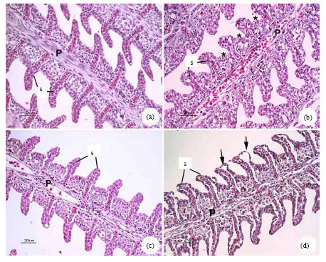 | |
| Fig. 1: | Gills of P. altus in (a) G1, showing normal arrangement of primary (P) and secondary (s) lamellae (b) G2, showing epithelial hyperplasia (*) and blood congestion (c) G3, no recognition change was observed (d) G4, showing edema or epithelial lifting (arrows) |
Treated groups: A variety of histological studies revealed that Fe-NTA affected gill tissue in G2. The lesions included hyperplasia, epithelial lifting or cell swelling and congestion. The lesion was started with the bending of the distal extremities of secondary lamellae, followed by a lifting of the outer layer of the lamellar epithelium, the formation of edematous spaces between the layers of epithelium which may become infiltrated with red blood cells and leukocytes. Finally, hyperplastic tissues were observed in the primary epithelial cells (Fig. 1b). Eventually, the whole epithelium sloughed off and the lamella lost its rigidity. Guava pre-obtained group (G4) showed similar but less severe alterations than those of G2 (Fig. 1d).
Liver
Control: Hepatocytes were polygonal and had a distinct central nucleus with densely staining chromatin margins and a prominent nucleolus. Sinusoids which were irregularly distributed between hepatocytes, were fewer in number and were lined by endothelial cells. No recognizable changes were observed in the hepatocytes of the control (G1) and guava supplement group (G3) throughout this experiment (Fig. 2a, c).
Treated groups: The hepatocytes in G2 were swelling and numerous vacuolization were also observed. Blood congestion and exhibited increasing size and pyknotic nuclei were seen (Fig. 2b). However, the histological alterations were less severe in G4 (Fig. 2d).
Kidney
Control: The kidney was divided into anterior and posterior portions. The nephron of the freshwater fish was composed of a well-vascularized glomerulus, proximal segments, distal segments and collecting duct system.
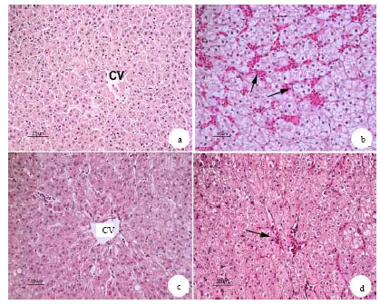 | |
| Fig. 2: | Liver of P. altus in (a) G1, showing normal hepatocytes and blood sinusoids (b) G2, showing severe blood congestion (arrows) (c) G3, no recognition change was observed (d) G4, showing mild blood congestion (arrow). CV: Central veins |
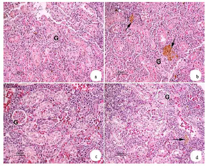 | |
| Fig. 3: | Kidney of P. altus in (a) G1, showing normal appearance of glomerulus (G) and renal tubules (b) G2, showing renal tubule necrosis in some area and numerous hemosiderin accumulation (arrows) (c) G3, no recognition change was observed (d) G4, showing mild hemosiderin accumulation |
No recognizable changes were observed in the glomerulus and renal tubule of the control (G1) and guava supplement group (G3) throughout this experiment (Fig. 3a, c).
Treated groups: There found to be some atrophy glomerulus and renal tubular necrosis in G2. Likewise, in G4 displayed similar alterations as those observed in G2 but they were less severe. Furthermore, there seemed to be a few hemosiderin accumulations in some area (Fig. 3b, d).
DISCUSSION
The result of this study has shown that the aqueous extract of P. guajava leaves were relatively very safe when taken at the tested doses. At a maximum dose of 60 mg kg-1, no death was recorded in the experimental fish. This safety was similar with the native fruit of Brazil, P. cattleyanum, its leaf extract was tested in mice at concentration of 1000, 1500 and 2000 mg kg-1, failed to induce a significant increase in cell DNA damage, in micronucleated cell frequency and in bone marrow toxicity (Costa et al., 2008). Teixeira et al. (2003) also evaluated the effects of the infusions of P. guajava in in vitro and in in vivo assays on chromosomes and the cell cycle. The two different concentrations of the infusions did not cause a statistically significant alteration in Allium cepa L. root-tip cells, in rat cells or in cultured human lymphocytes.
Intraperitoneally injected Fe-NTA was absorbed into the portal vein through mesothelium and passed into circulation via the liver (Umemura et al., 1990). The present study was tested the efficiency of guava leave supplement dietary to reduce the allied damage in vivo by Fe-NTA toxicity. No recognition change in the histopathological analysis in the guava supplement group (G3). Fe-NTA caused the histopathological alterations in the gills, liver and kidney (G2 and G4). The gills were seen edema, lamellar cell hyperplasia and epithelial lifting. There was blood congestion in sinusoids and hepatocytes necrosis. Renal tubular swelling and also necrosis were seen. However, fish with guava pre-obtained (G4) showed slightly alteration when compare the only Fe-NTA treatment group (G2). These obtained results agree with our previously study in the protective influence of ascorbic acid against the toxicity of waterborne cadmium exposure in P. altus and lead exposure in O. niloticus (Jiraungkoorskul et al., 2006, 2008).
The anti-inflammatory property of the aqueous leaf extract was investigated in rats, using fresh egg albumin induced pedal edema. P. guajava aqueous extract (50-800 mg kg-1, i.p.) produced dose dependent and significant inhibition of fresh egg albumin-induced acute inflammation in rats (Ojewole, 2006). The hepatoprotective effect of an aqueous leaf extract of P. guajava was studied on rat liver damage induced by carbon tetrachloride by monitoring serum transaminase, alkaline posphatase, cholesterol, total lipids and histopathological alterations. The leaf extract at doses of 500 mg kg-1 produced significant hepatoprotection (Roy et al., 2006). Pretreatment with asiatic acid (a triterpenoid extracted from P. guajava leaves and fruit) at doses of 25, 50 or 100 mg kg-1 significantly blocked the lipopolysaccharide and D-galactosamine-induced increases in both serum aspartate and alanine aminotransferase levels, showing improved nuclear condensation, ameliorated proliferation and less lipid deposition (Gao et al., 2006). Several studies have indicated the ability of guava to reduce several parameters associated with liver injury. Another hypothesis of cellular damage may arise from free radicals or reactive oxygen species. Guava leaf extracts are a potential source of natural antioxidants. These antioxidant properties are associated with its phenolic compounds such as protocatechuic acid, ferulic acid, quercetin and guavin B, ascorbic acid, gallic acid and caffeic acid (Jimenez et al., 2001; Thaipong et al., 2005). Morales et al. (1994) showed that quercetin acts as a smooth muscle calcium antagonist. Therefore, the protection activity of PG may be due to the presence of these compounds.
In conclusion, the results presented in this study show that the efficiency of dietary supplement of Psidium guajava leaf reducing histopathological alterations associated with exposed to ferric nitrilotriacetate uptake in fish which are cultured near heavy metal contaminated areas.
ACKNOWLEDGMENTS
This study was funded by the Thailand Research Fund and the Commission on Higher Education: Research Grant for Mid-Career University Faculty 2008 (RMU5180001) and in part by Mahidol University International College and Faculty of Science, Mahidol University.
REFERENCES
- APHA., 2005. Standard Methods for the Examination of Water and Wastewater. 21st Edn., American Public Health Association, Washington, DC., USA., ISBN-13: 978-0875530475, Pages: 1200.
Direct Link - Awai, M., M. Narasaki, Y. Yamanoi and S. Seno, 1979. Induction of diabetes in animals by parenteral administration of ferric nitrilotriacetate. A model of experimental hemochromatosis. Am. J. Pathol., 95: 663-672.
Direct Link - Begum, S., S.I. Hassan, S.N. Ali and B.S. Siddiqui, 2004. Chemical constituents from the leaves of Psidium guajava. Nat. Prod. Res., 18: 135-140.
CrossRefPubMedDirect Link - Costa, T.D.A., S. Vieira, S.F. Andrade and E.L. Maestro, 2008. Absence of mutagenicity effects of Psidium cattleyanum Sabine (Myrtaceae) extract on peripheral blood and bone marrow cells of mice. Genet. Mol. Res., 7: 679-686.
Direct Link - Fracalossi, D.M., M.E. Allen, L.K. Yuyama and O.T. Oftedal, 2001. Ascorbic acid biosynthesis in Amazonian fishes. Aquaculture, 192: 321-332.
CrossRefDirect Link - Gao, J., J. Chen, X. Tang, L. Pan, L. Xu, L. Zhao and Q. Xu, 2006. Mechanism underlying mitochondrial protection of asiatic acid against hepatotoxicity in mice. J. Pharm. Pharmacol., 58: 227-233.
Direct Link - Gutierrez, R.M.P., S. Mitchell and R.V. Solis, 2008. Psidium guajava: A review of its traditional uses, phytochemistry and pharmacology. J. Ethnopharmacol., 117: 1-27.
CrossRefPubMedDirect Link - Iqbal, M., U. Giri and M. Athar, 1995. Ferric nitrilotriacetate (Fe-NTA) is a potent hepatic tumor promoter and acts through the generation of oxidative stress. Biochem. Biophy. Res. Commun., 212: 557-563.
Direct Link - Jaiarj, P., P. Khoohaswan, Y. Wongkrajang, P. Peungvicha, P. Suriyawong, M.L.S. Saraya and O. Ruangsomboom, 1999. Anticough and antimicrobial activities of Psidium guajava Linn. leaf extract. J. Ethopharmacol., 67: 203-212.
CrossRefDirect Link - Jimenez-Escrig, A., M. Rincon, R. Pulido and F. Saura-Calixto, 2001. Guava fruit (Psidium guajava L.) as a new source of antioxidant dietary fiber. J. Agric. Food Chem., 49: 5489-5493.
CrossRefPubMedDirect Link - Jiraungkoorskul, W., S. Sahaphong, N. Kangwanrangsan and M.H. Kim, 2006. Histopathological study: The effect of ascorbic acid on cadmium exposure in fish (Puntius altus). J. Fish. Aquatic Sci., 1: 191-199.
CrossRefDirect Link - Jiraungkoorskul, W. and S. Sahaphong, 2007. Efficacy of ascorbic acid reducing waterborne copper toxicity in butterfish (Poronotus triacanthus). J. Boil. Sci., 7: 620-625.
CrossRefDirect Link - Jiraungkoorskul, W., S. Sahaphong, P. Kosai and M.H. Kim, 2007. Micronucleus test: The effect of ascorbic acid on cadmium exposure in fish (Puntius altus). Res. J. Environ. Toxicol., 1: 27-36.
CrossRefDirect Link - Jiraungkoorskul, W., S. Sahaphong, N. Kangwanrangsan and S. Zakaria, 2008. The protective influence of ascorbic acid against the genotoxicity of waterborne lead exposure in Nile tilapia (Oreochromis niloticus). J. Fish Biol., 73: 355-366.
CrossRefDirect Link - Liu, M., S. Okada and T. Kawabata, 1991. Radical promoting free iron level in the serum of rats treated with ferric nitrilotriacetate, comparison with iron chelate complexes. Acta Med. Okayama, 45: 401-408.
PubMed - Morales, M.A., J. Tortoriello, M. Meckes, D. Paz and X. Lozoya, 1994. Calcium-antagonist effect of quercetin and its relation with the spasmolytic properties of Psidium guajava L. Arch. Med. Res., 25: 17-21.
PubMed - Ojewole, J.A., 2006. Antiinflamatory and analgesic effects of Psidium guajava Linn (Myrtaceae) leaf aqueous extract in rats and mice. Method. Find. Exp. Clin. Pharmacol., 28: 441-446.
PubMed - Rattanachaikunsopon, P. and P. Phumkhachorn, 2007. Bacteriostatic effect of flavonoids isolated from leaves of Psidium guajava on fish pathogens. Fitoterapia, 78: 434-436.
PubMed - Roy, C.K., J.V. Kamath and M. Asad, 2006. Hepatoprotective activity of Psidium guajava Linn. leaf extract. Indian J. Exp. Biol., 44: 305-311.
PubMedDirect Link - Teixeira, R.O., M.L. Camparoto, M.S. Mantovani and V.E.P. Vicentini, 2003. Assessment of two medicinal plants, Psidium guajava L. and Achillea millefolium L. in in vivo assays. Gen. Mol. Biol., 26: 551-555.
Direct Link - Thaipong, K., U. Boonprakob, L. Cisneros-Zevallos and D.H. Byrne, 2005. Hydrophilic and lipophilic antioxidant activities of guava fruits. South. Asian J. Trop. Med. Pub. Health, 36: 254-257.
PubMed - Umemura, T.Y., K. Sai, A. Takagi, R. Hasegawa and Y. Kurakawa, 1990. Oxidative DNA damage lipid peroxidation and nephrotoxicity induced in the rat kidney after ferric nitrilotriacetate administration. Cancer Lett., 54: 95-100.
CrossRef - Winkaler, E.U., T.R.M. Santos, J.G. Machado-Neto and C.B.R. Martinez, 2007. Acute lethal and sublethal effects of neem leaf extract on the neotropical freshwater fish Prochilodus lineatus. Comp. Biochem. Physiol. Part C: Toxicol. Pharmacol., 145: 236-244.
CrossRefDirect Link