
Keivan Beheshti Maal
Department of Microbiology, Faculty of Biological Sciences, Falavarjan Branch, Islamic Azad University, Isfahan, Iran
Abbas Soleimani Delfan
Department of Microbiology, Faculty of Biological Sciences, Falavarjan Branch, Islamic Azad University, Isfahan, Iran
Sharareh Salmanizadeh
Department of Microbiology, Faculty of Biological Sciences, Falavarjan Branch, Islamic Azad University, Isfahan, Iran
Research Journal of Environmental Sciences
Year: 2014 | Volume: 8 | Issue: 3 | Page No.: 123-133


ABSTRACT
Recently prevention of bacterial infections using their specific viruses, bacteriophages, is challenging. In this research we isolated and identified a native strain of Klebsiella pneumoniae from Isfahan municipal wastewater treatment plant we named K. pneumoniae SBSWIF3. Also we isolated and characterized two novel bacteriophages related to Myoviridae and Podoviridae families of bacteriophages from Zayandehrood River, Isfahan, Iran that had lytic effects on K. pneumoniae SBSWIF3, K. pneumonia PTCC1290 and K. oxytoca PTCC1402 as coliform's index. The Myovirus had a hexagonal head measuring 27.28 nm and a noncontractile tail measuring 204.5x13.63 nm. The Podovirus had an oval head measuring 98x35 nm and a tail with 14 nm in diameter. The treatment of municipal sewage with the coliphage mixture resulted in 22 folds decrease of the coliform’s MPN from 2400-110 after 2 h incubation. We suggested that the use of these lytic coliphages for reduction of coliform’s population in sewage could be considered as an effective and simple alternative for costly replacement of instruments and establishments of the old wastewater treatment plants.
PDF Abstract XML References Citation
Received: October 13, 2013;
Accepted: December 25, 2013;
Published: March 28, 2014
How to cite this article
Keivan Beheshti Maal, Abbas Soleimani Delfan and Sharareh Salmanizadeh, 2014. Isolation and Identification of Klebsiella pneumonia and Klebsiella oxytoca Bacteriophages and their Application in Wastewater Treatment and Coliform’s Phage Therapy. Research Journal of Environmental Sciences, 8: 123-133.
DOI: 10.3923/rjes.2014.123.133
URL: https://scialert.net/abstract/?doi=rjes.2014.123.133
DOI: 10.3923/rjes.2014.123.133
URL: https://scialert.net/abstract/?doi=rjes.2014.123.133
INTRODUCTION
Most of the potential pathogens are distributed to body waters, agricultural products and foods through sewage and ineffective-treated public wastewater. The main reasons for establishing the wastewater treatment plants have been providing resources of clean water for industrial uses, protecting of stream waters from sewage-contaminating microorganisms and controlling of sewage-borne pathogens from spreading infectious diseases (Bitton, 1994). Among sewage pathogens, coliforms as facultative aerobic gram negative rods that could ferment lactose at 35°C are responsible for frequent infectious diseases in human and animals (Baggi et al., 2001). From coliforms, Klebsiella spp. with extensive capsular polysaccharide are members of order Enterobacterials and family Enterobacteriaceae. These microorganisms are the normal flora of gastrointestinal tract in human and animals but could contaminate other body regions such as eyes, respiratory tract, genitourinary tract and soft tissues. While they are opportunistic pathogens but can result in severe infectious diseases e.g., pneumonia, urinary infections and septicemia (Wendt et al., 2010). The progressive distribution of antibiotic-resistant strains of Klebsiella spp. specially K. pneumonia and K. oxytoca is an alarming problem in the medical and environmental microbiology (Won et al., 2011). Klebsiella spp. are one of the valuable contamination indexes of water, food and agricultural products to coliforms and wastewater (Reid et al., 2001). Bacteriophages are the viruses that significantly distributed in the nature and attack specifically to their bacterial hosts (Waldor et al., 2005; Beheshti Maal et al., 2012). The phage therapy or use of bacteriophages as therapeutic agents for eliminating bacterial infections has its root in preliminary studies of Twort and D’Herelle at the first of 20th century (Marks and Sharp, 2000; Chanishvili et al., 2001). Several studies have proved the usefulness of phage therapy for controlling infectious diseases caused by pathogenic bacteria such as E. coli in human children and adults (Drozdova et al., 1998), E. coli infections in mice and calves (Smith et al., 1987), Pseudomonas aeroginosa and Acinetobacter baumanii in skin infections (Soothill, 1994), oral streptococci in dental infections (Beheshti Maal et al., 2010, 2012), Staphylococcus aureus and Staphyococcus epidermidis in skin infections (Chanishvili et al., 2001) and Vibrio cholera infections (Marks and Sharp, 2000). While most of the studies have focused on the bacteriophage potential for controlling bacterial pathogens in medical microbiology, there are few reports indicating the application of phages in wastewater treatment and environmental microbiology (Hennes and Simon, 1995; Withey et al., 2005). The aims of this research were isolation and identification of native strains of K. pneumoniae from wastewater, isolation and identification of specific lytic coliphages from water resources and investigation of their effects on native and standard strains of K. pneumonia and K. oxytoca as well as coliforms population in municipal wastewater.
MATERIALS AND METHODS
Bacterial strain, culture media and chemicals: The bacterial strains we used in this research were Klebsiella pneumonia PTCC1290 and Klebsiella oxytoca PTCC1402 we provided from Iranian Research Organization for Science and Technology (IROST), Tehran, Iran as well as a native strain of Klebsiella pneumoniae we isolated in this research. The main culture media we used were Eosin Methylene Blue (EMB), Lactose Broth (LB), Brilliant Green Lactose Bile Broth (BGLBB), Brain Heart Infusion Broth (BHI) and Brain Heart Infusion Agar (BHA) all from Merck, Germany. The chemicals and materials were glycine, MgSO4, phenol red, uranyl acetate, phosphotungstic acid (all from Sigma, USA), DNA extraction kit and PCR kit (both from Cinnagen, Iran), forward and reverse primers (RW01 and DG71, TAG Copenhagen, Denmark) and syringe 0.45 μm membrane filters (Millipore, UK and Albet, Spain).
Wastewater sampling and bacterial isolation: The sewage sample was gathered from sewage effluent before treatment at Isfahan southern wastewater treatment plant, Isfahan, Iran using sterile 1000 mL capped bottles under aseptic condition. The samples were transferred to microbiology laboratory in Falavarjan Branch, Islamic Azad University, Falavarjan, Iran at 4°C. One millilitre of sewage effluent was cultured in LB and incubated at 37°C for 24 h. One standard loop from positive LB medium was cultured in EMB using streak plate method and incubated at 37°C for 24 h. A well isolated violet colony with metallic greenish reflection was collected and cultured on EMB and incubated for 24 h at 37°C for purification purposes. Specified pure colonies were stained using gram method.
DNA extraction and molecular identification of isolated bacterium: A well isolated colony in EMB was cultured in 100 mL of BHI and incubated at 37°C with 120 rpm (round min-1) aeration speed for 24 h. Forty millilitres of bacteria in BHI was transferred to 50 mL sterile falcon and centrifuged at 8000 g for 10 min. The supernatant was discarded and 10 mg of bacterial biomass was used for DNA extraction using DNA extraction kit for gram negative bacteria (Cinnagen, Iran, Cat. No. DN8115C) according to instructions. Then the DNA purity was measured using UV spectrophotometer in 280 nm (Sambrook and Russell, 2001). The primers used for molecular identification of isolated coliform strain were high performance universal primers for bacterial identification (Leong and Greisen, 1993). These primers and their characteristics were summarized in Table 1. The PCR was performed in 20 μL of a reaction mixture containing 2 μL (<20 ng μL-1) of DNA template, 1 μL of each forward and reverse primers (55 μg mL-1), 10 mM of MgCl2, 0.5 mM of dNTP mixture, 0.5 μL Smar-Taq DNA polymerase (10 mM) and 2.5 μL of 10x PCR buffer (5 mM). Amplification was done using a gradient thermal cycler (eppendorph mastercycler) with following program, initial denaturation at 95°C for 5 min, 30 cycles for denaturing step at 94°C for 45 sec, primer annealing step at 54°C for 30 sec, extension at 72°C for 45 sec and final extension at 72°C for 5 min. The PCR final product, 370 bp, was run in electrophoresis in 85 V, 40 mA, for 60 min using 1.5% agarose gel (Invitrogene, USA). The PCR product and primers were sent to Macrogene Co., South Korea for DNA sequencing. The DNA sequence was reviewed using Finch TV and Mega4 softwares and its similarity to GenBank genomic sequences was investigated using BLAST software (http://blast.ncbi.nlm.nih.gov). The isolated strain was identified after bioinformatics analysis.
Isolation of bacteriophages from possible resources: The water of Zayandehrood River, Isfahan, Iran as a probable origin of bacteriophages was used. This water was gathered using 500 mL sterile capped bottles. The bottles were opened in 30 cm of inframarine surface and sampling was taken for 5 min. The bottles were closed under the water and transferred at 4°C to Microbiology laboratory, R and D center, Falavarjan Branch, Islamic Azad University, Falavarjan, Isfahan, Iran for next experiments.
Bacteriophage enrichment and isolation: The water of Zayandehrood River was centrifuged at 8000 g for 10 min and the supernatant was filter sterilized using 0.45 μm membrane filter. Ten mililitter of filtrate was added to each 40 mL sterile BHI and then the flasks were contaminated with 100 μL of isolated coliform, Klebsiella pneumoniae SBSWIF3 and standard strains of Klebsiella pneumonia PTCC1290 and Klebsiella oxytoca PTCC1402, respectively. The three BHI flasks were cultured using 100 μL of the same strains without adding river filtrates as negative controls. The BHI media were incubated at 37°C for 24 h with aeration speed of 120 rpm for bacteriophage enrichment. Ten millilitres of each flask were transferred to 15 mL sterile falcons and centrifuged at 8000 g for 10 min. The supernatant was passed through 0.45 μm syringe filters and stored at 4°C for next steps.
| Table 1: | Universal primers for amplification of bacterial 16S-rDNA were used in this research |
 | |
One hundred microliters of each Klebsiella pneumoniae SBSWIF3, Klebsiella pneumonia PTCC1290 and Klebsiella oxytoca PTCC1402 were cultured on BHA media using spread plate method and incubated at 37°C for 2 h. Then 20 μL of phage filtrate with dilution of 10-10-7 were dropped on specified zones in bacterial lawns and incubated at 37°C for 24 h.
Purification of specific lytic bacteriophages of K. pneumoniae SBSWIF3, K. pneumonia PTCC1290 and K. oxytoca PTCC1402: One sterile swab was touched with each phage plaque on Klebsiella culture media and transferred to 10 mL of SM buffer [NaCl, 5.8 g L-1; MgSO4 7H2O, 2 g L-1; Tris 1M, 50 mL; gelatin solution 2% (w/v), 5 mL; distilled water, up to 1000 mL] and diluted to 10-10 using SM buffer. One hundred microliters of diluted phage buffer was mixed to 100 μL of overnight Klebsiella culture and the mixture was added to 5 mL of 50°C pre-melted BHA top agar with 0.7% agar, rapidly vortexed and cultured on BHA using overlay method. After solidification of top agar, the plates were incubated at 37°C overnight until the appearance of individual separated phage plaques. The purification of plaques was done using same method for 3 times.
Identification of coliphages using transmission electron microscopy: One BHA plate containing the phage plaques was selected and the top agar was crashed using sterile tip. The 10 mL of SM buffer were added to plate and washed carefully. All of the crashed top agar and SM buffer transferred to a 15 mL sterile falcon and centrifuged at 8000 g for 10 min. The supernatant was passed through the 0.45 μm syringe filter. One drop of filtrate was transferred to a formvar grid (EM standard, 3.2 mm) and negatively stained with uranyl acetate 2% (w/v) solution (pH: 4-4.5). The grid was observed using transmission electron microscope (Philips, CM10) with 78K magnification.
Bacterial growth curve before and after treating with lytic bacteriophages For determining the growth curve of K. pneumoniae SBSWIF3, K. pneumonia PTCC1290 and K. oxytoca PTCC1402, 500 μL of each overnight culture was added to 50 mL BHI and incubated at 37°C with aeration speed of 120 rpm for 30 h. The Optical Density (OD) of medium was measured at 600 nm in 3 h intervals. For determining the growth curve of bacteria in the presence of lytic bacteriophages, 500 μL of overnight culture was added to 50 mL BHI. Then 500 μL of purified phage with the titration of 3.3x10⁹ mL-1 was added to BHI and incubated at the same condition. The OD of medium was measured as the same as previously described.
Effect of coliphages on sewage coliform’s load: The Most Probability Number (MPN) of 100 mL of Isfahan municipal wastewater was measured using LB media containing Durham tubes in 3 sets of 5 tubes after 120 min incubation at 37°C. Then the MPN of a mixture of 100 mL of Isfahan municipal wastewater and 1 mL purified coliphages with the titration of 3.3x10⁹ mL-1 was measured using the same condition and method.
RESULTS
The lactose broth media after 24 h incubation at 37°C showed positive turbidity and gas accumulation. Streaking of positive LB media on EMB after incubation at 37°C for 24 h confirmed the presence of coliforms in the sewage sample.
| Table 2: | Macroscopic and microscopic examinations of isolated bacteria from Isfahan wastewater after 24 h incubation at 37°C on EMB |
 | |
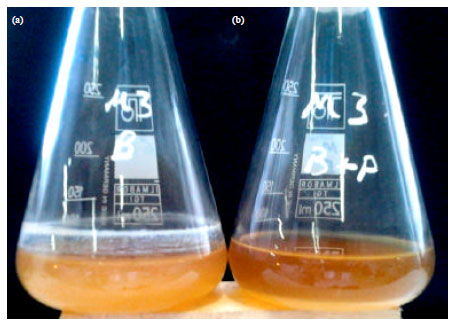 | |
| Fig. 1(a-b): | Enrichment of coliphages isolated from Zayandehrood river, Isfahan, Iran (a) Positive control: the growth of K. pneumoniae SBSWIF3, K. pneumonia PTCC1290 and K. oxytoca PTCC1402 mixture in BHI after 24 h incubation at 37°C and 120 rpm aeration speed and (b) The mixture of three Klebsiella strains and Zayandehrood river filtrate after 24 h incubation in the same condition |
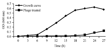
The macroscopic and microscopic characterizations of isolated bacteria on EMB was shown in Table 2. The PCR of purified bacterial DNA with universal primers showed the 370 bp band in gel electrophoresis. The analysis of genomic sequence of 16s-rDNA using Finch TV and Mega4 and then BLAST confirmed that the isolated strain from Isfahan wastewater sample was related to species K. pneumoniae. This strain was named K. pneumoniae SBSWIF3 and its genomic sequence has been deposited in GenBank with accession No. JN836932.1. The addition of Zayandehrood River filtrate to K. pneumoniae SBSWIF3, K. pneumonia PTCC1290 and K. oxytoca PTCC1402 in BHI culture media was resulted in complete clearance of BHI after 24 h incubation at 37°C (Fig. 1a-b). Spotting the enriched bacteriophage filtrate on the BHA culture of K. pneumoniae SBSWIF3, K. pneumonia PTCC1290 and K. oxytoca PTCC1402 showed big phage plaques after overnight incubation at 37°C (Fig. 2a-c). The growth curve of K. pneumonia SBSWIF3 before and after treatment with specific lytic bacteriophages during 30 h incubation at 37°C showed that the isolated specific coliphages could inhibit the normal growth of bacterium. Figure 3 shows the effect of coliphages on the growth curve of K. pneumonia SBSWIF3. The growth curve of K. pneumonia PTCC1290 after treating with isolated coliphages during 30 h incubation period at 37°C showed that the coliphages prevented the normal growth of K. pneumonia PTCC1290 (Fig. 4).
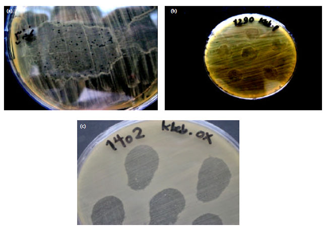 | |
| Fig. 2(a-c): | Spotting of enriched coliphages isolated from Zayandehrood river on the BHA spreading culture of (a) K. pneumoniae SBSWIF3, (b) K. pneumonia PTCC1290 and (c) K. oxytoca PTCC1402 after 24 h incubation at 37°C. The bacterial lawn was inoculated with different phage dilutions of 10-1-10-7 |
 | |
| Fig. 3: | Growth curve of K. pneumonia SBSWIF3 in the absence and presence of lytic specific bacteriophages isolated from Zayandehrood river during 30 h incubation at 37°C |
Also the growth curve of K. oxytoca PTCC1402 after treating first lytic coliphage had a small hexagonal head with diameter of 27.28 nm and a long noncontractile tail measuring 204.5 nm in length and 13.63 nm in diameter. According to its size and morphology, it was suggested that the first isolated coliphage was most probably related to Myoviridae family of bacteriophages (Fig. 6). The second lytic bacteriophage isolated from Zayandehrood River, Isfahan, Iran that had lytic effects on K. pneumoniae SBSWIF3, with isolated coliphages during 30 h incubation period at 37°C showed that the coliphages prevented the normal growth of K. oxytoca PTCC1402 (Fig. 5).
 | |
| Fig. 4: | Comparison between normal growth curve of K. pneumonia PTCC1290 and after treatment with specific coliphaes isolated from Zayandehrood River during 30 h incubation at 37°C |
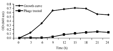 | |
| Fig. 5: | Comparison between normal growth curve of K. oxytoca PTCC1402 and after treatment with specific coliphaes isolated from Zayandehrood River during 30 h incubation at 37°C |
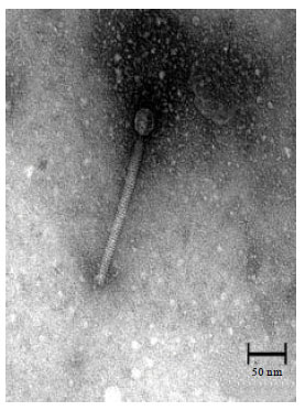 | |
| Fig. 6: | TEM micrograph of the first specific lytic bacteriophage of K. pneumoniae SBSWIF3, K. pneumonia PTCC1290 and K. oxytoca PTCC1402 isolated from Zayandehrood river, Isfahan, Iran related to Myoviridae family of bacteriophages. Head diameter: 27.28 nm, tail length: 204.5 nm, Tail diameter: 13.63 nm (Bar: 50 nm) |
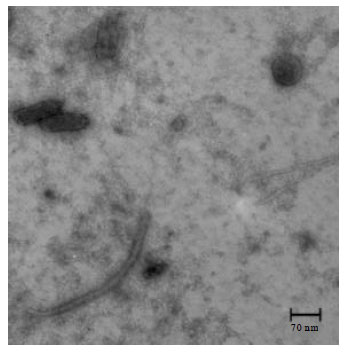 | |
| Fig. 7: | TEM micrograph of the second specific lytic bacteriophage of K. pneumoniae SBSWIF3, K. pneumonia PTCC1290 and K. oxytoca PTCC1402 isolated from Zayandehrood river, Isfahan, Iran related to Podoviridae family of bacteriophages. Head: 98x35 nm, Tail: 14x14 nm (Bar: 70 nm) |
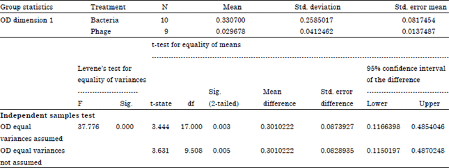
The transmission electron microscopy of purified phage solution isolated from the mix culture of K. pneumoniae SBSWIF3, K. pneumonia PTCC1290 and K. oxytoca PTCC1402 showed two types of lytic bacteriophages. The K. pneumonia PTCC1290 and K. oxytoca PTCC1402 had oval head measuring 98x35 nm and a small tail measuring 14x14 nm. According to TEM characterizations, it was suggested that second coliphage was most probably related to Podoviridae family of bacteriophages (Fig. 7). The coliforms MPN test of Isfahan municipal wastewater using 3 sets of 5 tubes containing LB culture media after 2 h incubation at 37°C showed that all of the 15 tubes were positive regarding turbidity and gas production and the MPN (5-5-5) was measured 2400 using MPN standard scale. Addition of phage solution includes both coliphages with the dilution of 10-7 (phage titer of 2.15x10⁹ mL-1) to MPN series of Isfahan wastewater after 2 h incubation at 37°C resulted in MPN significant decline. The new MPN (5-3-1) was measured 110 using MPN standard scale. The comparison of MPN index of wastewater coliforms before and after treatment with coliphages showed that after 2 h incubation, MPN from 2400 has been reduced to 110 i.e., 22 folds reduction of coliform’s load in wastewater. The statistical t test of two independent samples confirmed the effect of bacteriophages on the reduction of coliform's load after treatment of wastewater with coliphage mixture (Table 3). According to data obtained from Leven’s test for equality of variances, the decision making criteria is 0.00 and of sig (2-tailed) test is 0.003 and is smaller than 5% so the Null hypothesis is not confirmed. This statistical test indicates that the MPN decline of wastewater coliforms after treatment with coliphages mixture is meaningful.
| Table 3: | Effect of coliphages isolated from Zayandehrood River on the reduction of coliform’s load using t test of two independent samples |
 | |
DISCUSSION AND CONCLUSION
The use of bacteriophages in controlling bacterial population has been distributed from medicine to agriculture and food industries but in environmental aspects including wastewater treatment has been poorly investigated (Marks and Sharp, 2000; Chanishvili et al., 2001; Withey et al., 2005). The effect of cyanobacterial specific bacteriophages in preventing bloom has been reported in a research (Mole et al., 1997). There are few reports suggesting that the normal presence of bacteriophages in sewage could be useful in wastewater treatment especially in the activated sludge procedure (Hantula et al., 1991; Hertwig et al., 1999; Khan et al., 2002) as well as focusing on the phages as biological tracers of pathogenic bacteria in water and wastewater treatment (Borrego et al., 1987; Abdulla et al., 2007). Zumstein et al. (2000) have studied the interactions of bacterial population and bacteriophages in anaerobic wastewater treatment using laboratorial anaerobic digesters. They suggested that bacteriophages could influence the dominance of bacterial strains during the process (Zumstein et al., 2000). Thomas et al. (2002) have reported the possible application of bacteriophages as antifoam agents in activated sludge system. They have isolated lytic phages related to family Siphoviridae that could destroy foam producing bacteria (Thomas et al., 2002). According to the latest ICTV (International Committee for Taxonomy of Viruses) classification of bacteriophages, the Enterobacteriaceae have 982 different bacteriophages among which 344 phages have been related to Myoviridae, 297 phages were members of Siphoviridae and 265 phages were related to Podoviridae (Ackermann, 2007). So far there is no report addressing the use of coliphages as useful biological control for eliminating or reducing the coliform's microbial load in wastewater treatment. In this research we isolated and identified two novel bacteriophages related to Myoviridae and Podoviridae families of bacteriophages from Zayandehrood River, Isfahan, Iran that had lytic effects on K. pneumoniae SBSWIF3, K. pneumonia PTCC1290 and K. oxytoca PTCC1402 as coliform’s index. These two isolated coliphages did not morphologically match to known Myoviruses and Podoviruses that specifically attack to K. pneumoniae and K. oxytoca strains (Ackermann, 2007). The treatment of municipal sewage with the coliphage mixture resulted in 22 folds decrease of the coliform’s MPN from 2400-110 after 2 h incubation, the period that could be accessible and amenable in a wastewater treatment plant. The reduction of coliform’s population as an important index of wastewater treatment efficacy should be monitored carefully during the wastewater treatment procedure (Greenberg et al., 1992). Many wastewater treatment plants with the age of 40-50 years especially in non-developing countries may lost their effectiveness to remove or decrease the number of pathogenic bacteria including the coliforms during the treatment process. So, we suggested that the use of bacteriophages for reduction of pathogenic bacteria in sewage along with other standard methods like activated sludge could be considered as an effective and simple alternative for costly replacement of instruments and establishments of the old wastewater treatment plants. In conclusion this is the first report of isolation and identification of two novel lytic Myovirus and Podovirus from Zayandehrood River in Isfahan, Iran that had lytic effects on K. pneumoniae SBSWIF3, K. pneumonia PTCC1290 and K. oxytoca PTCC1402 strains as well as coliform’s population of Isfahan municipal wastewater. Phage therapy of coliforms using these specific coliphages could be an asset in wastewater treatment and appointing approach in the area of modern environmental biotechnology.
ACKNOWLEDGMENTS
This research funded by an operating grant of the Vice Chancellor of Research of Falavarjan Branch, Islamic Azad University, Isfahan, Iran. We thank Ali Safavi from the School of Veterinary Sciences, University of Shiraz, Iran for his technical assistance in preparing the TEM micrographs.
REFERENCES
- Abdulla, H., I. Khafagi, M. Abd El-Kareem and A. Dewedar, 2007. Bacteriophages in engineered wetland for domestic wastewater treatment. Res. J. Microbiol., 2: 889-899.
CrossRefDirect Link - Ackermann, H.W., 2007. 5500 phages examined in the electron microscope. Arch. Virol., 152: 227-243.
CrossRefDirect Link - Baggi, F., A. Demarta and R. Peduzzi, 2001. Persistence of viral pathogens and bacteriophages during sewage treatment: Lack of correlation with indicator bacteria. Res. Microbiol., 152: 743-751.
Direct Link - Maal, K.B., M. Bouzari and F.A. Zavareh, 2012. Characterization of two lytic bacteriophages of Streptococcus sobrinus isolated from Caspian sea. Asian J. Biol. Sci., 5: 138-147.
CrossRefDirect Link - Maal, K.B., M. Bouzari and F.A. Zavareh, 2010. Identification of Streptococcus salivarius bacteriophage isolated from Persian Gulf as a potential agent for dental caries phage therapy. Afr. J. Microbiol. Res., 20: 2127-2132.
Direct Link - Borrego, J.J., M.A. Morinigo, A. Devicente, R. Cornax and P. Romero, 1987. Coliphages as an indicator of faecal pollution in water. Its relationship with indicator and pathogenic micoorganisms. J. Water Res., 21: 1473-1480.
Direct Link - Chanishvili, N., T. Chanishvili, M. Tediashvili and P.A. Barrow, 2001. Phages and their application against drug-resistant bacteria. J. Chem. Technol. Biotechnol., 76: 689-699.
CrossRefDirect Link - Drozdova, O.M., R.N. An, T.G. Chanishvili and M.L. Livshits, 1988. [Experimental study of the interaction of phages and bacteria in the environment]. Zhurnal Mikrobiologii Epidemiologii Immunobiologii, 7: 35-39, (In Russian).
PubMed - Hantula, J, A. Kurki, P. Vuoriranta and D.H. Bamford, 1991. Ecology of bacteriophages infecting activated sludge bacteria. Applied Environ. Microbiol., 57: 2147-2151.
Direct Link - Hennes, K.P. and M. Simon, 1995. Significance of bacteriophages for controlling bacterioplankton growth in a mesotrophic lake. Applied Environ. Microbiol., 61: 333-340.
Direct Link - Hertwig, S., A. Popp, B. Freytag, R. Lurz and B. Appel, 1999. Generalized transduction of small Yersinia enterocolitica plasmids. Applied Environ. Microbiol., 65: 3862-3866.
Direct Link - Khan, M.A., H. Satoh, T. Mino, H. Katayama, F. Kurisu and T. Matsuo, 2002. Bacteriophage-host interaction in the enhanced biological phosphate removing activated sludge system. Water Sci. Technol., 46: 39-43.
Direct Link - Marks, T. and R. Sharp, 2000. Bacteriophages and biotechnology: A review. J. Chem. Technol. Biotechnol., 75: 6-17.
CrossRefDirect Link - Mole, R., D. Meredith and D.G. Adams, 1997. Growth and phage resistance of Anabaena sp. strain PCC 7120 in the presence of cyanophage AN-15. J. Applied Phycol., 9: 339-345.
CrossRefDirect Link - Reid, G., J. Howard and B.S. Gan, 2001. Can bacterial interference prevent infection? Trends Microbiol., 9: 424-428.
CrossRef - Sambrook, J. and D.W. Russell, 2001. Molecular Cloning: A Laboratory Manual. 3rd Edn., Cold Spring Harbor Laboratory Press, New York, USA., ISBN-13: 9780879695774, Pages: 2344.
Direct Link - Soothill, J.S., 1994. Bacteriophage prevents destruction of skin grafts by Pseudomonas aeruginosa. Bruns, 20: 209-211.
CrossRefDirect Link - Smith, H.W., M.B. Huggins and K.M. Shaw, 1987. The control of experimental Escherichia coli diarrhoea in calves by means of bacteriophages. J. Genet. Microbiol., 133: 1111-1126.
CrossRef - Thomas, J., J. Soddell and D. Kurtbke, 2002. Fighting foam with phages? Water Sci. Technol., 46: 511-518.
Direct Link - Wendt, C., S. Schutt, A.H. Dalpke, M. Konrad and M. Mieth et al., 2010. First outbreak of Klebsiella pneumonia carbapenemase (KPC)-producing K. pneumonia in Germany. Eur. J. Clin. Microbiol. Infect. Dis., 29: 563-570.
CrossRefDirect Link - Withey, S., E. Cartmell, L.M. Avery and T. Stephenson, 2005. Bacteriophages-potential for application in wastewater treatment processes. Sci. Total Environ., 339: 1-18.
Direct Link - Won, S.Y., L.S. Munoz-Price, K. Lolans, B. Hota, R.A. Weinstein and M.K. Hayden, 2011. Emergence and rapid regional spread of Klebsiella pneumoniae carbapenemase-producing Enterobacteriaceae. Clin. Infect. Dis., 53: 532-540.
CrossRefDirect Link - Zumstein, E., R. Moletta and J.J. Godon, 2000. Examination of two years of community dynamics in an anaerobic bioreactor using fluorescence Polymerase Chain Reaction (PCR) single‐strand conformation polymorphism analysis. Environ. Microbiol., 2: 69-78.
CrossRefDirect Link