
Radwa Wahid Mohamed
Department of Biochemistry and Nutrition, Women�s College for Art Science and Education, Ain Shams University, Cairo, Egypt
LiveDNA: 20.38786
ORCID: 0000-0002-9044-4507
Marwa Sharaky
Pharmacology Unit, Department of Cancer Biology, Cairo University, Giza, Egypt
Sameh Hamed Ismail
Faculty of Nanotechnology for Postgraduate Studies, Cairo University, Sheikh Zayed Campus, 6th of October City, Giza 12588, Egypt
Nehad Naem Hamed Shosha
Department of Biochemistry and Nutrition, Women�s College for Art Science and Education, Ain Shams University, Cairo, Egypt
Pakistan Journal of Biological Sciences
Year: 2022 | Volume: 25 | Issue: 10 | Page No.: 952-960


ABSTRACT
Background and Objective: Cancer is a complex interaction among multiple signalling pathways involving a variety of target molecules. Nanoparticles were used in cancer treatment because of their intrinsic anticancer properties. The use of plant extracts in the preparation of metallic nanoparticles as a convenient substitute has been proposed. This study assessed the cytotoxic, antioxidant and apoptotic effects of copper nanoparticles shelled with either turmeric or sumac biosynthesized as core-shell nanostructures on the liver tumour cell line (Huh-7). Materials and Methods: The nanostructures were synthesized by sonochemical method and characterization was done to confirm the successful synthesis within the nanoscale. Cytotoxicity of nanostructures was investigated on Huh-7 and normal kidney epithelial cell lines (VERO). Malondialdehyde, nitric oxide, reduced glutathione and superoxide dismutase were estimated in cell lysate to assess the antioxidant properties of nanostructures. Caspase-3 was also measured as an apoptotic marker. Results: Both nanostructures had low IC50 on Huh-7 cells and a non-toxic effect on VERO cells. The cytotoxic effect was coupled with a significant increase in antioxidant activities and apoptotic efficiency compared to control. Conclusion: The findings summarized here support the utilization of biosynthesized copper with turmeric or sumac as core-shell nanostructures as a novel chemotherapeutic drug for cancer treatment that improves antioxidant effect that modulates the side effect of cytotoxicity. Also, it is obvious that copper nanostructure biosynthesized with turmeric has a more advanced effect than that of sumac.
PDF Abstract XML References Citation
Copyright: © 2022. This is an open access article distributed under the terms of the creative commons attribution License, which permits unrestricted use, distribution and reproduction in any medium, provided the original author and source are credited.
How to cite this article
Radwa Wahid Mohamed, Marwa Sharaky, Sameh Hamed Ismail and Nehad Naem Hamed Shosha, 2022. Pivotal Role of Copper Nanoparticles Shelled by Turmeric or Sumac on Huh-7 Cell Line Cytotoxicity, Apoptosis and Antioxidant Capacity. Pakistan Journal of Biological Sciences, 25: 952-960.
DOI: 10.3923/pjbs.2022.952.960
URL: https://scialert.net/abstract/?doi=pjbs.2022.952.960
DOI: 10.3923/pjbs.2022.952.960
URL: https://scialert.net/abstract/?doi=pjbs.2022.952.960
INTRODUCTION
Cancer is the most common disease that causes a growing health problem globally. It affects millions of people and significantly continues to increase rapidly in the following years. Every year, many people die due to cancer all over the world1. Hepatocellular carcinoma is the third leading cause of cancer mortality worldwide, with more than 500,000 people affected annually2. Therapeutic strategies of plant origin are a better choice as both dietary plant products and their isolated active constituents are against the development and progression of cancer1.
One of these plants is Rhus coriaria L. commonly known as sumac (Sum), used as a spice, condiment and flavouring agent, especially in the Mediterranean region3. Owing to its generous beneficial values, Sum has been used in traditional medicine for the management and treatment of many ailments. This plant is rich in various classes of phytochemicals including flavonoids, tannins, polyphenolic compounds, organic acids and many others. Sumac possesses powerful antioxidant capacities that have ameliorative and therapeutic benefits for many common diseases including cardiovascular disease, diabetes and cancer4. Probably, the remedial properties of Sum are related to these active compounds3.
The turmeric (Tur) (Curcuma longa L.) plant is a member of the ginger family (Zingiberaceae). Although it has more than 300 active components, turmeric yellow or orange pigmentation related to curcumin (Cur) that is obtained from its root is the main biologically active component constituting the basis for the medicinal properties of this plant. Laboratory studies have presented some valuable results in terms of curcumin’s antioxidant, anti-inflammatory and anticancer properties in particular5.
Copper nanoparticles (CuNPs) indicated high toxicity against numerous types of tumour cells such as human pulmonary adenocarcinoma (A549)6. Therefore, it seems that the Cu NPs-based products on the nanoscale have the potential to be used as the chemotherapy drug. On the other hand, it is considered as a rule that apoptosis-inducing agents are the only cytotoxic molecules that can be used as chemotherapeutic drugs7. In vitro anticancer results of Nagajyothi et al.8, indicated that Copper oxide nanoparticles (CuO NPs) induced intracellular ROS generation in a dose-dependent manner and significantly reduced cervical carcinoma colonies.
Synthesis of metal nanoparticles for improving therapeutic index and drug delivery is coming up as an attractive strategy in mainstream cancer therapeutic research9. The size, morphology and stability of NPs can also be easily optimized for medicinal and pharmaceutical usage using biosynthetic methods. Biosynthesis of CuNPs and copper oxide (CuO NPs) is more advantageous than chemical and physical synthesis as it is a clean, nontoxic, cost-effective and environmentally friendly approach. It bypasses the use of harsh, toxic and expensive chemicals and, instead, utilizes natural biological entities. Among these natural elements used in the biosynthesis of Cu and CuO NPs are plants that serve as a reducing, stabilizing and capping agent during NP synthesis. This makes them safer and more effective10.
Biosynthesized metallic nanoparticles have a wide range of pharmaceutical applications that contributed to many pharmaceutical products11. Previously, biosynthesis of silver nanoparticles (AgNPs) was accomplished using the aqueous extract of Tur or Sum, in which plant biomaterials were used as a reducing as well as a capping agent12,13.
The synthesis of CuNPs from plant extracts had contributed to numerous industries and pharmaceutical applications14. Thus, in the current study, CuNPs were synthesized using Tur or Sum to improve their antitumor effects. Cytotoxicity of turmeric or sumac-shelled CuNPs was explored in Huh-7 and VERO cell lines using in vitro assays. Antioxidant and apoptotic properties were also conducted to reveal the inhibitory effects of these nanostructures.
MATERIALS AND METHODS
Study area: The study was carried out at the Department of Pharmacology, National Cancer Institute (NCI), Egypt in January, 2022.
Materials: Copper sulfate and vitamin C were purchased from Sigma Aldrich Company, Egypt. Turmeric (Tur) and sumac (Sum) were purchased from the Ministry of Agriculture (Giza, Egypt).
Synthesis and characterization of nanostructures
Synthesis of copper nanoparticles: Copper nanoparticles (CuNPs) were synthesized by the precipitation method assisted by the sonochemical method in which CuNPs are precipitated from copper sulfate (source material) by vitamin C (reducing agent) under subjection to ultrasonication (Sono-chemical) according to Ismail et al.15. Copper with Tur core-shell nanostructure (Tur+Cu) was synthesized by adding CuNPs to Tur solution, then the solution was subjected to ultrasonic irradiation (60 kHz with cycle 5, plus every 3 sec and amplitude of 100% for 2 hrs). On the other hand, Copper with Sum core-shell nanostructure (Sum+Cu) was synthesized in two steps. The first step was the preparation of Sum water extract then, the extract was freeze-dried. In the second step, CuNPs were added to the dried water extract of Sum and subjected to ultrasonic irradiation (60 kHz with cycle 1, plus every 6 sec and amplitude of 100% for 4 hrs) before it was precipitated and air dried for 24 hrs as previously described by Shosha et al.16.
Characterization of nanostructures: Core-shell nanostructures were investigated to confirm the crystalline phase and chemical composition of synthesis nanomaterials using an X-ray diffractometer (XRD, D8-Discover, Bruker, German) working at a current of 35 mA and voltage of 35 kV and transmission electron microscope TEM (JEOL, TEM-2100, Japan) worked at a potential of 25 kV and were used to study the morphology, shape and surface topography of the core-shell nanostructure. Atomic Force Microscope (AFM, 5600LS, Agilent) was performed to study 2D and 3D surface topographic images of Core-shell nanostructure.
Human cancer cell lines: In this study, different concentrations of Tur+Cu and Sum+Cu nanostructures were in vitro scanned for their anticancer activity on the human liver tumour (Huh7) cell line and normal (VERO) cell line. The cell lines were obtained from the American Type Culture Collection (ATCC, Minnesota, USA) and maintained at NCI, Cairo, Egypt.
Cytotoxic assay: The antitumor activities of Tur+Cu and Sum+Cu nanostructures were evaluated by sulforhodamine-B (SRB) assay17 using human liver tumour cells (Huh-7). Briefly, cells were seeded at a density of 3×10³ cells/well in 96-well microtiter plates. They were left to attach for 24 hrs. Next, Huh-7 and VERO cell lines were treated with nanostructures at different concentrations of 0, 62.5, 125, 250 and 500 μg mL–1. For each concentration, three wells were used and incubation was continued for 48 hrs. Dimethyl sulfoxide (DMSO) was used as a control vehicle (1% v/v). At the end of incubation, cells were fixed with 20% trichloroacetic acid and stained with 0.4% SRB dye. The optical density (O.D.) of each well was measured spectrophotometrically at 570 nm using an ELISA microplate reader (TECAN sunrise™, Germany). The mean survival fraction at each nanostructure was calculated as follows: O.D. of the treated cells/O.D. of the control cells. The IC50 (concentration that produces 50% of cell growth inhibition) value of either Tur+Cu or Sum+Cu was calculated using sigmoidal dose-response curve-fitting models (Graph Pad Prism software, version 5).
Determination of oxidative stress markers
Determination of malondialdehyde content (MDA): Lipid peroxidation products were quantified by measuring malondialdehyde MDA level in cell culture lysate of control and treated cells using lipid peroxidation (MDA) assay kit (Sigma Aldrich Chemical Co., St. Louis, USA) following the manufacturer’s instructions. The MDA level was calculated as nmol of MDA/mg protein. The absorbance was determined at 532 nm using a spectrophotometer (Spectronic, Milton Roy Co., USA).
Determination of superoxide dismutase (SOD): Superoxide dismutase (SOD) was measured in cell culture lysate of control and treated cells by SOD determination kit (Sigma Aldrich Chemical Co., St. Louis, USA) following the manufacturer’s instructions. SOD activity was calculated relative to the corresponding protein content. The absorbance of the supernatant was determined at 450 nm using a spectrophotometer (Spectronic, Milton Roy Co., USA). The experiment was carried out 3 independent times.
Determination of reduced glutathione (GSH) content: Reduced glutathione was determined by adopting Ellman’s method. Huh-7 cells were harvested, the protein was precipitated with trichloroacetic acid and Ellman’s reagent [5,5-dithiobis-(2-nitrobenzoic acid)] (Sigma Aldrich Chemical Co., St. Louis, USA) was added to the supernatant. The absorbance was read at 405 nm and total GSH was calculated as μM of GSH/mg protein.
Determination of nitric oxide (NO) content: Nitric oxide was determined in culture media of the control and treated cells spectrophotometrically at 540 nm using an ELISA microplate reader (TECAN Sunrise™, Germany)18 following an incubation period of 30 min. The level of total nitrite/nitrate was expressed as mM supernatant media and determined using a standard curve.
Determination of caspase-3 activity as an apoptotic marker: Activity of caspase-3 was measured in cell lysate following kit instruction (Biovision, USA) (Cat. No K106-25) spectrophotometrically at 450 nm. The experiment was carried out 3 independent times.
Statistical analysis: Data were analyzed by Statistical Package for Social Science (SPSS) version 16.0. Statistical differences between groups were performed using a One-way Analysis of Variance (ANOVA). Values are presented as Mean±Standard Deviation (SD) the mean difference was considered significant at (p<0.05)19.
RESULTS
Synthesis and characterization of Tur+Cu and Sum+Cu nanostructures: TEM images illustrated in Fig. 1 revealed the formation of core-shell nanostructure with shell (Tur Fig.1a or Sum Fig.1b) is smaller than the core (Copper nanoparticles). Core had rough edges because of metal corrosion of Cu NPs from Sum or Tur nanostructures. The size of Tur+Cu nanostructures was about 85 nm, while Sum+Cu nanostructure was about 75 nm. The data in Fig. 2 showed an XRD pattern that illustrated the main characteristic peaks of copper at 2 thetas 43.3 and 50.4° while Sum and Tur have no XRD characteristic peaks (amorphous nature). The 2D and 3D AFM images of Fig. 3a-b confirmed with TEM image of control shape and size of synthesis method. However, both core-shell nanostructures have spherical shapes without any agglomeration or aggregation in a certain area.
Cytotoxic activities of Tur+Cu and Sum+Cu nanostructures: The cytotoxic activities of Tur+Cu and Sum+Cu nanostructures, were evaluated against normal VERO Fig. 4a and liver tumour Huh-7 cell lines at different concentrations (0, 62.5, 125, 250 and 500 μg mL–1). Results of the present study revealed that the Huh-7 cell line was affected by different concentrations of both nanostructures. Data shown in Fig. 4b revealed that Tur+Cu nanostructure has IC50 of 217 μg mL–1, whereas Sum+Cu has IC50 of 354 μg mL–1.
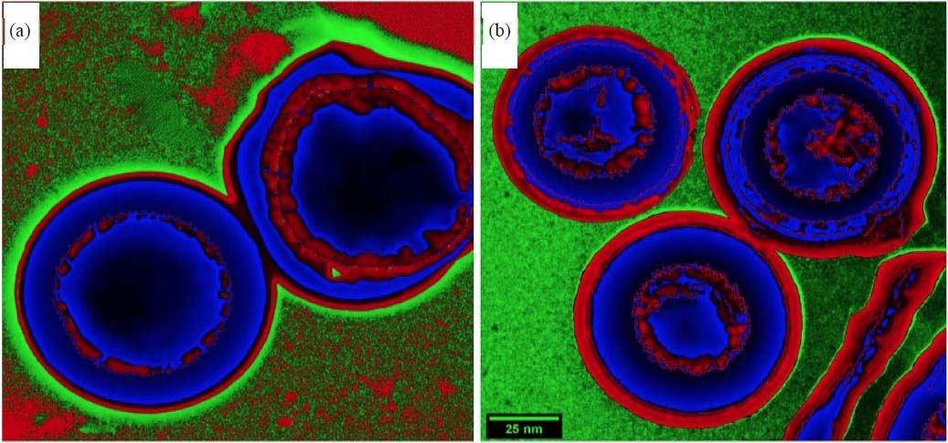 |
| Fig. 1(a-b): | TEM image, (a) Tur+Cu and (b) Sum+Cu nanostructures |
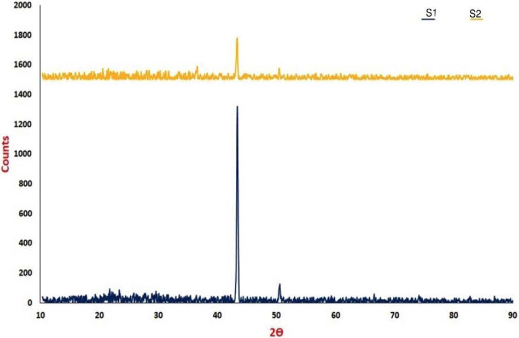 |
| Fig. 2: | XRD pattern of S1 (Tur+Cu) and S2 (Sum+Cu) nanostructures |
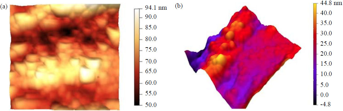 |
| Fig. 3(a-b): | AMF image, (a) 2D image of Tur+Cu and (b) 3D image of Sum+Cu |
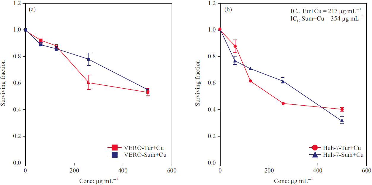 |
| Fig. 4(a-b): | IC50 of Tur+Cu and Sum+Cu nanostructures on (a) normal (VERO) and (b) liver tumour (HuH-7) cell lines |
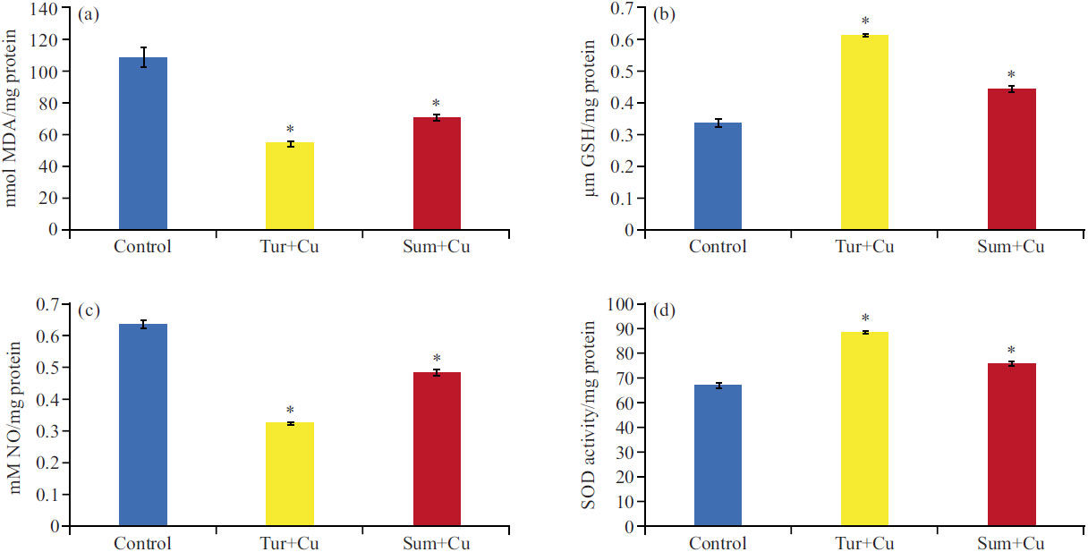 |
| Fig. 5(a-d): | Antioxidant activity of Tur+Cu and Sum+Cu nanostructures compared to control, (a) Malondialdehyde (MDA) levels, (b) Reduced glutathione (GSH) levels, (c) Nitric oxide (NO) levels and (d) Superoxide dismutase (SOD) activities Values are represented as mean±SD, (p<0.05), *indicated a significant difference compared to the control group and x-axis represents treated Huh-7 cell lines with Tur+Cu or Sum+Cu compared to untreated cell lines (treated groups) |
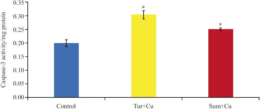 |
| Fig. 6: | Caspase-3 activity of Tur+Cu and Sum+Cu nanostructures compared to control Values are represented as mean±SD, (p<0.05), *indicated significant difference compared to control group and x-axis represents treated Huh-7 cell lines with Tur+Cu or Sum+Cu compared to untreated cell lines (treated groups) |
This indicated a clear-concentration response relationship and a higher anticancer activity of Tur+Cu more than Sum+Cu. The biosynthesized Tur+Cu and Sum+Cu showed anticancer activity in the treated Huh-7 liver tumour cell line and as well as the commendable non-toxic effect on the normal VERO cell line.
Antioxidant activity of Tur+Cu and Sum+Cu nanostructures: The results presented in Fig. 5a-d, in general, indicated that both Tur+Cu and Sum+Cu showed excellent antioxidant activity with a significant reduction in MDA by (50.13%, 34.93) and NO levels by (49.21%, 23.34%) with significant elevation in GSH level by (43.44%, 31.55%) and SOD activities by (38.01%, 15.53%), respectively compared to control. Using Tur adds more antioxidant power to CuNPs than using Sum.
Antiapoptotic properties of Tur+Cu and Sum+Cu nanostructures: Based on the obtained results, Fig. 6 showed that both Tur+Cu and Sum+Cu nanostructures significantly increase caspase-3 activity in Huh-7 by 52.27 and 25.63%, respectively compared to control cells. In addition to its higher efficiency to improve antioxidant properties, Tur+Cu nanostructure also showed an advanced effect indicated by higher caspase-3 activities compared to Sum+Cu. nanostructure.
DISCUSSION
Cancer cells are characterized by an increase in the rate of reactive oxygen species (ROS) production and an altered redox environment compared to normal cells. Most chemotherapeutics agents induced elevation of ROS and disrupt redox homeostasis of cancer cells20.
Additionally, it has been reported that the cytotoxicity of nanomaterials is related to intracellular ROS increment. The apoptotic mechanism of heavy metal nanoparticles may correlate with the elevation of ROS7. Upon looking at the results of Sammar et al.21, who showed that cytotoxic activities of 57 plant extracts are not mainly accredited to the level of free radical scavenging but could also be associated with the inhibitory effects via other signalling pathways. However, an in depth analysis of the results from it indicated that the extracts of plants that had free radical scavenging exhibited a certain enrichment toward more cytotoxicity.
Recently the biosynthesis of metal nanoparticles using plant extract earn much attention as the presence of plant active component improve the action of NPs and modulate their side effects. So, this study investigated the antitumor, antioxidant and apoptotic effects of biosynthesized CuNPs as core-shell nanostructure with either Tur or Sum.
Previously, the toxicity of Cu/CuO NPs was evaluated in human lung normal cell lines (WI-38) and human lung carcinoma cell (A549) showing that Cu/CuO NPs induced suppression of normal and carcinoma lung cells viability. Treatment of both cell types with their IC50 of Cu/CuO NPs resulted in DNA damage besides the generation of ROS and consequently the generation of a state of oxidative stress22. Furthermore, CuO NPs were found to induce cytotoxicity in (HepG2) cells in a dose-dependent manner, which was likely to be mediated through tumour suppressor gene p53 and apoptotic gene caspase-3 were up-regulated due to CuO NPs exposure. A decrease in mitochondrial membrane potential with a concomitant increase in the gene expression of the Bax/bcl2 ratio suggested that the mitochondria-mediated pathway involved in CuO NPs induced apoptosis23.
Taking into account, the in vitro cytotoxicity of the biosynthesized Tur+Cu and Sum+Cu tested on normal and liver carcinoma cell lines that exhibited a non-toxic effect on the normal cell line. On the other hand, shelling CuNPs with either Tur or Sum added the properties of their active component to the core CuNPs that reflect a positive effect on the cytotoxic activities of CuNPs indicated by its low cytotoxicity to normal cell lines. The current work indicated that Tur or Sum nanostructures improve the antioxidant status and enhance the apoptotic properties in tumour cells compared to control.
The current results were in agreement with Selvan et al.24, who showed that AgNPs biosynthesized with Tur extract caused high cytotoxicity activity against many cancer cell lines and interestingly induced low cytotoxicity to normal cell lines because Tur extract contains a huge amount of nutrients and phytocompounds including the phenolic compound Cur. The balance between the therapeutic potential and toxic side effects o f a compound is very important when evaluating its usefulness as a pharmacological drug25. The AgNPs biosynthesized with Tur showed comparable antioxidant activity to the standard antioxidant ascorbic acid, although the chemically synthesized AgNPs exhibited lower antioxidant activity24. This indicated the direct role of Tur phytochemicals particularly flavonoids and phenolic compounds in free radical scavenging.
In mice, Cur- the main active component of Tur- administration was used to overcome AgNPs side effects and provided significant antioxidant effects for the treatment of the ehrlich ascites carcinoma cells (EAC). Curcumin may decrease the production of free radicals which could lead to decreasing hepatic antioxidant enzyme activities catalase, glutathione peroxidase and glutathione reductase in all groups treated with AgNPs that resulted in a significant reduction in lipid peroxidation26.
Curcumin coadministration with chemotherapeutic drugs increased the drug sensitivity. Moreover, it hinders the colonization of cancer cells, which further provides better protection27. However, due to its low bioavailability, the benefits are not utilized efficiently in the tumour site and thus the importance of nano curcumin is more focused due to its ability to enhance the benefits of Cur as it increases the binding of the phytoconstituents in the target site and helps in treating the tumours28. The effect of nano curcumin on breast cancer cell lines was investigated and it was deduced that nano curcumin was found to be an effective antitumor agent along with low toxicity29. A previous study demonstrated that curcumin nano-complexes turned out to exhibit the highest selective cytotoxicity and also showed a significant reduction in solid tumour volume in ascites tumour-bearing mice without harmful effects on liver and kidney function30. Furthermore, Nguyen et al.31 found that using nano curcumin along with anticancer drugs showed promising antiproliferative and antitumor effects in cancer cells which further helps in other cancer therapies.
Various studies reported that Cur induces apoptosis through intrinsic signalling pathways by depolarizing the mitochondrial membrane and triggering the release of cytochrome c followed by cleavage of caspase-9 and caspase-332,33.
The present results go hand in hand with many previous works that studied the antioxidant and apoptotic effects of Sum extract on many types of cancer cells. The extraction of Sum plant was used for the ecological preparation of nanoparticles with four metal ions: Zn2+, Cu2+, Ag+ and evaluated against known cancer cell lines, e.g., human colorectal adenocarcinoma cells (Caco-2), human liver cancer cell line (HEPG2) and human breast cancer cell line (T47D) using the SRB assay. CuNPs and FeNPs from Sum extract showed good cytotoxicity against HEPG2 and T47D cell lines34. The study of Ghorbani et al.35, was conducted to determine the cytotoxic and apoptotic effects of AgNPs synthesised from Sum extract on human breast cancer cells (MCF-7). The apoptosis of MCF-7 cells was induced via upregulation of Bax and downregulation of Bcl-2.
On the other hand, microglia cells as in vitro model for neuroinflammation in Khalil et al.36 study, found that treatment with Sum fruits ethanolic extract significantly decreased the release of NO due to the inhibition of mRNA of the Inducible Nitric Oxide Synthase (iNOS) enzyme.
El Hasasna et al.37, found that Sum inhibited angiogenesis and reduced Vascular Endothelial Growth Factor (VEGF) production in both triple-negative breast cancer cell line (MDA-MB-231) and Human Umbilical Vein Endothelial Cells (HUVECs) and downregulated the inflammatory cytokines TNF-α, IL-6 and IL-8. The underlying mechanism for Sum effects appears to be through inhibiting NO pathways.
Sumac treatment in MCF-7 and MDA-MB-231 cell lines showed a time-dependent trend of caspase-3 activation. Sumac treatment induced mitochondrial stress that causes the release of antiapoptotic Bcl-2 protein complexes from mitochondria to cytosol38.
The most important results described in the present study identified Tur or Sum nanostructures as the promising therapeutic candidate that modulate tumour cell viability while improving the antioxidant capacity and apoptotic activity.
CONCLUSION
This study proved that biosynthesized CuNPs as core-shell nanostructures with Tur or Sum were successfully synthesized. It was obvious that Cu-shelled nanostructures with either Tur or Sum had a cytotoxic effect on the liver tumour cell line and a non-toxic effect on the normal VERO cell line. The cytotoxic effect of both nanostructures was coupled with enhancement of antioxidant effect and induction of apoptotic activities. Although the two tested nanostructures induced cytotoxic effects, the Tur core-shell nanostructure displayed higher cytotoxic, antioxidant and apoptotic effects than its counterpart synthesized by Sum.
SIGNIFICANCE STATEMENT
Nanoparticles synthesized using plant extracts exhibit various pharmaceutical and therapeutic effects. This study designed novel copper nanostructures combined with turmeric, or sumac in core-shell forms and investigate their cytotoxic, antioxidant and apoptotic effects on Human Liver Tumor Cell Line (Huh-7). This work concluded that using turmeric, or sumac in the biosynthesis of Cu nanostructures increases their use as tumour therapeutic agents as they have a cytotoxic and apoptotic effect on the tumour cell. Additionally, they showed an advanced antioxidant effect that reduced the side effect of chemotherapeutic agents. Turmeric core-shell nanostructure exhibited higher cytotoxic, antioxidant and apoptotic effects than the sumac nanostructure did.
REFERENCES
- Gabr, S.A. and A.H. Alghadir, 2021. Potential anticancer activities of Rhus coriaria (sumac) extract against human cancer cell lines. Biosci. Rep., Vol. 41.
CrossRefDirect Link - El-Ahwany, E.G.E., L. Mourad, M.M.K. Zoheiry, H. Abu-Taleb and M. Hassan et al., 2019. MicroRNA-122a as a non-invasive biomarker for HCV genotype 4-related hepatocellular carcinoma in Egyptian patients. Arch. Med. Sci., 15: 1454-1461.
CrossRefDirect Link - Liu, J., A. Zangeneh, M.M. Zangeneh and B. Guo, 2020. Antioxidant, cytotoxicity, anti-human esophageal squamous cell carcinoma, anti-human Caucasian esophageal carcinoma, anti-adenocarcinoma of the gastroesophageal junction, and anti-distal esophageal adenocarcinoma properties of gold nanoparticles green synthesized by Rhus coriaria L. fruit aqueous extract. J. Exp. Nanosci., 15: 202-216.
CrossRefDirect Link - Alsamri, H., K. Athamneh, G. Pintus, A.H. Eid and R. Iratni, 2021. Pharmacological and antioxidant activities of Rhus coriaria L. (sumac). Antioxidants, Vol. 10.
CrossRefDirect Link - Unlu, A., E. Nayir, M.D. Kalenderoglu, O. Kirca and M. Ozdogan, 2016. Curcumin (turmeric) and cancer. JBUON, 21: 1050-1060.
PubMedDirect Link - Lanone, S., F. Rogerieux, J. Geys, A. Dupont and E. Maillot-Marechal et al., 2009. Comparative toxicity of 24 manufactured nanoparticles in human alveolar epithelial and macrophage cell lines. Part. Fibre Toxicol., Vol. 6.
CrossRefDirect Link - Azizi, M., H. Ghourchian, F. Yazdian, F. Dashtestani and H.A. Zeinabad, 2017. Cytotoxic effect of albumin coated copper nanoparticle on human breast cancer cells of MDA-MB 231. PLoS ONE, Vol. 12.
CrossRefDirect Link - Nagajyothi, P.C., P. Muthuraman, T.V.M. Sreekanth, D.H. Kim and J. Shim, 2017. Green synthesis: In-vitro anticancer activity of copper oxide nanoparticles against human cervical carcinoma cells. Arabian J. Chem., 10: 215-225.
CrossRefDirect Link - Kamble, S., B. Utage, P. Mogle, R. Kamble, S. Hese, B. Dawane and R. Gacche, 2016. Evaluation of curcumin capped copper nanoparticles as possible inhibitors of human breast cancer cells and angiogenesis: A comparative study with native curcumin. AAPS PharmSciTech, 17: 1030-1041.
CrossRefDirect Link - Letchumanan, D., S.P.M. Sok, S. Ibrahim, N.H. Nagoor and N.M. Arshad, 2021. Plant-based biosynthesis of copper/copper oxide nanoparticles: An update on their applications in biomedicine, mechanisms, and toxicity. Biomolecules, Vol. 11.
CrossRefDirect Link - Zhang, D., X.L. Ma, Y. Gu, H. Huang and G.W. Zhang, 2020. Green synthesis of metallic nanoparticles and their potential applications to treat cancer. Front. Chem., Vol. 8.
CrossRefDirect Link - Alsammarraie, F.K., W. Wang, P. Zhou, A. Mustapha and M. Lin, 2018. Green synthesis of silver nanoparticles using turmeric extracts and investigation of their antibacterial activities. Colloids Surf. B: Biointerfaces, 171: 398-405.
CrossRefDirect Link - Patra, J.K. and K.H. Baek, 2016. Biosynthesis of silver nanoparticles using aqueous extract of silky hairs of corn and investigation of its antibacterial and anticandidal synergistic activity and antioxidant potential. IET Nanobiotechnol., 10: 326-333.
CrossRefDirect Link - Akintelu, S.A., A.K. Oyebamiji, S.C. Olugbeko and D.F. Latona, 2021. Green chemistry approach towards the synthesis of copper nanoparticles and its potential applications as therapeutic agents and environmental control. Curr. Res. Green Sustainable Chem., Vol. 4.
CrossRefDirect Link - Ismail, S., G. Mohamed, A. Amer and M. Amer, 2020. Comparative killing activity of different nanoparticles and nano-composites based on Dermanyssus gallinae. Nano Biomed. Eng., 12: 338-350.
CrossRefDirect Link - Shosha, N.N.H., R.W. Mohamed, S.H. Ismail and O.M.S.A. Sadek, 2022. Amelioration of potential hazard of copper nanoparticles by bio-synthetic methods using novel turmeric (Curcuma longa), sumac (Rhus coriaria L.), and vitamin B12 shelled nanostructures. Teikyo Med. J., 45: 6445-6460.
Direct Link - Skehan, P., R. Storeng, D. Scudiero, A. Monks and J. McMahon et al., 1990. New colorimetric cytotoxicity assay for anticancer-drug screening. J. Nat. Cancer Inst., 82: 1107-1112.
CrossRefPubMedDirect Link - Miranda, K.M., M.G. Espey and D.A. Wink, 2001. A rapid, simple spectrophotometric method for simultaneous detection of nitrate and nitrite. Nitric Oxide, 5: 62-71.
CrossRefDirect Link - Levesque, R., 2006. SPSS Programming and Data Management: A Guide for SPSS and SAS Users. 3rd Edn., SPSS Inc., Chicago, Illinois, ISBN: 1568273746, Pages: 388.
Direct Link - Kirtonia, A., G. Sethi and M. Garg, 2020. The multifaceted role of reactive oxygen species in tumorigenesis. Cell. Mol. Life Sci., 77: 4459-4483.
CrossRefDirect Link - Sammar, M., B. Abu‑Farich, I. Rayan, M. Falah and A. Rayan, 2019. Correlation between cytotoxicity in cancer cells and free radical‑scavenging activity: In vitro evaluation of 57 medicinal and edible plant extracts. Oncol. Lett., 18: 6563-6571.
CrossRefDirect Link - Fahmy, H.M., N.M. Ebrahim and M.H. Gaber, 2020. In-vitro evaluation of copper/copper oxide nanoparticles cytotoxicity and genotoxicity in normal and cancer lung cell lines. J. Trace Elem. Med. Biol., Vol. 60.
CrossRefDirect Link - Siddiqui, M.A., H.A. Alhadlaq, J. Ahmad, A.A. Al-Khedhairy, J. Musarrat and M. Ahamed, 2013. Copper oxide nanoparticles induced mitochondria mediated apoptosis in human hepatocarcinoma cells. PLoS ONE, Vol. 8.
CrossRefDirect Link - Selvan, D.A., D. Mahendiran, R.S. Kumar and A.K. Rahiman, 2018. Garlic, green tea and turmeric extracts-mediated green synthesis of silver nanoparticles: Phytochemical, antioxidant and in vitro cytotoxicity studies. J. Photochem. Photobiol. B: Biol., 180: 243-252.
CrossRefDirect Link - Yang, H., R.M. Villani, H. Wang, M.J. Simpson, M.S. Roberts, M. Tang and X. Liang, 2018. The role of cellular reactive oxygen species in cancer chemotherapy. J. Exp. Clin. Cancer Res., Vol. 37.
CrossRefDirect Link - El-Fotoh, M.F.A., A.H.A. Hadeed and E.K.G. Kotb, 2017. Toxicological effects on silver nanoparticles as anticarcinogenic agent and its treatment with curcumin. Zagazig Vet. J., 45: 296-304.
CrossRefDirect Link - Ashrafizadeh, M., A. Zarrabi, F. Hashemi, A. Zabolian and H. Saleki et al., 2020. Polychemotherapy with curcumin and doxorubicin via biological nanoplatforms: Enhancing antitumor activity. Pharmaceutics, Vol. 12.
CrossRefDirect Link - Sheikh, E., M.L.B. Bhatt and M. Tripathi, 2017. Role of nano-curcumin: A treatment for cancer. J. Med. Plants Stud., 5: 394-397.
Direct Link - Chopra, H., P.S. Dey, D. Das, T. Bhattacharya and M. Shah et al., 2021. Curcumin nanoparticles as promising therapeutic agents for drug targets. Molecules, Vol. 26.
CrossRefDirect Link - Mohamed, R.W., T.E. Mohamed, G.S.E. Moram, A.M. Badawy and M.F. Zaky, 2018. Comparative study on the effect of nano-curcumin complexes as antitumor in mice. World J. Pharm. Res., 7: 1404-1436.
CrossRefDirect Link - Nguyen, N.T., N.N.T. Nguyen, N.T.N. Tran, P.N. Le and T.B.T. Nguyen et al., 2018. Synergic activity against MCF-7 breast cancer cell growth of nanocurcumin-encapsulated and cisplatin-complexed nanogels. Molecules, Vol. 23.
CrossRefDirect Link - Laubach, V., R. Kaufmann, A. Bernd, S. Kippenberger and N. Zöller, 2019. Extrinsic or intrinsic apoptosis by curcumin and light: Still a mystery. Int. J. Mol. Sci., Vol. 20.
CrossRefDirect Link - Chang, P.Y., S.F. Peng, C.Y. Lee, C.C. Lu and S.C. Tsai et al., 2013. Curcumin-loaded nanoparticles induce apoptotic cell death through regulation of the function of MDR1 and reactive oxygen species in cisplatin-resistant CAR human oral cancer cells. Int. J. Oncol., 43: 1141-1150.
CrossRefDirect Link - Ibrahim, F.Y., A.Y. El-Khateeb and A.H. Mohamed, 2019. Rhus and safflower extracts as potential novel food antioxidant, anticancer, and antimicrobial agents using nanotechnology. Foods, Vol. 8.
CrossRefDirect Link - Ghorbani, P., F. Namvar, M. Homayouni‐Tabrizi, M. Soltani, E. Karimi and P. Yaghmaei, 2018. Apoptotic efficacy and antiproliferative potential of silver nanoparticles synthesised from aqueous extract of sumac (Rhus coriaria L.). IET Nanobiotechnol., 12: 600-603.
CrossRefDirect Link - Khalil, M., A. Bazzi, D. Zeineddine, W. Jomaa, A. Daher and R. Awada, 2021. Repressive effect of Rhus coriaria L. fruit extracts on microglial cells-mediated inflammatory and oxidative stress responses. J. Ethnopharmacol., Vol. 269.
CrossRefDirect Link - El Hasasna, H., A. Saleh, H. Al Samri, K. Athamneh and S. Attoub et al., 2016. Rhus coriaria suppresses angiogenesis, metastasis and tumor growth of breast cancer through inhibition of STAT3, NFκB and nitric oxide pathways. Sci. Rep., Vol. 6.
CrossRefDirect Link - Kubatka, P., M. Kello, K. Kajo, M. Samec and A. Liskova et al., 2021. Rhus coriaria L. (sumac) demonstrates oncostatic activity in the therapeutic and preventive model of breast carcinoma. Int. J. Mol. Sci., Vol. 22.
CrossRefDirect Link