
Maryamalsadat Jalali
Physiology Research Center, School of Medicine, Ahvaz Jundishapur University of Medical Sciences, Ahvaz, Iran
Masoud Hemadi
Fertility, Infertility and Perinatology Research Center, School of Medicine, Ahvaz Jundishapur University of Medical Sciences, Ahvaz, Iran
Ghasem Saki
Physiology Research Center, School of Medicine, Ahvaz Jundishapur University of Medical Sciences, Ahvaz, Iran
Alireza Sarkaki
Physiology Research Center, School of Medicine, Ahvaz Jundishapur University of Medical Sciences, Ahvaz, Iran
Pakistan Journal of Biological Sciences
Year: 2013 | Volume: 16 | Issue: 19 | Page No.: 1010-1015


ABSTRACT
Noise stress is dangerous natural contaminant that produces harmful physiological, psychological and morphological outcomes to the body. So this study was conducted in order to investigate the effects of noise stress on the parenchyma of testis. Healthy mature females rats (n = 20) were mated with the mature male rats and then randomly allocated equally either to experimental or control groups. Experimental group has given daily noise stress up to birth their child. In the second step, the child's pregnant rats of experimental group were distributed to three subgroups as follow: group I (without exposure to noise stress), group II (exposure to noise for 8 weeks) and group III (exposure to noise for 14 weeks) for morphometric analysis of their child's testicles by sacrificing of them at weeks 14. In general, the testes of non-exposed group were grown larger than ones in the noise exposed groups. Moreover, the testes of the experimental group 1 were larger than the other experimental groups. Indeed, the rate of atrophic seminiferous tubules and jumbled appearance of the interstitial space were more observed in the noise stress exposed group than non-exposed ones. In addition, seminiferous tubules analysis revealed that the characteristics of interstitial space cells and epithelial germinative cells of the seminiferous tubules in the control group were better than the noise exposed groups. It seems that the noise stress has negative influences on the fertility of male based on enhancing of the apoptotic process induced by pathogenesis stress and suppressing the kinetics spermatogenesis.
PDF Abstract XML References Citation
Received: December 15, 2012;
Accepted: January 22, 2013;
Published: April 16, 2013
How to cite this article
Maryamalsadat Jalali, Masoud Hemadi, Ghasem Saki and Alireza Sarkaki, 2013. Study of Spermatogenesis Fetal Testis Exposed Noise Stress During and after Natal Period in Rat. Pakistan Journal of Biological Sciences, 16: 1010-1015.
DOI: 10.3923/pjbs.2013.1010.1015
URL: https://scialert.net/abstract/?doi=pjbs.2013.1010.1015
DOI: 10.3923/pjbs.2013.1010.1015
URL: https://scialert.net/abstract/?doi=pjbs.2013.1010.1015
INTRODUCTION
Noise stress, of all variety of environmental contaminants, is the most widespread and dangerous natural contaminant that produces harmful physiological, psychological and morphological outcomes to the body. Noise stress is also known to reduce the reproductive function so that this impairment in reproduction were observed as decreasing the rate of the pregnancy and increased neonatal mortality and teratogeny in the cases that exposed to high frequencies of noise (Swami et al., 2007; Saki et al., 2010). It's noteworthy to mention that the teratogenic and embryo toxic properties of noise stress with an extensive band of high frequency (16-42 kHz) and high-intensity (110 dB) were assessed (Cosa and Cosa, 1989). Moreover, it was reported that noise stress can affect the neuroendocrine gonadal response (Shenaieva and Reznikov, 2003). In addition, some of concentrations of the stress hormones, i.e., somatropin, adrenocorticosterone and nor-epinephrine and as well as testosterone, Follicular Stimulating Hormone (FSH) and Luteinizing Hormone (LH) were impaired (Crino et al., 2013). Instance, the FSH and LH concentrations and with subsequent the testosterone concentration were impaired through dehydration, excessive heat exposure and immobilization (Ganesh and Yajurvedi, 2002; Saki et al., 2010; Naraghi et al., 2010; Dong et al., 2004). Indeed, acute noise stress with intensity of 80 dB impaired the hypothalamic-pituitary-gonadal axis (HPG axis) (Sasagawa et al., 2001). Also, the hypothalamic-pituitary-adrenal activity (HPA axis) was affected by intensity of 85 dB chronic noise stress (Armario and Castellanos, 1984).
It is believed that the noise stress may have negative influences on male reproductive activity (Jalali et al., 2012; Kobegenova et al., 1985). Yu et al. (2004) reported that High Power Microwave (HPM) radiation during few minutes could be affected the germinative and somatic cells of testes parenchyma by increasing apoptotic and necrotic cells which in turn might influence the reproductive activity of the males (Shokri et al., 2013; Lue et al., 2000).
The side effects of noise stress such as dehydration stress, heat stress and immobilization stress, on the fertility of males of different species were also tested in some studies (Ganesh and Yajurvedi, 2002; Saki et al., 2009; Yazawa et al., 1999). Generally, it was suggested that noise stress can affect the male sex hormones and in turn produced changes in the reproductive organs and glands (Lue et al., 2000; Saki et al., 2009). To our knowledge, there has been no research regarding the effects of noise stress on the testes of fetus rats that their mothers placing to high levels of noise from fetal life up to weeks 14 of the worldly life. Therefore, the main objective of the present study is to investigate the effect of noise stress on the parenchyma of testes.
MATERIALS AND METHODS
Study design 1: Healthy Mature females (Albino Wistar) rats (n = 20, 200-250 g) were obtained from the Laboratory Animal Reproduction and Breeding Center, Ahvaz, Iran for doing this study. All animals were kept under a 12/12 h dark/light cycle at a 22±1°C temperature with free access to food and water. In the first step, females rats were mated with the 10-12 weeks old of the same strain male rats and then randomly allocated equally either to experimental (n = 10) or control (n = 10) groups. Experimental group has given daily noise stress up to birth their child.
Study design 2: In the second step, the pregnant rats of experimental group were randomly distributed to three subgroups for morphometric analysis of their child's testicles by sacrificing of them at weeks 14 (Fig. 1).
Experimental group I (group I) was subjected to neonatal male rats that growing up to maturity period without exposure to noise stress; experimental group II (group II), in which the neonatal male rats were exposure to noise for 8 weeks and after that, to maturing time were not exposure to noise; experimental group III (group III) served as the neonatal male rats received noise stress continually for 14 weeks. The control group and their fetuses were maintained like experimental groups to maturing time in all aspects except exposure to noise. These experiments were carried out in accordance with national guidelines and protocols as approved by the National Institute of Health Guide.
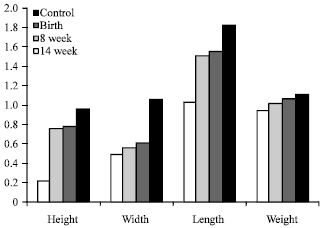 | |
| Fig. 1: | The mean of weight, length, width and height of testicles in experimental and control groups |
Method of noise exposure: The cage of experimental group transported to the room which has dimensions of 3x4x3 and was lagged by wood and acoustic segments (anti loud voice). In the room that experimental group is located, the set which produce noise of WHITE NOISE was being prepared for 19 O’clock in the case of the frequency of 300-350 HZ and intensity of 90-120 db (Helmstetter and Bellgowan, 1994) Turning on the aperture in 19 O’clock and turning off it on the 7 O’clock of morning for 21 day was continuing which was during pregnancy of rats. Then this condition was continued for experimental groups 2 and 3 for 8 weeks and 14 weeks, respectively.
Preparation of testes: The 14 weeks old of the male rats of the three experimental and control groups were anaesthetized by excessive doses of ketamine HCl (80 mg kg-1) and xylazine (10 mg kg-1) (Pharmacia and Upiohn, Erlangen, Germany) in accordance with the protocol approved by the Animal Care and Use Committee. Every effort was made to minimize the number of animals used and their suffering. Then, the right testes of the rats were collected.
Histology staining: Samples were placed overnight in 4% buffered formaldehyde (37% formaldehyde, Merck, Germany). After that, the fixed ovaries were embedded in paraffin blocks and then sectioned serially at 4 μm thickness. Thirty sequential sections were put on slides. The sections were stained with hematoxylin-eosin (Sigma-Aldrich, USA).
Morphometric analysis of the testes: Histological sample were assessed by light microscopy. The examined sections (approximately every 10th section) that have been photographed then were scored by NIH Image software. Further, the following parameters were determined for each testis: general shape (weight, length, width and height), seminiferous tubule, interstitial space, germinal epithelium thickness and most advanced germ cell type present in each tubule. The tubular thickness was also determined on micrographs using the NIH Image software. One section per testes was used and, if present, at least 50 tubule cross-sections in stage VI-X [9] were evaluated. All sections were examined and assessed blindly by the same observer.
Statistics analysis: All data were expressed as Mean±SEM the paired sample t-test was employed to compare the average testis weight of the experimental vs. control groups and to compare the average length, width and height of testes from the exposed and non-exposed group. Quantitative results obtained from two groups were assessed for statistical differences by One Way Analysis of Variance (ANOVA). Multiple comparison tests (Tukey test) were employed to compare data from all groups. p≤0.05 was considered as significant.
RESULTS
General observations and morphometric findings of the testes: The morphometric findings such as weight, length, width and height of testicles of both non-/exposed of noise stress groups are outlined in Fig. 1. It's worthing to mention that the rate of alive fetuses obtained from pregnant female rats that were lived in the high noise stress condition, were reduced when compared with ones that were not been on the exposure of loud voice. In general, after 14 weeks, all samples in control group were grown larger in comparison with all of the experimental groups. Moreover, the mean testes weight of the experimental group 1 was larger than the other experimental groups. Also this variable in testes of the experimental group 2 was larger than those from experimental group 3. Moreover, in comparison to ones of the non exposed group the mean length, width and height of testicles in the noise exposed animals group had decreased significantly.
Histological analysis of the testes: In general, histological analysis of testicles of the experimental groups 1, 2 and 3 at 14 weeks showed various degrees of spermatogesis process in the seminiferous tubules, displaying from total atrophy to full spermatogenesis progression of the seminiferous tubules (Fig. 2-4). However, the majority of these atrophic tubules with a severe decrease in germ cells were presented in the rats testicles of the experimental group 2 and especially, in the experimental group 3 when compared with the control and experimental group 1 (Fig. 2-4).
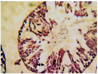 | |
| Fig. 2: | Histological architecture of hematoxylin stained sections of the testis in experimental group 3 at 14 weeks: H and E stain of an area of section selected from testis (stage VI) to show the arrangement of sertoli cells and germinal cells with disruption morphology (arrow) and random arrangement of the cell due to loss of cell-cell interactions (arrow) are marked and as well as the presence of degradation as a separation between the germ cells (arrow) are marked. Magnification 400 |
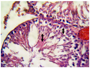 | |
| Fig. 3: | Histological architecture of hematoxylin stained sections of the testis in experimental group 2 at 14 weeks: H and E stain of an area of section selected from testis (stage VII) in which a decreased germ cells (arrow) and arrested spermatogenesis (arrow) are marked. Magnification 200 |
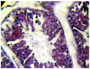 | |
| Fig. 4: | Histological architecture of hematoxylin stained sections of the testis in experimental group 1 at 14 weeks: H and E stain of an area of section selected from testis (stage VII) in which a decreased germ cells (arrow) and arrested spermatogenesis (arrow) are marked. The space of lumen of tubule increased as well. Magnification 200 |
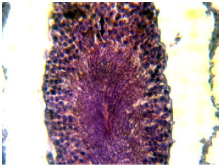 | |
| Fig. 5: | Histological architecture of hematoxylin stained sections of testis in the control group at 14 weeks: H and E stain of an area of section selected from testis (stage VI) to show the arrangement of germinatvie epithelium with normal morphology (arrow) and designed arrangement of the cells with complete kinetics spermatogenesis. Magnification 200 |
Besides, the spermatozoids were rarely and the complete kinetics of spermatogenic progression was lower marked in the experimental group 3 than in control, experimental group 1 and even in experimental group 2 (Fig. 2-4).
In addition, in both experimental groups 2 and 3, the germinative cells of seminiferous tubules were decreased which in overall contain more primitive germ cells than advanced germ cells (Fig. 2, 3). A dilation of the lumen accompanied by a disorganized epithelium and premature sloughing of postmeiotic germ cells was higher in these groups compared to non exposed group (Fig. 2-4). In some testes of experimental groups 2 and 3 especially in those that have a well kinetic of spermatogenic progression, the morphologically normal appearance interstitial space with interstitial cells was observed (Fig. 2, 3). However, noise stress exposing could be worsening the light microscopic morphology of interstitial tissue of the testes. Meanwhile, in case of the control specimens, no histopathological changes were seen (Fig. 5).
Seminiferous tubules of the testes: Overall, the mean proportion number of damaged seminiferous tubules in each histological section of testis was lower in non exposed group (42±6%) than noise stress exposed group (62.6±11%). The mean proportion thickness of seminiferous tubules was larger in non-exposed groups in comparison to exposed ones (214.8±46 vs. 148.8±26 μm, p>0.001). Indeed, the mean proportion thickness of the germinal epithelium was significantly different between controls (49.2±16 μm) and noise stress exposed testes (31±11 μm) (Fig. 1, 2) (p>0.05). Moreover, in experimental group 3, simple morphological examination showed that the signs of disorganization, degradation and separation between the sertoli and germinal cells were increased in comparison with the non-exposed ones (Fig. 2).
DISCUSSION
Today, noise pollution is known as one of the problems of human societies and reviewing the effect on human beings life seems very necessary (Paunovic, 2013). The role of noise pollution as one stress in terms of different diseases was studied and its effects on the hormones secretion, pregnancy rate, abnormal child birth, preterm birth and even the weight and the number of children is being reviewed (Saki et al., 2010; Jalali et al., 2012). Since the noise pollution in the pregnancy period is noticed in previously, the effect of it on the pregnancy is shown by more details in this research. The gain data from the present study shows that in pregnant mother which exposure to high levels of noise, by intensity of 90-120 and frequency of 300-350 Hz, the general appearance of the their child's testicles is worse than in the non-exposed of noise stress group. Moreover, it is obvious that the rate of atrophic seminiferous tubules, jumbled appearance in the interstitial space and also germinative epithelium were more observed in the testicles of noise stress exposed group than in the non-exposed of noise ones. Additionally, if the placing of the rats in the place noisy take for a longtime, this impair is increasing as well.
In keeping line with the above-mentioned suggestions, testes morphometric analysis of the current study indicated that impairments in the testicular tissue (i.e., weight, length, width and height of the testes and spermatogenesis dynamic) remarkably get worse if the cases were placed, especially for a long time, in the high noise stress situation. In addition this study declared that number of alive fetuses obtained from pregnant female rats that were lived in the high noise stress condition, were reduced when compared with ones that were not been on the exposure of loud voice.
Noguchi et al. (1993) showed that the traffic voice, by intensity of 100 db, could be reduced significantly testosterone hormones concentration and subsequent the interstitial space cells, i.e., leydig cells in the murine species. Therefore, it seems that every stress that can cause, reduce or even inhibit gonadotropin and sex hormones secretion, normally can lead to induction of damage in germinative and somatic cells of testes parenchyma (Mylchreest et al., 2002). Indeed, noise stress may disrupt steroid hormones concentration and neuroendocrine gonadal axis in turn is resulted in increased rate of cellular damage by expressing, as mentioned for testicular tissue, apoptosis-related proteins and genes in testicular cells (Swami et al., 2007).
Additionally, Mylchreest et al. (2002) showed that placing of the rats in one place noisy for a longtime can be impaired the spermatogenesis process and also reducing of sex hormones secretion that in turn cause increasing the activity of leydig cells by compensation mechanism for increasing the testicular steroid hormone.
In accordance with the above suggestion, Swami et al. (2007) also reported that the testosterone serum level in rats under noise stress (100 dB) was reduced. Also, more structural changes in the testis tissue were observed following the noise stress (Swami et al., 2007). In addition, it was reported that apoptosis ratio is negatively correlated with normal morphology and motility of sperm and conversely positively correlated with sperm tail defects (Aziz et al., 2007; Chen et al., 2006; Saki et al., 2010).
Studies shows that the reduction of testosterone level is accompany by significant reduction of the number of sperms of epididymis (Mylchreest et al., 2002). In addition in histology studies it was cleared that epididymal sperms in a group of mice that for a long time were under effect of loud voice, the number of dead cells was increased and the maturation in sexual cells was stopped (Ozguner et al., 2005; Saki et al., 2011).
CONCLUSION
Taken together can be said that it seems that the noise stress has negative influences on the fertility of male based on enhancing of the apoptotic process induced by pathogenesis stress and suppressing the kinetics spermatogenesis through exposing in the noise stress.
ACKNOWLEDGMENT
This project was financially supported by the research deputy of Ahvaz Jundishapur University of Medical Sciences (AJUMS) (grant n = PRc-70). We would like to express our great appreciation for their support.
REFERENCES
- Chen, Z., R. Hauser, A.M. Trbovich, J.L. Shifren, D.J. Dorer, L. Godfrey-Bailey and N.P. Singh, 2006. The relationship between human semen characteristics and sperm apoptosis: A pilot study. J. Androl., 27: 112-120.
PubMed - Swami, C.G., J. Ramanathan and C.C. Jeganath, 2007. Noise exposure effect on testicular histology, morphology and on male steroidogenic hormone. Malaysian J. Med. Sci., 14: 28-35.
Direct Link - Cosa, M. and G. Cosa, 1989. Annoyance, disturbance and damage caused by noise and vibration. Ann. Lg, 1: 133-156.
PubMedDirect Link - Crino, O.L., E.E. Johnson, J.L. Blickley, G.L. Patricelli and C.W. Breuner, 2013. The effects of experimentally elevated traffic noise on nestling white-crowned sparrow stress physiology, immune function and life-history. J. Exp. Biol., (In Press).
CrossRefDirect Link - Dong, Q., A. Salva, C.M. Sottas, E. Niu, M. Holmes and M.P. Hardy, 2004. Rapid glucocorticoid mediation of suppressed testosterone biosynthesis in male mice subjected to stress. J. Androl., 25: 973-981.
PubMed - Ganesh, C.B. and H.N. Yajurvedi, 2002. Stress inhibits seasonal and FSH-induced ovarian recrudescence in the lizard, Mabuya carinata. J. Exp. Zool., 292: 640-648.
CrossRef - Jalali, M., G. Saki, A.R. Sarkaki, K. Karami and S. Nasri, 2012. Effect of noise stress on count, progressive and non-progressive sperm motility, body and genital organ weights of adult male rats. J. Hum. Reprod. Sci., 5: 48-51.
CrossRefDirect Link - Helmstetter, F.J. and P.S. Bellgowan, 1994. Hypoalgesia in response to sensitization during acute noise stress. Behav. Neurosci., 108: 177-185.
PubMedDirect Link - Lue, Y., A.P. Hikim, C. Wang, M. Im, A. Leung and R.S. Swerdloff, 2000. Testicular heat exposure enhances the suppression of spermatogenesis by testosterone in rats: The two-hit approach to male contraceptive development. Endocrinology, 141: 1414-1424.
PubMed - Mylchreest, E., M. Sar, D.G. Wallace and P.M.D. Foster, 2002. Fetal testosterone insufficiency and abnormal proliferation of Leydig cells and gonocytes in rats exposed to di(n-butyl) phthalate. Reprod. Toxicol., 16: 19-28.
CrossRefPubMedDirect Link - Aziz, N., T. Said, U. Paasch and A. Agarwal, 2007. The relationship between human sperm apoptosis, morphology and the sperm deformity index. Hum. Reprod., 5: 1413-1419.
CrossRef - Naraghi, M.A., F. Abolhasani, I. Kashani, I.J. Anarkooli and M. Hemadi et al., 2010. The effects of swimming exercise and supraphysiological doses of nandrolone decanoate on the testis in adult male rats: a transmission electron microscope study. Folia Morphol. (Warsz), 69: 138-146.
PubMed - Ozguner, M., A. Koyu, G. Cesur, M. Ural, F. Ozguner, A. Gokcimen and N. Delibas, 2005. Biological and morphological effects on the reproductive organ of rats after exposure to electromagnetic field. Saudi Med. J., 26: 405-410.
PubMedDirect Link - Noguchi, J., M. Yoshida, H. Ikadai, T. Imamichi, G. Watanabe and K. Taya, 1993. Age-related changes in blood concentrations of FSH, LH and testosterone and testicular morphology in a new rat sterile mutant with hereditary aspermia. J. Reprod Fertil., 97: 433-439.
PubMed - Paunovic, K., 2013. Noise and children's health: Research in Central, Eastern and South-Eastern Europe and newly independent states. Noise Health, 15: 32-41.
CrossRefDirect Link - Saki, G., F. Rahim and K. Alizadeh, 2009. Effect of forced swimming stress on count, motility and fertilization capacity of the sperm in adult rats. J. Hum. Reprod. Sci., 2: 72-75.
PubMed - Saki, G., F. Rahim and O.A. Vaysi, 2010. Effect of forced swimming stress on in-vivo fertilization capacity of rat and subsequent offspring quality. J. Human Reprod. Sci., 3: 32-34.
CrossRefPubMedDirect Link - Saki, G., S. Razie and S. Amirpoor, 2011. Pregnancy rate in female mice exposed to forced swimming stress. Asian J. Biol. Sci., 4: 266-271.
CrossRefDirect Link - Sasagawa, I., H. Yazawa, Y. Suzuki and T. Nakada, 2001. Stress and testicular germ cell apoptosis. Syst. Biol. Reprod. Med., 47: 211-216.
CrossRef - Shenaieva, T.O. and O.H. Reznikov, 2003. The hypothalamic-hypophyseal-gonadal system and its functional reserve during long-term vibration and noise. Fiziol. Zh., 49: 56-62.
PubMedDirect Link - Shokri, S., M. Hemadi, G. Bayat, M. Bahmanzadeh, I. Jafari-Anarkooli and B. Mashkani, 2013. Combination of running exercise and high dose of anabolic androgenic steroid, nandrolone decanoate, increases protamine deficiency and DNA damage in rat spermatozoa. Andrologia (In Press).
CrossRefDirect Link - Yu, C., Y. Yao, Y. Yang and D. Li, 2004. Changes of rat testicular germ cell apoptosis after high power microwave radiation. Zhonghua Nan Ke Xue, 10: 407-410.
PubMed