

Rohaya Megat Abdul Wahab
Department of Orthodontics, Faculty of Dentistry, National University of Malaysia,
Jalan Raja Muda Abdul Aziz, 50300 Kuala Lumpur, Malaysia
Noor Ayuni Ahmad Shafiai
Department of Orthodontics, Faculty of Dentistry, National University of Malaysia,
Jalan Raja Muda Abdul Aziz, 50300 Kuala Lumpur, Malaysia
Shahrul Hisham Zainal Ariffin
School of Biosciences and Biotechnology, Faculty of Science and Technology,
National University of Malaysia, 43600 Bangi, Selangor, Malaysia
Journal of Medical Sciences
Year: 2017 | Volume: 17 | Issue: 1 | Page No.: 1-9
DOI: 10.3923/jms.2017.1.9





ABSTRACT
The aim of this study is to cover the literature review on the risk of orthodontically induced inflammatory root resorption during orthodontic treatment. Reported studies related on root resorption in orthodontics until 2016 were searched electronically through PubMed, Scopus and ScienceDirect using key words such as ‘risk’,‘root’,‘resorption’ and ‘orthodontic’. In the findings, there is a wide range of prevalence of root resorption (4-91%) caused by orthodontic treatment and this was mainly due to various assessment methods. The risk of root resorption was further divided into host and treatment mechanic factors. Host risk factors that were found significant for root resorption during orthodontic treatment are teeth with existing root resorption during pretreatment, anterior openbite, abnormal root shape and teeth with root canal therapy. Significant risk factors for treatment mechanics include longer duration of treatment, heavy force, intrusion and extraction of premolars. This study also discusses ongoing studies towards finding biological markers for root resorption for periodic monitoring, which essentially provides a safer method, overcoming the limitation of radiograph. These latest 5 years of findings on the studies of risk factors of orthodontically induced inflammatory root resorption have not changed much from the old studies. Orthodontically induced inflammatory root resorption is inevitable, however it can be prevented from progressing into severe stage; provided that the orthodontists taking early measures and aware of the host and treatment risk factors plus biomarkers involved.
PDF Abstract XML References Citation
Article History
Received: September 26, 2016;
Accepted: December 14, 2016;
Published: February 21, 2017
Copyright: © 2017. This is an open access article distributed under the terms of the Creative Commons Attribution License , which permits unrestricted use, distribution and reproduction in any medium, provided the original author and source are credited.
How to cite this article
Rohaya Megat Abdul Wahab, Noor Ayuni Ahmad Shafiai and Shahrul Hisham Zainal Ariffin, 2017. An Insight into Risk Factors for Root Resorption During
Orthodontic Treatment. Journal of Medical Sciences, 17: 1-9.
DOI: 10.3923/jms.2017.1.9
URL: https://scialert.net/abstract/?doi=jms.2017.1.9
DOI: 10.3923/jms.2017.1.9
URL: https://scialert.net/abstract/?doi=jms.2017.1.9
INTRODUCTION
The term ‘Orthodontically induced inflammatory root resorption’ (OIIRR) is mainly used in orthodontic to differentiate from any other causes of root resorption in permanent teeth1. It is inevitable as the force applied during orthodontic treatment induces inflammation which is essential for tooth movement, however it is also a fundamental component for root resorption.
In order to minimize the degree of root resorption, optimum force level is suggested to be within 20-150 g tooth‾1 for desired tooth movement2. Force level exceeding this threshold can cause periodontal ischaemia especially in adults as they induce more apical stress due to thicker cementum when compared to adolescents3. Apical region is more susceptible to root resorption than the cervical region, since it is softer and less mineralized and contains fewer sharpey’s fibers. Furthermore, the region where periodontal ligaments are compressed during tooth movement increases the risk for root resorption.
Root resorption is a common side effect during orthodontic treatment, however it usually ceases and repaired when the force is removed. Although, OIIRR is unavoidable in orthodontics, it usually occurs at mild degree with radiographic mean resorption4 of less than 2.5 mm. Although, the event where root resorption becomes progressive is minimal, it will reduce the prognosis of the tooth and in severe cases, may become mobile and consequently jeopardized the initial treatment plan. Therefore, it is important for orthodontists to identify risk factors for root resorption even before starting orthodontic treatment5,6. Modification on treatment mechanics may be suggested to minimize the occurrence of root resorption during orthodontic treatment.
The OIIRR is a complex and sterile inflammatory process of which many aspects still remain unclear. It has been a debate for many years on certain patient characteristics and treatment modalities as risk factors for OIIRR. Justus4highlighted genetic predisposition as the main aetiologic risk factor for severe form of OIIRR4. This study provides the most current view, over the past 6 years (2010-2015), on the multiple risk factors of root resorption so that the clinical management for minimizing root resorption during orthodontic treatment can be addressed appropriately.
All reported studies were identified electronically through PubMed, Scopus and ScienceDirect with the key words of ‘risk’,‘root’,‘resorption’ and ‘orthodontic’ covers until year 2015. The findings of the risk factors were divided further into host and treatment mechanic factors.
PREVALENCE OF ROOT RESORPTION DURING ORTHODONTIC TREATMENT
Prevalence of root resorption during orthodontic treatment reported5,7 variation from 4-91%. It is found that almost all patients and up to 91% of all teeth showed some degree of root shortening but few patients and teeth had root shortening7 of >4 mm. Pereira et al.8 reported percentage in external apical root resorption varies on each anterior teeth ranging from 8.5% for upper right canines, to 12.6% for upper right lateral incisors, with significant higher root resorption in incisors than canines. Makedonas et al.5 reported 4% of patients were diagnosed with root resorption after 6 months of fixed appliances with only 3.1% of maxillary incisors involved. Marques et al.9 found 14.5% prevalence of severe root resorption while Motokawa et al.10 found high incidence of 78.2% in their study. Matsuda et al.11 also reported 78% patients with root resorption at the end of orthodontic treatment.
Reports of these prevalence of root resorption have to be closely monitored as they can be mistakenly interpreted. Some studies reported their prevalence in relation to number of patients and others in the number of teeth5,7,11. It does differs as proven by Motokawa et al.10, where the researchers separated the results for evaluation of prevalence of root resorption by number of teeth and patients. It is found that for severe root resorption, no significant differences were found in the number of patients but when evaluated by the number of teeth, the prevalence was significantly greater in the extraction than in the non-extraction group.
Other factor that can be contributed to the variation of prevalence of root resorption during orthodontic treatment is the assessment methods. There are two techniques in discovering the presence of root resorption which are by histological or radiographically. Histological finding was performed either on animal teeth or human premolar extracted teeth due to the necessity to observe the teeth in vitro12-21. The advantage of investigating root resorption histologically is that it can be observed directly on the root surface and even the smallest craters can be noted. The measurement of root resorption was usually presented in the reduction of root length (millimeters) or increased in volume of resorption craters (cube root scale-millimeters).
Evaluating root resorption through radiograph is more related to clinical practise and many researchers had favored 3D images such as cone beam CT and micro-CT in detecting root resorption during orthodontic treatment as compared to 2D images such as periapical, panoramic and lateral cephalometric5-8,10,18,21-30. Higher prevalence of root resorption is expected in 3D images as the view eliminates overlapping structure and allow for better visualization of specific tissue volume7. However, the disadvantages of 3D imaging are more on the technical aspect; where it has higher radiation compared to 2D images and access for view is only through its software. Nevertheless, the advent of CBCT has reduced its radiation, making it an advantageous tool in dentistry.
MONITORING OF ROOT RESORPTION AND POTENTIAL BIOMARKERS DURING ORTHODONTIC TREATMENT
It is difficult to detect root resorption early as the event is usually noted only when radiograph is taken. However, radiographs have the limitation of radiation exposure, the way it is handled, standardization and limited points of view. Several protocols suggest that radiographic examination should take place in the first 3-6 months and then every year after appliance placement. The practical usage of the first radiographic evaluation in the first 6 months to diagnose and predict OIIRR has not yet been studied thoroughly5.
Given these limitations, hence the current trends of finding biological markers for root resorption which essentially provides sensitive, safer and more prognostic diagnostic methods for detecting root resorption31. Protein markers such as aspartate aminotransferase, salivary sIgA and serum IgG as well as biomarkers representing biological changes during specific phenomenon during orthodontic tooth movement such as ALP (bone formation), TRAP5a (bone resorption), LDH (inflammation) and DSP (root resorption) was found to have potential for detecting root resorption32,33.
George and Evans34 tested the hypothesis that during root resorption, organic matrix proteins and cytokines from the surrounding bone and dentin are released into the gingival crevice and the preliminary results confirm of their presence. The OPG was locally present in excess amounts over RANKL and an increased RANKL/OPG in the study groups could be correlated with an increased bone resorption activity during orthodontic tooth movement.
Mah and Prasad31 and Kereshanan et al.35 found high level of DSP (dentine siolaprotein) and DPP (dentine phosphoproteins) respectively on sites undergoing physiological root resorption. These proteins are parts of 10% of the organic matrix in dentine composition as non-collagenous proteins36. Although, DSP is found in some control sample, it was in reduced level, suggesting that it is dentine specific. Therefore, there is a strong indication that the presence of DSP or DPP is an indication of undergoing root resorption35.
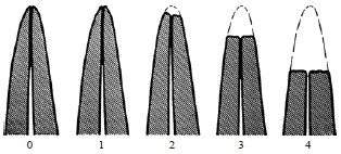 | |
| Fig. 1: | Score system categorizing the severity of root resorption by Levander and Malmgren37 based on index score from 0-4 |
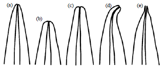 | |
| Fig. 2(a-e): | Subjective scoring of root form by Malmgren et al.39. The shape of the roots was classified as, (a) Normal, (b) Short, (c) Blunt, (d) Bent at the apex and (e) Pipette form |
CLASSIFICATION OF ROOT RESORPTION SEVERITY
The categorization of root resorption severity also differs in studies which would vary the prevalence of root resorption. Levander and Malmgren37 root resorption scoring system (Fig. 1) with index scores from 0-4 is the most commonly used to classify the severity of root resorption5,6,9,27,29,38. However, some studies reported their findings in reduction of the root length without categorizing the severity of root loss12,13,15,16,18,19,21. In the study of abnormal root shape as the risk factor for root resorption, most studies follow guidelines which was introduced by Malmgren et al.39 (Fig. 2), where the roots were differentiated between short, blunt, apical bent and pipette shaped.
Treatments received by the subjects in the study also had contributed to wide range of prevalence of root resorption and this will be further discussed in the later part of this review. Types of appliances, the forces used and duration of treatment which were set as the control in the studies may somehow increase or decrease the percentage of prevalence as they are proven to commit as risk factors for root resorption.
RISK FACTORS FOR ROOT RESORPTION
Risks factors are divided into host and treatment mechanics. Host factor means that it is patients-originated; for example gender, race, history or the condition and location of the teeth and it is beyond control of the clinician. Clinician can only screen for high-risk patients and the risks of root resorption from host factor need to be fully considered when planning for an orthodontic treatment. Treatments mechanics factors are more of a concern by the clinician as these can be manipulated and amenable via modifications during orthodontic treatment to reduce progressive root resorption.
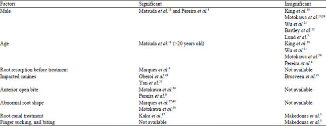
Host factors: Gender differences in the risk of root resorption were investigated and male was found to have a higher chance of root resorption as compared to women8,11. Nevertheless, there were studies that proved that there was insignificant difference in gender in relation to root resorption7,10,13,18,21,38. As far as age concern, patients above 20 years old may have significant relation to root resorption but most of the studies found age is not a risk factor for root resorption8,11,18,21,38.
Teeth with root resorption even before orthodontic treatment, whether of physiological or pathological reasons, have proven to be associated with progressive root resorption during orthodontic treatment9. Physiological root resorption can occur as a consequence of an adjacent impacted tooth. The most common problem with impacted canines is resorption of the roots of the adjacent lateral incisors or premolars. Oberoi and Knueppel28, on the study of 3D assessment of impacted canines and root resorption reported 40.4% of lateral incisors have no resorption, 35.7% with slight root resorption, 14.2% moderate root resorption and 4.0% with severe root resorption. Another study on Chinese population reported that eruptive movement or migration of the impacted canines during its root development process is likely to increase the risk of root resorption. The eruptive movement of canines during root development might cause mechanical and molecular changes to the local environment involving adjacent roots. This was proven with significant increase in root resorption of adjacent root on closed apex canine when compared to open apex canines. Physical proximity of less than 1 mm between impacted canine and adjacent roots is also the most important predictor for root resorption at the incisors and first premolars30. Root resorption which occurs at the adjacent roots may be caused by direct physical damage, increased pressure at local root cementum and dentin or concentrated resorptive molecules from the canine eruption follicle.
Brusveen et al.23 had demonstrated in order to disrupt the resorptive process, the impacted canine has to be distanced from the adjacent roots. In the study, canine retraction during orthodontic treatment was done by pulling it away from the adjacent incisor roots via a specially constructed transpalatal arch with a finger spring. Fixed appliances on the incisors were only applied as late as possible, after canine eruption. The protocol was done as such, that they could not support the hypothesis that an impacted canine is a risk factor for apical root resorption of incisors. Even after 13-28 years post-treatment, most cases of incisor root resorption induced by ectopic maxillary canine did not progress and showed no clinically relevant symptoms22.
Anterior open bite was also a significant factor for Orthodontically Induced Inflammatory Root Resorption (OIIRR) with the speculation of the jiggling force caused by orthodontic force and tongue pressure to move the teeth in opposite direction8,38. Most cases with an anterior open bite may end up with extraction of premolars as the treatment plan but Pereira et al.8 found that these two factors, anterior open bite and extractions of premolars were independent of each other. In the study, patients with open bite only had 26.1% of OIIRR, approximately similar to open bite patients submitted to premolar extractions which had 26.0% of OIIRR.
Abnormal root shapes had always been associated with root resorption and it is in agreement with recent studies27,38,40. Root forms are usually classified as normal, short, blunt, bent at the apex and pipette form (Fig. 2). It was speculated that abnormal root shape causes root resorption due to reduced resistance of the root to mechanical stimuli. The prevalence of root resorption is greater in abnormal root shape with hypofunctional teeth than in functional teeth. It was suggested that the reason might be that non-occlusion in hypofunctional teeth did not cause physiological root remodeling after teeth eruption hence development of root resorption38. A case study on rare condition of short root anomaly on maxillary and mandibular incisors noted of a girl aged 10 years old that had a treatment of functional appliance27. There was significant mobility in the mandibular incisors at the termination of wearing functional appliances which was then decided to be retained with fixed lingual retainer. Although it is reported that no change in the amount of root shortening, caution must be taken in deciding which treatment is the best for each patient.
Other host factors in the risk of root resorption such as root canal treatment and history of trauma were also reported. Kaku et al.17 showed that teeth with root canal treatment may be a predictor for progressive severe inflammatory root resorption during orthodontic tooth movement while Makedonas et al.5 found no statistically significant relationship of root resorption with the risk of trauma. Makedonas et al.5 also found no statistically significant on relationship of root resorption with the risk factors of finger sucking, finger sucking history, nail biting and nail biting history after 6 months of wearing fixed appliances. The significant and insignificant finding for host risk factors in root resorption during orthodontic treatment is presented in Table 1.
Treatment mechanics factors: Some of the orthodontic treatment mechanics that are applied in the clinic are inevitably related for increasing the risk of root resorption.
| Table 1: | Significant and insignificant studies of host risk factors for root resorption during orthodontic treatment |
 | |
Longer duration of orthodontic treatment had been proven to be one of them6,10,19,29. Motokawa et al.10 studied on the effect of duration of multiloop edgewise archwire (MEAW) treatment, elastics and total treatment time on root resorption. For the duration of MEAW, there is significantly higher prevalence of severe root resorption, evaluated by number of teeth and in long term use of elastics (>6 months). Total treatment time of more than 30 months was proven to have increased the risk of severe root resorption when evaluated both by number of patients and teeth. Long duration of orthodontic treatment exposes the teeth to long term jiggling force causes an increase in root resorption.
The duration of force applied at one time, whether continuous or intermittent forces was also in the investigation for the effect in root resorption. Aras et al.12 found that although the continuous force produces faster tooth movement than intermittent forces, it brings higher risk in root resorption. Intermittent force allows time for reparative mechanism thus produces less root resorption. Paetyangkul et al.19 studied the root resorption craters histologically with 4 or 8 weeks of retention after application of continuous light or heavy orthodontic force. New cementum layer had almost repaired resorption cavity of root in 8 weeks retention group, unlike the 4 weeks retention group, resorption craters were still repairing with new cementum and presence of inflammatory infiltrates. From these studies it can be concluded that the reparative cementum is time-dependent and tooth movement is efficient when the force is given intermittently, for longer period of time. The method of applying intermittent force was mentioned by Justus4 in his book as one of the treatment options if root resorption is pronounced in the radiograph during the middle of orthodontic treatment. Activation of archwire once every 2-3 months are preferable instead of monthly.
Depending on the force direction and root morphology of the tooth, the compression area on root surface is more susceptible to root resorption than in tension area21. Study on tipping tooth movement and buccal root torque of 2.5 and 15° on extracted premolars also agreed with increase of root resorption on root area of compression13,18. During tipping movement, periodontal ligament compression was more pronounced in the apical and cervical thirds of the tooth whereas in buccal root torque, the root resorption was seen more on the apical. Intrusion was indicated as the most deleterious tooth movement as the force is directed and compressed on the small apical region of the tooth. A comparison between two groups which had en masse retraction of upper incisors with and without intrusion with minis crews showed a significant apical root resorption of lateral incisors, not in central incisors, in retraction group with minis crews26. Mechanics of intrusion with micro implant when comparing to J-hook during retraction of upper anterior segment also proved significant root resorption41.
New approach for reducing the amount of root resorption during intrusion was proposed by introducing lighter and more continuous force as well as control of force vector and labiolingual position of the tooth24. This was proven in several studies that heavy force applied beyond its optimum for tooth movement will increase the risk of root resorption15,16,18,19,21. One study proved contradictly, where histological evaluation of mandibular teeth in beagle dogs found that constant intrusive forces between 50-200 g produced similar degree of root resorption, regardless of the force magnitude20.
During functional appliances, teeth that are used for anchorage will sustain great amount of force as patient is expected to wear the appliance full time. This, theoretically, will favor the development of root resorption. However, in teeth with incomplete root formation, this was proved otherwise, Kinzinger et al.25 focused on growing patients of class II malocclusion wearing Herbst appliance.
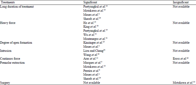
| Table 2: | Significant and insignificant findings of risk factors for root resorption during orthodontic treatment (Treatment mechanics factors) |
 | |
From this study there was a tendency towards root length decrease in teeth with complete root formation but significant increase in root length of teeth with incomplete root formation at the start of the treatment. Teeth with incomplete root formation has wide open apical foramen which ensure the pulp still retain its vitality as fewer blood vessels are compressed, otherwise might be involved in resorptive developments even at the strong forces. Therefore, it is suggested that if Herbst appliance or any types of functional appliance are indicated in the treatment plan, the treatment should starts early when the root is still developing as they have higher biologic tolerance. This statement was also supported by Maues et al.6, in which they find teeth with complete root formation were more likely to develop severe root resorption compared to teeth with incomplete root formation at the onset of treatment.
Nevertheless, optimum force during intrusive movement is important to balance the benefit of intrusion movement and disadvantage of root resorption. It was assumed that the optimum force for intrusion is similar whether the technique is conventional or using minis crews24. Extrusion movement also cause root resorption but the results showed insignificant difference in the cervical, middle and apical thirds in relation to root resorption after light and heavy force16.
Teeth extraction is usually a treatment option for relieving crowding and reduces overset in malocclusion however it does come with consequences. Premolar extraction was found to have significant risk for root resorption and this could be related to distance that canines and incisors are moved6,8,10,27,29,40. The anterior segment is retracted several millimeters after premolar extraction, especially in the case of high incisor protrusion. Motokawa et al.10 found significant difference in the prevalence of severe root resorption evaluated by number of teeth in extraction than in non-extraction group. Extraction of premolars increases the amount of tooth movement and also requires longer duration of treatment to close the extraction space. Motokawa et al.10 also found that displacements of maxillary central incisors and changes in tooth inclination were significantly larger in the group with severe root resorption than in the group without resorption. Therefore it can be concluded that apical vertical movements and incisor proclination are strong predictors of external apical root resorption.
Motokawa et al.10 also found no significant difference in the prevalence of root resorption between two groups with and without surgery. This is subjected to evaluation of both by the number of patients and teeth. This study was performed based on the believe of increase in chronic tongue pressure and circumoral muscle tonus after surgical treatment might rise the prevalence of root resorption. All these latest findings of treatment mechanics risk factors during root resorption in orthodontic treatment patients are presented at Table 2.
CONCLUSION
Orthodontically Induced Inflammatory Root Resorption (OIIRR) is inevitable, however, it can be prevented from progressing into severe stage; provided that the orthodontists taking early measures and aware of the host and treatment risk factors. Understanding the severity of root resorption and system to measure the events is important to monitor the progress. Therefore, determining the potential biomarkers during root resorption are important to detect early thus can be prevented to progress into severe stage. However, as precaution approach patient can also be informed about the risk of root resorption prior to treatment.
Root resorption is one of the effects that can jeopardize the prognosis of orthodontic treatment. It is usually monitored when there is history of trauma or after root canal treatment by using radiograph. However, there are other factors that can contribute to root resorption such as blunted root, long treatment time and usage of heavy force in orthodontic treatment. Multiple studies had focus on incidence of root resorption and this review highlights the risk factors that are importance in promoting root resorptions. The techniques in identifying root resorption are also reviewed to highlights the advantage and disadvantage of radiographs usage for monitoring process. Optimistically the usage of biomarker for detection of root resorption can reduce repeated radiographs exposure in monitoring root resorption.
ACKNOWLEDGMENTS
The authors would like to thanks the Ministry of Higher Education (MOHE) for the financial grants (ERGS/1/2012/ SKK11/UKM/02/5 and FRGS/1/2015/SG05/UKM/02/2) and the National University of Malaysia (UKM-DLP-2012-025 and UKM-DPP-2015-033) for supporting this study.
REFERENCES
- Brezniak, N. and A. Wasserstein, 2016. Orthodontitis: The Inflammation Behind Tooth Movement and Orthodontic Root Resorption. In: Biology of Orthodontic Tooth Movement: Current Concepts and Applications in Orthodontic Practice, Shroff, B. (Ed.). Springer International Publishing, Switzerland, ISBN: 978-3-319-26607-7, pp: 67-101.
- Vikram, N.R., K.S.S. Kumar, K.S. Nagachandran and Y.M. Hashir, 2012. Apical stress distribution on maxillary central incisor during various orthodontic tooth movements by varying cemental and two different periodontal ligament thicknesses: A FEM study. Indian J. Dent Res., 23: 213-220.
CrossRefPubMedDirect Link - Makedonas, D., H. Lund, K. Grondahl and K. Hansen, 2012. Root resorption diagnosed with cone beam computed tomography after 6 months of orthodontic treatment with fixed appliance and the relation to risk factors. Angle Orthodontist, 82: 196-201.
CrossRefPubMedDirect Link - Maues, C.P.R., R.R.D. Nascimento and O.D.V. Vilella, 2015. Severe root resorption resulting from orthodontic treatment: Prevalence and risk factors. Dental Press J. Orthodontics, 20: 52-58.
CrossRefDirect Link - Lund, H., K. Grondahl, K. Hansen and H.G. Grondahl, 2012. Apical root resorption during orthodontic treatment. A prospective study using cone beam CT. Angle Orthodontist, 82: 480-487.
CrossRefPubMedDirect Link - Pereira, S.A., M. Lopez, N. Lavado, J.M. Abreu and H. Silva, 2014. A clinical risk prediction model of orthodontic-induced external apical root resorption. Revista Portuguesa Estomatologia Medicina Dentaria Cirurgia Maxilofacial, 55: 66-72.
CrossRefDirect Link - Marques, L.S., M.L. Ramos-Jorge, A.C. Rey, M.C. Armond and A. de Oliveira Ruellas, 2010. Severe root resorption in orthodontic patients treated with the edgewise method: Prevalence and predictive factors. Am. J. Orthodontics Dentofacial Orthopedics, 137: 384-388.
CrossRefDirect Link - Motokawa, M., T. Sasamoto, M. Kaku, T. Kawata, Y. Matsuda, A. Terao and K. Tanne, 2012. Association between root resorption incident to orthodontic treatment and treatment factors. Eur. J. Orthodontics, 34: 350-356.
CrossRefPubMedDirect Link - Matsuda, Y., M. Motokawa, M. Kaku, T. Kawata and R. Yamamoto et al., 2012. Clinical survey of the association between root resorption incident to orthodontic treatment and host factors. Orthodontic Waves, 71: 40-40.
CrossRefDirect Link - Aras, B., L.L. Cheng, T. Turk, S. Elekdag-Turk, A.S. Jones and M.A. Darendeliler, 2012. Physical properties of root cementum: Part 23. Effects of 2 or 3 weekly reactivated continuous or intermittent orthodontic forces on root resorption and tooth movement: A microcomputed tomography study. Am. J. Orthodontics Dentofacial Orthopedics, 141: e29-e37.
CrossRefDirect Link - Bartley, N., T. Turk, C. Colak, S. Elekdag-Turk, A. Jones, P. Petocz and M.A. Darendeliler, 2011. Physical properties of root cementum: Part 17. Root resorption after the application of 2.5° and 15° of buccal root torque for 4 weeks: A microcomputed tomography study. Am. J. Orthodontics Dentofacial Orthopedics, 139: e353-e360.
CrossRefDirect Link - Cheng, L.L., T. Turk, S. Elekdag-Turk, A.S. Jones, P. Petocz and M.A. Darendeliler, 2009. Physical properties of root cementum: Part 13. Repair of root resorption 4 and 8 weeks after the application of continuous light and heavy forces for 4 weeks: A microcomputed-tomography study. Am. J. Orthodontics Dentofacial Orthopedics, 136: 320.e1-320.e10.
CrossRefDirect Link - Ho, C., T. Turk, S. Elekdag-Turk, A.S. Jones, P. Petocz, L.L. Cheng and M.A. Darendeliler, 2011. Physical properties of root cementum: Part 19. Comparison of the amounts of root resorption between the right and left first premolars after application of buccally directed heavy orthodontic tipping forces. Am. J. Orthodontics Dentofacial Orthopedics, 140: e49-e52.
CrossRefDirect Link - Montenegro, V.C.J., A. Jones, P. Petocz, C. Gonzales and M.A. Darendeliler, 2012. Physical properties of root cementum: Part 22. Root resorption after the application of light and heavy extrusive orthodontic forces: A microcomputed tomography study. Am. J. Orthodontics Dentofacial Orthopedics, 141: e1-e9.
CrossRefDirect Link - Kaku, M., H. Sumi, H. Shikata, S. Kojima and M. Motokawa et al., 2014. Effects of pulpectomy on the amount of root resorption during orthodontic tooth movement. J. Endodontics, 40: 372-378.
CrossRefDirect Link - King, A.D., T. Turk, C. Colak, S. Elekdag-Turk, A.S. Jones, P. Petocz and M.A. Darendeliler, 2011. Physical properties of root cementum: Part 21. Extent of root resorption after the application of 2.5° and 15° tips for 4 weeks: A microcomputed tomography study. Am. J. Orthodontics Dentofacial Orthopedics, 140: e299-e305.
CrossRefDirect Link - Paetyangkul, A., T. Turk, S. Elekdag-Turk, A.S. Jones, P. Petocz, L.L. Cheng and M.A. Darendeliler, 2011. Physical properties of root cementum: Part 16. Comparisons of root resorption and resorption craters after the application of light and heavy continuous and controlled orthodontic forces for 4, 8 and 12 weeks. Am. J. Orthod. Dentofacial Orthopedics, 139: e279-e284.
CrossRefDirect Link - Ramirez-Echave, J.I., P.H. Buschang, R. Carrillo, P.E. Rossouw, W.W. Nagy and L.A. Opperman, 2011. Histologic evaluation of root response to intrusion in mandibular teeth in beagle dogs. Am. J. Orthodontics Dentofacial Orthopedics, 139: 60-69.
CrossRefDirect Link - Wu, A.T.J., T. Turk, C. Colak, S. Elekdag-Turk, A.S. Jones, P. Petocz and M.A. Darendeliler, 2011. Physical properties of root cementum: Part 18. The extent of root resorption after the application of light and heavy controlled rotational orthodontic forces for 4 weeks: A microcomputed tomography study. Am. J. Orthodontics Dentofacial Orthopedics, 139: e495-e503.
CrossRefDirect Link - Bjerklin, K. and C.H. Guitirokh, 2011. Maxillary incisor root resorption induced by ectopic canines: A follow-up study, 13 to 28 years posttreatment. Angle Orthodontist, 81: 800-806.
CrossRefDirect Link - Brusveen, E.M.G., P. Brudvik, O.E. Boe and M. Mavragani, 2012. Apical root resorption of incisors after orthodontic treatment of impacted maxillary canines: A radiographic study. Am. J. Orthodontics Dentofacial Orthopedics, 141: 427-435.
CrossRefDirect Link - Heravi, F., S. Bayani, A.S. Madani, M. Radvar and N. Anbiaee, 2011. Intrusion of supra-erupted molars using miniscrews: Clinical success and root resorption. Am. J. Orthodontics Dentofacial Orthopedics, 139: S170-S175.
CrossRefDirect Link - Kinzinger, G.S.M., S. Savvaidis, U. Gross, N. Gulden, N. Ludwig and J. Lisson, 2011. Effects of class II treatment with a banded herbst appliance on root lengths in the posterior dentition. Am. J. Orthodontics Dentofacial Orthopedics, 139: 465-469.
CrossRefDirect Link - Liou, E.J.W. and P.M.H. Chang, 2010. Apical root resorption in orthodontic patients with en-masse maxillary anterior retraction and intrusion with miniscrews. Am. J. Orthodontics Dentofacial Orthopedics, 137: 207-212.
CrossRefDirect Link - Marques, L.S., R. Generoso, M.C. Armond and C.A. Pazzini, 2010. Short-root anomaly in an orthodontic patient. Am. J. Orthodontics Dentofacial Orthopedics, 138: 346-348.
CrossRefDirect Link - Oberoi, S. and S. Knueppel, 2012. Three-dimensional assessment of impacted canines and root resorption using cone beam computed tomography. Oral Surg. Oral Med. Oral Pathol. Oral Radiol., 113: 260-267.
CrossRefDirect Link - Sharab, L.Y., L.A. Morford, J. Dempsey, G. Falcao-Alencar and A. Mason et al., 2015. Genetic and treatment-related risk factors associated with External Apical Root Resorption (EARR) concurrent with orthodontia. J. Orthodontics Craniofacial Res., 18: 71-82.
CrossRefDirect Link - Yan, B., Z. Sun, H. Fields and L. Wang, 2012. Maxillary canine impaction increases root resorption risk of adjacent teeth: A problem of physical proximity. Am. J. Orthodontics Dentofacial Orthopedics, 142: 750-757.
CrossRefDirect Link - Mah, J. and N. Prasad, 2004. Dentine phosphoproteins in gingival crevicular fluid during root resorption. Eur. J. Orthodontics, 26: 25-30.
CrossRefDirect Link - Ramos, S.P., G.O. Ortolan, L.M.D. Santos, P.L. Tobouti, M.M. Hidalgo A. Consolaro and E.N. Itano, 2011. Anti-dentine antibodies with root resorption during orthodontic treatment. Eur. J. Orthodontics, 33: 584-591.
CrossRefDirect Link - Ariffin, S.H.Z., Z. Yamamoto, L.Z.Z. Abidin, R.M.A. Wahab and Z.Z. Ariffin, 2011. Cellular and molecular changes in orthodontic tooth movement. Sci. World J., 11: 1788-1803.
CrossRefDirect Link - George, A. and C.A. Evans, 2009. Detection of root resorption using dentin and bone markers. J. Orthodontics Craniofacial Res., 12: 229-235.
CrossRefDirect Link - Kereshanan, S., P. Stephenson and R. Waddington, 2008. Identification of dentine sialoprotein in gingival crevicular fluid during physiological root resorption and orthodontic tooth movement. Eur. J. Orthodontics, 30: 307-314.
CrossRefPubMedDirect Link - Levander, E. and O. Malmgren, 1988. Evaluation of the risk of root resorption during orthodontic treatment: A study of upper incisors. Eur. J. Orthodontics, 10: 30-38.
CrossRefDirect Link - Motokawa, M., A. Terao, M. Kaku, T. Kawata, C. Gonzales, M.A. Darendeliler and K. Tanne, 2013. Open bite as a risk factor for orthodontic root resorption. Eur. J. Orthodontics, 35: 790-795.
CrossRefPubMedDirect Link - Malmgren, O., L. Goldson, C. Hill, A. Orwin, L. Petrini and M. Lundberg, 1982. Root resorption after orthodontic treatment of traumatized teeth. Am. J. Orthodontics, 82: 487-491.
CrossRefDirect Link - Marques, L.S., K.C. Chaves, A.C. Rey, L.J. Pereira and A.C. Ruellas, 2011. Severe root resorption and orthodontic treatment: Clinical implications after 25 years of follow-up. Am. J. Orthodontics Dentofacial Orthopedics, 139: S166-S169.
CrossRefPubMedDirect Link - Wang, Q., W. Chen, R.J. Smales, H. Peng, X. Hu and L. Yin, 2012. Apical root resorption in maxillary incisors when employing micro-implant and J-hook headgear anchorage: A 4-month radiographic study. J. Huazhong Univ. Sci. Technol. Med. Sci., 32: 767-773.
CrossRefPubMedDirect Link - Eross, E., T. Turk, S. Elekdag-Turk, F. Cakmak and A.S. Jones et al., 2015. Physical properties of root cementum: Part 25. Extent of root resorption after the application of light and heavy buccopalatal jiggling forces for 12 weeks: A microcomputed tomography study. Am. J. Orthodontics Dentofacial Orthopedics, 147: 738-746.
CrossRefPubMedDirect Link