
Idris Abdullahi Nasir
Department of Medical Microbiology, University of Abuja Teaching Hospital, PMB 228, Gwagwalada, FCT Abuja, Nigeria
Yahaya Usman
Department of Medical Laboratory Science, Faculty of Medicine, Ahmadu Bello University, PMB 05, Zaria, Kaduna State, Nigeria
Adamu Babayo
Department of Medical Microbiology, Abubakar Tafawa Balewa University Tecahing Hospital, PMB 0117, Bauchi, Bauchi State, Nigeria
Research Journal of Microbiology
Year: 2015 | Volume: 10 | Issue: 7 | Page No.: 336-342


ABSTRACT
Cytomegalovirus (CMV) screening in pregnancy has not been recommended during antenatal clinic days in Nigeria and most countries of the world. However, CMV has been widely accepted as, the viral etiology with the greatest propensity for congenital transmission. Due to CMV ubiquity, seronegative women are highly susceptible to CMV infection and thus, has increased risk of maternal infections and possibly congenital transmission. In view of this, this study aimed to determine the seroprevalence of women, who are anti-CMV IgG seronegative, thus susceptible to CMV infections. We made use of NovalisaTM anti-CMV IgG ELISA kit to screen 182 blood samples of pregnant women attending antenatal clinics of University of Maiduguri Teaching Hospital (UMTH), Maiduguri, Nigeria. Structured questionnaire was used to collect participants’ sociodemographic data. A total of 38 out of 182 subjects were anti-CMV IgG seronegative making a seroprevalence of 20.9%. There was significant statistical association between seronegativity and subjects’ education level and history of previous blood transfusion (p<0.05) but not with age, parity, gravida and gestation age (p>0.05). Findings from our evaluation indicated that many pregnant women were anti-CMV IgG seronegative and thus susceptible to maternal CMV infections. These women have high risk of contacting primary CMV infections and might eventually pose danger to their unborn fetuses in the absence of appropriate preventive measures.
PDF Abstract XML References Citation
Received: June 23, 2015;
Accepted: July 31, 2015;
Published: August 17, 2015
How to cite this article
Idris Abdullahi Nasir, Yahaya Usman and Adamu Babayo, 2015. Evaluation of Pregnant Women Susceptible to Cytomegalovirus Infection in Maiduguri, Nigeria. Research Journal of Microbiology, 10: 336-342.
URL: https://scialert.net/abstract/?doi=jm.2015.336.342
URL: https://scialert.net/abstract/?doi=jm.2015.336.342
INTRODUCTION
Cytomegalovirus (CMV) has been established, as a common viral infection in pregnancy and a leading etiology with the greatest propensity for recurrent pregnancy lose (Sherkat et al., 2014) and sensorineural disabilities in children, who contracted it congenitally. Although, CMV has a world-wide distribution, infection with CMV is more common in developing nations, especially in areas with poor socioeconomic conditions (Kenneson and Cannon, 2007).
In epidemiology of acute viral infections, three parameters determine incidence; the proportion of the population susceptible, the proportion of the population that are infected and infectious and the rate of contact between susceptible and infectious individuals, with contact defined as an encounter sufficient for transmission (Nathanson and Moss, 2013).
Serological surveys may be used to detect the footprints that a virus leaves in a population. They are particularly useful for viruses because most viral infections leave an imprint on all infected individuals, the presence of immunoglobulin G (IgG) antibody, which is often lifelong (Nathanson and Moss, 2013). Because many viruses cause asymptomatic infections or nondescript illnesses in addition to diagnosable diseases, serological surveys identify inapparent as well as apparent infections. Moreover, IgG antibody negativity indicates non-infected, non-immuned and susceptibility to the viral infection. When a population is exposed to an infectious individual with a specific virus, the susceptible part of the population will determine the spread of the agent and account for all new cases (Nathanson and Moss, 2013).
Primary infection is defined as CMV infection in a previously seronegative person whereas secondary infection is defined as intermittent excretion of the virus in the presence of host immunity and may be due to either reactivation of an endogenous virus (Vauloup-Fellous et al., 2013; Rahav et al., 2007) or exposure to a new virus strain from an exogenous source (Rahav et al., 2007).
The CMV can be transmitted to the fetus when a CMV seronegative woman contracted primary CMV infection during pregnancy or from latent virus reactivation from maternal CMV infection acquired prior to pregnancy or re-infection with a new CMV strain during pregnancy. The risk of CMV transmission to the fetus is higher among pregnant women with primary infection compared to those who were IgG positive prior to pregnancy, IgG positive at their first pregnancy visit or IgM positive with low IgG avidity are therefore presumed to have primary maternal CMV infection (Hyde et al., 2010).
Hygienic measures constitute an effective way to lower the rate of primary CMV infection in pregnant women (Vauloup-Fellous et al., 2009). As latently infected individuals may shed CMV in urine or saliva over years, they represent the most common source of expectant mothers’ infection (Manicklal et al., 2013). Therefore, the Center for Disease Control (CDC) and prevention recommends to seronegative pregnant women very simple and manageable hygienic precaution measures like frequent hand washing after contact with body fluids of a child whose CMV status is positive or unknown and avoidance of too intimate contact with such a child, to reduce their risk of infection. However, according to surveys conducted in the United States between 2005 and 2007, only 14-22% of female respondents out of the general population ever had heard of CMV (Jeon et al., 2006; Ross et al., 2008) and only about a half of obstetricians and gynecologists routinely counseled their patients about CMV and infection-prevention measures (Jeon et al., 2006; Centers for Disease Control and Prevention, 2008). Much would not be expected among medical officers in developing countries. This is because, they are hardly aware of the presence of this virus and the damage it could to the unborn foetuses, nor do they consider CMV screening for their women during antenatal clinics. Because CMV is ubiquitous, seronegative women contract infections through personal contact with people who excrete the virus in body fluids. In view of this we sort to evaluate the proportion of pregnant women seronegative and susceptible to primary CMV infections at Maiduguri, Nigeria.
MATERIALS AND METHODS
Study area: This is a descriptive study which was carried out in the WHO National Polio Reference Laboratory, University of Maiduguri Teaching Hospital, Borno State, Nigeria. The study was approved by the Ethical Research Committee, University of Maiduguri Teaching Hospital, Maiduguri, the capital city of Borno state, located in northeastern Nigeria shares borders with neighboring countries, such as Niger Republic, Chad and Cameroon within Nigeria, Maiduguri shares borders with other states, such as Adamawa, Yobe and Gombe and has Sahel savannah vegetation. The annual average temperature of Maiduguri ranges from 19.1-34.7°C and average annual precipitation is 562 mm. In Nigeria, there has been no consensus of opinion regarding, conducting CMV screening or other "TORCH" panel tests for pregnant women during their antenatal visits.
Participants and settings: This prospective study was conducted on 182 blood samples of pregnant women attending antenatal clinics of the UMTH, who consented to participate in the study and excluded those, who declined to participate in the study or refused to consent. The Median age of the women was 28 years with range of 16-40 years.
Sample size calculation: The sample size was determined using data from a prevalence rate conducted by Okwori et al. (2008) in Nigeria. Seroprevalence of 84.2%, therefore, the minimum ample size at 95% confidence level was 204. However, only 182 consented to voluntarily participate in the study.
Data collection: Structured Questionnaires were used to collect demographic data, such as; age, place of residence, gravida, gestational age, educational status, occupation, marital status, history of blood transfusion.
Sample collection and preparation: Samples were collected between December, 2013 and March, 2014. 5 mL of blood was collected aseptically into plain vacutainer tubes. The tubes were then appropriately labelled with patients’ laboratory number. Sera from these blood samples were separated by allowing the blood to clot at room temperature and centrifuged at 2500 rpm for 10 min. The sera were then separated using clean Pasteur pipettes, transferred into serum containers and stored at -70°C until laboratory analysis.
Laboratory investigations: Serum samples were analyzed by enzyme-linked immunosorbent assay (ELISA) using anti-CMV IgG kits (NovaLisaTM Immunodiagnostica, Germany) with product number CMVG0110.
Analytical procedure: All samples and reagents were brought to room temperature. The test was performed according to manufacturer’s instructions. The Optical Density (OD) was read using a GF-M3000 microplate reader at 450 nm wavelength. Samples were considered positive if the absorbance value was higher than 10% over the cut-off and negative if the absorbance value was lower than 10% below the cut-off. The cut-off is the mean absorbance value of the cut-off control determinations.
Ethical clearance and informed consent: This study was conducted in accordance with the Declaration of Helsinki and the protocol was approved by the human research ethical committees of University of Maiduguri Teaching Hospital. All the subjects gave their written informed consent for inclusion before they participated in the study. All data were analyzed anonymously throughout the study.
Statistical analysis: The data obtained from the questionnaire and the results of the laboratory analysis were entered into Microsoft excel and analyzed using SPSS (statistical package for social sciences Version 20, California Inc., USA). Results obtained were reduced to percentages and figures. The Pearson Chi square at 95% confidence interval and 0.05 level of significance was used to determine the relationships between demographic and proportions.
RESULTS
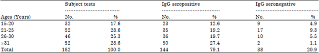
From Table 1, age group 21-25 had the highest seronegative subjects, 17 (9.3%), while IgG seronegativity was least among pregnant women greater than 31 years, 2 (1.1%). There was no statistical relationship between age distributions of anti-CMV IgG seronegativity among pregnant women (p-value = 0.135).
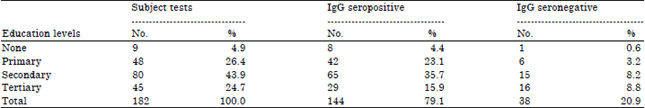
From Table 2, pregnant women with tertiary education had the most CMV IgG seronegativity, 16 (8.8%), while only 1 (0.6%) of those with no formal education was seronegative. There was no statistical relationship between education level and distributions of anti-CMV IgG seronegativity among the pregnant women (p-value = 0.033).
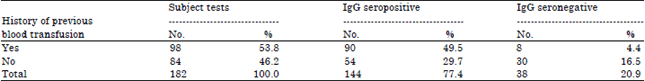
From Table 3, anti-CMV IgG seronegativity among pregnant women who had no history of previous blood transfusion were relatively higher than those, who had history of previous blood transfusion, 30 (16.5%). There was no statistical relationship between history of previous blood transfusion and anti-CMV IgG seronegativity among the pregnant women (p-value = 0.002).
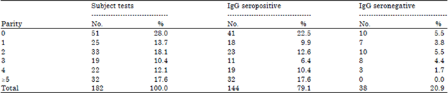
From Table 4, pregnant women with no parity and those with 2 parities had the highest anti-CMV IgG seronegativity, 10 (5.5%), while none of the pregnant women with more than 5 parities was seronegative, 0 (0.0%). There was no statistical relationship between age distributions of anti-CMV IgG seronegativity among pregnant women (p-value = 0.788).
| Table 1: | Age distribution of anti-CMV IgG seronegativity among pregnant women |
 | |
| Table 2: | Anti-CMV IgG seronegativity across education level of pregnant women |
 | |
| Table 3: | Distribution of anti-CMV IgG seronegativity with history of previous blood transfusion among pregnant women |
 | |
| Table 4: | Distribution of anti-CMV-specific IgG seronegativity across parity of pregnant women |
 | |
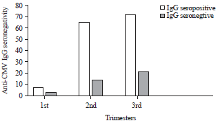 | |
| Fig. 1: | Anti-CMV IgG seronegativity across gestation age of pregnant women |
From Fig. 1, pregnant women at third trimester had the highest anti-CMV IgG seronegativity, while those at first trimester of pregnancy had the least seronegative. There was no statistical relationship between age distributions of anti-CMV IgG seronegativity among pregnant women (p-value = 0.556).
DISCUSSION
As part of effort to evaluate a crucial determinant of incidence of acute CMV infections, this study exclusively examined the proportion of pregnant women susceptible to CMV infection by evaluating their seronegativity to anti-CMV IgG antibodies. The overall seronegativity in the pregnant women was 20.9% (95% CI: 15.2-26.68), without any significant significance between age classes, gestation age and parity. However, there was statistical association between seronegativity with education level and history of previous blood transfusion. These findings are in consonance with previous studies (Hamdan et al., 2011; Matos et al., 2010; Yeroh et al., 2015; Akinbami et al., 2011).
In this study, 3 (1.6%), 14 (7.7%) and 21 (11.5 %) of the pregnant women were non-immune (IgG-negative) during their first, second and third trimesters, respectively. They are at risk of acquiring primary infection, which increases the chances of intrauterine transmission to fetus (Forsgren, 2009). Women who will be infected with CMV during late gestation are most likely to transmit the virus to their unborn child than women infected early in gestation. This might explain why vertical transmission rates are higher in the late gestation as compared to the first or second trimester (Malek et al., 1996; Revello et al., 2011). Indeed, the transmission rates among studies are remarkably consistent ranging from 30-42, 38-44 and 59-73% for the first, second and third trimester, respectively (Feldman et al., 2011; Revello et al., 2011; Bodeus et al., 2010; Enders et al., 2011).
Findings from our study showed that seronegativity rate increased significantly with increase in education level. This increase agrees with a previous report by Hamdan et al. (2011), who showed that illiterate women are at higher risk of CMV infection due to contact with contagious secretions from their own children and poor hygienic practice, fortunately they are immune against CMV except in rare case of reactivation or reinfection. Paradoxically, the seronegative women with higher level of education are susceptible to primary CMV, because of non-functional immunity against CMV.
There was statistical association between women, who had history of prior blood transfusion and CMV IgG seronegativity. This was higher among those that were never transfused than those that had been transfused. This result agrees with the report that blood transfusion was shown to be a risk factor for transmission of CMV infection (Matos et al., 2010). This is true because CMV testing is not part of laboratory screening in blood transfusion services, coupled with the high IgM seropositivity in African societies, recipients are either endangered with contracting CMV infections or acquire passive immunity from IgG seropositive donors (Juckstock et al., 2015).
Finally some limitations of the study must be taken into account as no outcome data for women after birth, their newborns and limited testing of IgG negative women for primary infections. However, for the latter case, as there are no official recommendations, the follow-up maybe performed at the discretion of the attending physician with compliance of women’ request.
CONCLUSION
Although, CMV screening during antennal days is not recommended by Nigeria’s health system probably, because of its cost/benefit ratio. However, some authors argued that screening cannot be justified on the grounds of its economic cost, the imperfect nature of congenital CMV prognostic criteria and few data concerning effective treatments during pregnancy. It is unfortunate to deny pregnant women appropriate information concerning their health and risks of contracting CMV, as this could raise a number of ethical and legal questions. CMV screening provides opportunity to identify seronegative women, who can be counselled about using appropriate hygienic measures to prevent infection. The proportion of IgG seronegativity and risk of CMV infection in pregnancy found in our study, therefore, support the use of serological screening, certainly in the first trimester when the risk of infection is higher.
ACKNOWLEDGMENTS
We will like to appreciate Professor Marycelin Baba, Department of Medical Laboratory Science, College of Medical Sciences, University of Maiduguri, Nigeria for proof-reading the final draft manuscript before submission. Idris Abdullahi Nasir conceptualized and designed the study; acquired, analyzed and interpreted the data; Yahaya Usman and Adamu Babayo drafted and critically revised the manuscript for important intellectual content; and approved the final manuscript as submitted.
REFERENCES
- Akinbami, A.A., K.A. Rabiu, A.A. Adewunmi, K.O. Wright and A.O. Dosunmu et al., 2011. Seroprevalence of cytomegalovirus antibodies amongst normal pregnant women in Nigeria. Int. J. Women's Health, 3: 423-428.
CrossRefDirect Link - Bodeus, M., B. Kabamba-Mukadi, F. Zech, C. Hubinont, P. Bernard and P. Goubau, 2010. Human cytomegalovirus in utero transmission: Follow-up of 524 maternal seroconversions. J. Clin. Virol., 47: 201-202.
CrossRefPubMedDirect Link - Centers for Disease Control and Prevention, 2008. Knowledge and practices of obstetricians and gynecologists regarding cytomegalovirus infection during pregnancy-United States, 2007. Morbidity Mortality Weekly Rep., 57: 65-68.
PubMedDirect Link - Enders, G., A. Daiminger, U. Bader, S. Exler and M. Enders, 2011. Intrauterine transmission and clinical outcome of 248 pregnancies with primary cytomegalovirus infection in relation to gestational age. J. Clin. Virol., 52: 244-246.
CrossRefDirect Link - Feldman, B., Y. Yinon, M.T. Oikawa, R. Yoeli, E. Schiff and S. Lipitz, 2011. Pregestational, periconceptional and gestational primary maternal cytomegalovirus infection: Prenatal diagnosis in 508 pregnancies. Am. J. Obstet. Gynecol., 205: 342-e1-342-e6.
CrossRefDirect Link - Forsgren, M., 2009. Prevention of congenital and perinatal infections. Euro Surveill, 14: 1-6.
Direct Link - Hamdan, H.Z., I.E. Abdelbagi, N.M. Nasser and I. Adam, 2011. Seroprevalence of cytomegalovirus and rubella among pregnant women in Western Sudan. Virol. J., Vol. 8.
CrossRefDirect Link - Hyde, T.B., D.S. Schmid and M.J. Cannon, 2010. Cytomegalovirus seroconversion rates and risk factors: Implications for congenital CMV. Rev. Med. Virol., 20: 311-326.
CrossRefDirect Link - Jeon, J., M. Victor, S.P. Adler, A. Arwady and G. Demmler et al., 2006. Knowledge and awareness of congenital cytomegalovirus among women. Infect. Dis. Obstet. Gynecol., Vol. 2006.
CrossRefDirect Link - Juckstock, J., M. Rothenburger, K. Friese and F. Traunmuller, 2015. Passive immunization against congenital cytomegalovirus infection: Current state of knowledge. Pharmacology, 95: 209-217.
CrossRefDirect Link - Kenneson, A. and M.J. Cannon, 2007. Review and meta-analysis of the epidemiology of congenital cytomegalovirus (CMV) infection. Rev. Med. Virol., 17: 253-276.
CrossRefDirect Link - Malek, A., R. Sager, P. Kuhn, K.H. Nicolaides and H. Schneider, 1996. Evolution of maternofetal transport of immunoglobulins during human pregnancy. Am. J. Reprod. Immunol., 36: 248-255.
CrossRefDirect Link - Manicklal, S., V.C. Emery, T. Lazzarotto, S.B. Boppana and R.K. Gupta, 2013. The silent global burden of congenital cytomegalovirus. Clin. Microbiol. Rev., 26: 86-102.
CrossRefDirect Link - Matos, S.B., R. Meyer and W.F.M. Lima, 2010. Seroprevalence of cytomegalovirus infection among healthy blood donors in Bahia State, Brazil. Revista Brasileira de Hematologia e Hemoterapia, 3: 1516-8484.
CrossRefDirect Link - Okwori, A., A. Olabode, E. Emumwen, G. Echeonwu and M. Lugos et al., 2008. Sero-epedemiological survey of cytomegalovirus infection among expectant mothers in Bida, Nigeria. Internet J. Infect. Dis., 7: 1-9.
Direct Link - Rahav, G., R. Gabbay, A. Ornoy, S. Shechtman, J. Arnon and O. Diav-Citrin, 2007. Primary versus nonprimary cytomegalovirus infection during pregnancy, Israel. Emerg. Infect. Dis., 13: 1791-1793.
Direct Link - Revello, M.G., E. Fabbri, M. Furione, M. Zavattoni and D. Lilleri et al., 2011. Role of prenatal diagnosis and counseling in the management of 735 pregnancies complicated by primary human cytomegalovirus infection: A 20-year experience. J. Clin. Virol., 50: 303-307.
CrossRefDirect Link - Ross, D.S., M. Victor, E. Sumartojo and M.J. Cannon, 2008. Women's knowledge of congenital cytomegalovirus: Results from the 2005 HealthStylesTM survey. J. Women's Health, 17: 849-858.
CrossRefDirect Link - Sherkat, R,. M. Meidani, H. Zarabian, A. Rezaei and A. Gholamrezaei, 2014. Seropositivity of cytomegalovirus in patients with recurrent pregnancy loss. J. Res. Med. Sci., 9: S22-S25.
Direct Link - Vauloup-Fellous, C., M. Berth, F. Heskia, J.M. Dugua and L. Grangeot-Keros, 2013. Re-evaluation of the VIDAS® cytomegalovirus (CMV) IgG avidity assay: Determination of new cut-off values based on the study of kinetics of CMV-IgG maturation. J. Clin. Virol., 56: 118-123.
CrossRef - Vauloup-Fellous, C., O. Picone, A.G. Cordier, I. Parent-du-Chatelet, M.V. Senat, R. Frydman and L. Grangeot-Keros, 2009. Does hygiene counseling have an impact on the rate of CMV primary infection during pregnancy?: Results of a 3-year prospective study in a French hospital. J. Clin. Virol., 46: S49-S53.
CrossRefDirect Link - Yeroh, M., M. Aminu and B.O.P. Musa, 2015. Seroprevalence of cytomegalovirus infection amongst pregnant women in Kaduna state, Nigeria. Afr. J. Cln. Exp. Microbiol., 16: 37-44.
Direct Link