
Mahino Fatima
Laboratory of Aquatic Toxicology Research, Department of Zoology, Faculty of Life Sciences, Aligarh Muslim University, Aligarh, India
Nazura Usmani
Laboratory of Aquatic Toxicology Research, Department of Zoology, Faculty of Life Sciences, Aligarh Muslim University, Aligarh, India
M. Mobarak Hossain
Interdisciplinary Brain Research Centre, Faculty of Medicine, Aligarh Muslim University, Aligarh, India
Journal of Environmental Science and Technology
Year: 2014 | Volume: 7 | Issue: 1 | Page No.: 1-15


ABSTRACT
From several decades environmental pollution is considered as a major global problem for both human and animals. The industrial effluents are the major source of pollution that are discharged into the water bodies posing serious threat to the aquatic animals like fishes. If the concentration of the metal is not in permissible limit as per World Health Organization (WHO) and Federal Environmental Protection Agency (FEPA) guidelines then these heavy metal accumulate in fishes and may cause serious human health hazard. Among the various toxic pollutants, heavy metals like Lead (Pb), Chromium (Cr), Nickel (Ni), Zinc (Zn), Cadmium (Cd), Cobalt (Co), Titanium (Ti), Iron (Fe) and several mixture of these heavy metals have severe action due to their tendency of bioaccumulation in fish tissues. In this review, a wide survey of bioaccumulation of heavy metals in fish tissue in relation to its effect on fish histopathology which is a sensitive biomarker of overall fish health and ecology of water bodies, has been studied. Fish being sensitive to xenobiotics can be used as ecological indicator of freshwater pollution and thus this review is useful in biomonitering studies. Also, some recent studies enlightened that fishes that live in polluted water bodies accumulate different concentration of heavy metals and thus is depleting the quality of major protein rich food item that is fish.
PDF Abstract XML References Citation
Received: November 04, 2013;
Accepted: November 11, 2013;
Published: March 10, 2014
How to cite this article
Mahino Fatima, Nazura Usmani and M. Mobarak Hossain, 2014. Heavy Metal in Aquatic Ecosystem Emphasizing its Effect on Tissue Bioaccumulation
and Histopathology: A Review. Journal of Environmental Science and Technology, 7: 1-15.
DOI: 10.3923/jest.2014.1.15
URL: https://scialert.net/abstract/?doi=jest.2014.1.15
DOI: 10.3923/jest.2014.1.15
URL: https://scialert.net/abstract/?doi=jest.2014.1.15
INTRODUCTION
Several water bodies present throughout the world have being contaminated by anthropogenic activities. The pollutants are in form of pesticides, heavy metals, personal care products and pharmaceuticals etc. However, among these pollutants heavy metal poses serious effect to the ecology of water body primarily influencing one of the major protein source that is, fish (Table 1). Recent studies on the biotransformation of xenobiotic chemicals in fish have been focused on the specific metabolites produced, since these metabolic reactions affect distribution, accumulation and toxicity of chemicals majorly heavy metals (Lech and Bend, 1980). Xenobiotic chemicals may also affect the distribution, accumulation and toxicity of other chemicals by modifying the activity of enzymes that carry out these biotransformation processes (Lech et al., 1982). These heavy metals bring about histopathological alterations, however, they take place at later stages. At initial stage the metabolic activity of the fish is disturbed for instance, the mechanism of action of several xenobiotics could initiate the formation of a specific enzyme that causes changes in metabolism, further leading to cellular intoxication and death at a cellular level.
| Table 1: | Assessment of heavy metal in different water bodies and its bioaccumulation and effect on different organ of fishes |
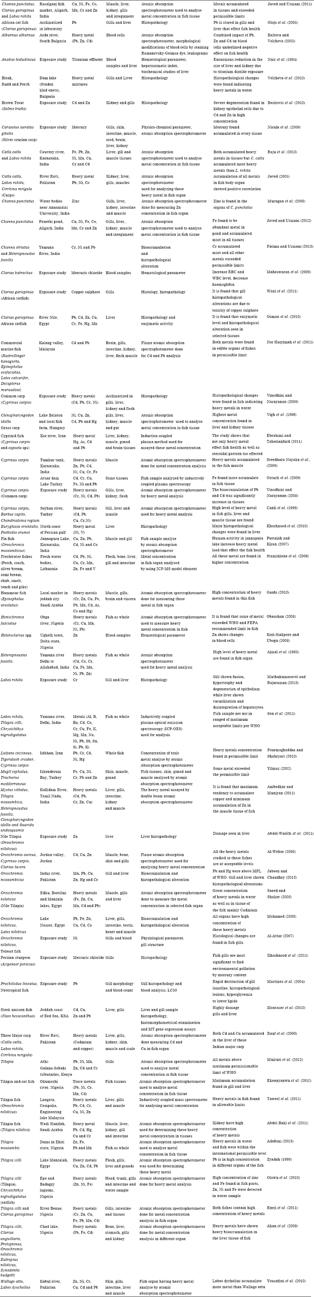 | |
This is manifested as necrosis at the tissue level (Bailey et al., 1996). Histological and histopathological changes produced by pollutants in organs and tissues can occur before they produce irreversible effects on the biota. Histological methods can be used in conjunction with other ecotoxicological bioindicators as an early warning system for the survival of the species, as well as for environmental protection (Khoshnood et al., 2010). Histopathological biomarkers are closely related to other biomarkers of stress since many pollutants have to undergo metabolic activation in order to be able to provoke cellular change in the affected organism. The simplest explanation for aquatic data showing greater concentrations at higher trophic levels (up to fish) is that of passive uptake by diffusion through body surfaces including gills, with elimination rates decreasing with increased body size (Gray, 2002).
Since field studies account for both waterborne and dietary metal exposures, bioaccumulation factor data compiled from field are more ecologically important as all the routes of exposure (e.g., dietary absorption, transport across the respiratory surface) are contributing in field data (Gobas and Morrison, 2000; DeForest et al., 2007).
MECHANISM OF METAL BIOACCUMULATION IN FISHES
Fishes are continuously exposed to waterborne and particulate heavy metals due to continuous flow of water through gills and through food sources. Metals bioaccumulated in different tissues follow different patterns of bioaccumulation factors (Fatima and Usmani, 2013). The mechanism of bioaccumulation of heavy metal in fish includes different processes in dynamic manner. Both physiological/biochemical responses and metal geochemistry are responsible for the differences in metal concentrations observed in different populations of aquatic species. It was confirmed that the internalization of metals into the cells of gills and internal epithelia follows similar mechanisms from different bioaccumulation studies (Noegrohati, 2006).
Since decades, study of metal bioaccumulation has led to the formulation of many models. The Free Ion Activity Model (FIAM), proposed in the 1980s, gives an insight in the study of metal uptake in different species of aquatic organisms (Campbell, 1995). Few years later, the Biotic Ligand Model (BLM), was introduced which is based on the interaction of the free metal ion with the proposed biological site of action, fish gill being the initial site of action (Paquin et al., 2002). Then came Subcellular Partitioning Model (SPM) which directly addresses toxicity within organisms in terms of subcellular components of accumulated metal, a variation of a tissue residue approach (Wang and Rainbow, 2007). A simple biokinetic model including all the processes that potentially leads to metal bioaccumulation, quantitatively given by Wang and Rainbow (2008). Thus, metal bioaccumulation in an organism is controlled by the balance between uptake and elimination:
dCt/dt = KuxCw+AExIRxCf-(ke+g)xCt
where, Ct is the metal concentration in an animal at time t, Ku is the uptake rate constant from the dissolved phase (L g-1 day-1), Cw is the metal concentration in the dissolved phase (μg L-1), AE is the metal assimilation efficiency from the dietary phase, IR is the weight-specific ingestion rate of the animal (g g-1 day-1), Cf is the metal concentration in the dietary phase (μg g-1), Ke is the efflux rate constant (day-1) and g is the growth rate constant of the animal (day-1).
A number of factors such as sex, age, season, spawning period, variability of food habitats and pollutant exposure and phylogenetic differences in regulatory mechanisms, may influence the uptake, retention and bioaccumulation of trace contaminants in fish tissues (Nesto et al., 2007). Zhao et al. (2012) shown correlation of heavy metals in the tissue of fish to their living environments both qualitatively and quantitatively and there was diverse metal bioaccumulation characteristics which was significantly affected by environment factors and living habits. The bioaccumulation model showed that Uptake Efficiency factor of essential heavy metals such as Cu and Zn decreases as exposure concentration increases, due to homeostasis regulation while for non essential heavy metal Hg, it is increases as the exposure concentration increases and excretion was observed as manifestation of homeostasis regulation (Noegrohati, 2006).
MECHANISM OF HISTOPATHOLOGICAL DAMAGE
Histopathological damage in tissues is outcome of various biochemical and physiological interactions within cell owing to exposure to various xenobiotics. Heavy metals generates Reactive Oxygen Species (ROS) which damages protein, lipid and DNA content of exposed animal which on gross level can be visualized through histopathology. Heavy metals grouped as Redox active (Fe, Cu, Cr etc) undergo redox cycling whereas redox inactive metals (such as Pb, Cd and Hg) undergo covalent electron sharing with cells major antioxidant enzymes (Thiols). Both types lead to the production of ROS as hydroxyl radical (OH), Superoxide radical (O2¯) or hydrogen peroxide (H2O2) which deplete cells intrinsic antioxidant defense. ROS lead to lesions to lipid, protein and DNA which can be visualized through cross index i.e., histopathology of tissues (Ercal et al., 2001). Histopathology is a broader term and mirror of effects of exposure to a variety of anthropogenic pollutants (Hinton et al., 1992). Histological responses are relatively easily recognized provided that proper reference and control data are available (Hinton, 1994). Histopathology thus is a long term and reliable biomarker of toxicant exposure. Heavy metals undergo metabolic activation that induces a cellular change in affected fish. The tissue lesions and apoptosis arises from bioaccumulation stimulate necrotic alterations in the fish with an inflammatory defensive reaction (Roganovic-Zafirova et al., 2003). Below are given few mechanistic insight of metal toxicity leading to microscopically visible alterations.
Heavy metal ions can enter blood vessels some of them are carried by proteins like albumin and can be taken up by endothelial cells lining the vessels. Heavy metal ions induce mechanisms of gene activation in endothelial cells as do pro inflammatory mediators, indicating that corroding metal ion containing biomaterials can provoke inflammatory reactions by known, as well as by yet unknown, intracellular signalling pathways (Wagner et al., 1998). And thus blood profile changes with respect to heavy metal exposure and become sensitive bioindicator of heavy metal pollution as also shown by many workers (Baltova and Velcheva, 2005; Kori-Siakpere and Ubogu, 2008; Maheswaran et al., 2008). Teleost liver is major organ for heavy metal metabolism thus frequently studied by many workers (Canli et al., 1998; Javed, 2005; Vinodhini and Narayanan, 2008) to observe different deformities. Fish hepatocyte has relatively more glycogen/lipid content which lead to hepatocytes more vacuolated (Weber and Gingerich, 1982). Macrophage aggregates act as repositories for product of cell membrane and erythrocyte breakdown include lipofuscin, ceroid, hemosiderin and melanin (Wolke, 1992). Reason behind hepatocellular enlargement is organelle proliferation (hypertrophy), failed mitotic divison of hepatocytes (megalocytosis) and vacuolar swelling of endoplasmic reticulum cisternae (hydropic degeneration) (Hinton et al., 1992). Toxic chemicals lead to increased number of organelles as myelinated bodies, mitochondria, glycogenosomes, peroxisomes and lysosomes and changes in rough endoplasmic reticulum. Due to toxicology of chemicals, hepatocytes hypertrophy is accompanied by basophilia (as result of loss in glycogenic vacuolization and increased mRNA content (Wester et al., 2003). Kidney is another target organ for metabolism and removal of waste from blood and studied for metal bioaccumulation (Akan et al., 2009; Ambedkar and Muniyan, 2011; Fatima and Usmani, 2013) followed by heavy metal exposure kidneys follow specific metabolic process. Macrophages are the key defensive cells dealing with foreign materials and debris (Blazer et al., 1994). The macrophage comprise lipofuscin, melanin and hemosiderin pigment in heavy metal toxicated kidney tissues i.e., contaminants influence macrophage pigment composition (Kruger et al., 1996).
Gills are another organ for the concern of heavy metal toxicology as it shows significantly high bioaccumulation factor owing to the fact that gills have larger surface area and comes in direct contact with heavy metal laden water. It has been studied by several workers that various deformities such as epithelial lifting, interstitial oedema, leucocytic infiltration, hyperplasia of the epithelial cells, lamellar fusion, vasodilatation and necrosis are caused due to heavy metals (Martinez et al., 2004; Al-Attar, 2007; Taweel et al., 2011; Fatima and Usmani, 2013). Muscle and integument are the least effected tissue in terms of bioaccumulation and BAF studied by many workers (Javed, 2005; Al-Weher, 2008; Nicula et al., 2009; Rauf et al., 2009; Javed and Usmani, 2011) as these are major edible parts and relished as protein diet. Other workers (Ajmal et al., 1985; Obasohan, 2008; Pourmoghaddas and Shahryari, 2010; Sen et al., 2011) estimated heavy metal in fish taken as whole. The muscle and integument are of prime concern as the fish may not only be consumed by local population but may be transported to other region for economy.
CONCLUSION
Every heavy metal has its own bioaccumulation dynamics, depend to the metal studied and environmental conditions. It is imperative to say that histological biomarkers are the indicators of pollutants in overall health of the entire population in a waterbody since all fishes are exposed to same physico chemical environment. In this review, we have illustrated possible mechanism of heavy metal bioaccumulation in fish tissues and its effect on histopathology because as different species have contrasting patterns of accumulating and eliminated metals. There remains a substantial need to understand the subcellular controls of metal accumulation and toxicity for different metals in different species of fishes as it form major source of protein all around the world. Fish bioaccumulated substantial amount of heavy metals in short exposures and thus form acute ecological indicator of water pollution and later, histopathology of fish tissues can be another tool for chronic effect of exposure used in biomonitering studies. Here, this data is also useful for the understanding and awareness as how we are contaminating our most natural resource of protein through anthropogenic activities and thus appropriate guidelines and policies should be laid down to protect our water resources. Further, human health hazard studies can be encouraged based on difference between fish species bioaccumulation mechanism.
REFERENCES
- Abdel-Warith, A.A., E.M. Younis, N.A. Al-Asgah and O.M. Wahbi, 2011. Effect of zinc toxicity on liver histology of nile tilapia, Oreochromis niloticus. Sci. Res. Essays, 6: 3760-3769.
Direct Link - Abdel-Baki, A.S., M.A. Dkhil and S. Al-Quraishy, 2011. Bioaccumulation of some heavy metals in tilapia fish relevant to their concentration in water and sediment of Wadi Hanifah, Saudi Arabia. Afr. J. Biotechnol., 10: 2541-2547.
CrossRefDirect Link - Ambedkar, G. and M. Muniyan, 2011. Bioaccumulation of some heavy metals in the selected five freshwater fish from Kollidam River, Tamilnadu, India. Adv. Applied Sci. Res., 2: 221-225.
Direct Link - Al-Weher, S.M., 2008. Levels of heavy metal Cd, Cu and Zn in three fish species collected from the Northern Jordan valley, Jordan. Jordan J. Biol. Sci., 1: 41-46.
Direct Link - Adefemi, S.O., 2013. Bio magnification factor of some heavy metals in sediment and fish samples from major dams in Ekiti state, Nigeria. Arch. Applied Sci. Res., 5: 140-145.
Direct Link - Al-Attar, A.M., 2007. The influences of nickel exposure on selected physiological parameters and gill structure in the teleost fish, Oreochromis niloticus. J. Biol. Sci., 7: 77-85.
CrossRefDirect Link - Ajmal, M., M.A. Khan and A.A. Nomani, 1985. Distribution of heavy metals in plants and fish of the Yamuna River (India). Environ. Monitor. Assess., 5: 361-367.
CrossRefDirect Link - Bailey, G.S., D.E. Williams and J.D. Hendricks, 1996. Fish models for envioronmental carcinogenesis: The rainbow trout. Environ. Health Perspect., 104: 5-21.
Direct Link - Baltova, S. and I. Velcheva, 2005. Some morphological and pathological modification of the blood cells from Alburnus alburnus in intoxication with heavy metals (Pb, Zn and Cd). Bulgarian J. Agric. Sci., 11: 577-582.
Direct Link - Besirovic, H., A. Alic, S. Prasovic and W. Drommer, 2010. Histopathological effects of chronic exposure to cadmium and zinc on kidneys and gills of brown trout (Salmo trutta m. fario). Turk. J. Fish. Aquat. Sci., 10: 255-262.
Direct Link - Canli, M., O. Ay and M. Kalay, 1998. Levels of heavy metals (Cd, Pb, Cu, Cr and Ni) in tissues of Cyprinus carpio, Barbus capito and Chondrostoma region from the seyhan river Turkey. Trurk. J. Zool., 22: 149-157.
Direct Link - DeForest, D.K., K.V. Brix and W.J. Adams, 2007. Assessing metal bioaccumulation in aquatic environments: The inverse relationship between bioaccumulation factors, trophic transfer factors and exposure concentration. Aquat. Toxicol., 84: 236-246.
CrossRef - Eneji, I.S., R. Sha'Ato and P.A. Annune, 2011. Bioaccumulation of heavy metals in fish (Tilapia zilli and Clarias gariepinus) organs from River Benue, North-Central Nigeria. Pak. J. Anal. Environ. Chem., 12: 25-31.
Direct Link - Ebrahimi, M. and M. Taherianfard, 2011. The effects of heavy metals exposure on reproductive systems of cyprinid fish from Kor River. Iran. J. Fish. Sci., 10: 13-26.
Direct Link - Ekeanyanwu, C.R. C.A. Ogbuinyi and O.F. Etienajirhevwe, 2010. Trace metals distribution in fish tissues, bottom sediments and water from Okumeshi river in Delta State, Nigeria. Ethiopian J. Environ. Stud. Manage., 3: 12-17.
Direct Link - Mohamed, F.A.S., 2008. Bioaccumulation of selected metals and histopathological alterations in tissues of Oreochromis niloticus and Lates niloticus from Lake Nasser, Egypt. Global Vet., 2: 205-218.
Direct Link - Fatima, M. and N. Usmani, 2013. Histopathology and bioaccumulation of heavy metals (Cr, Ni and Pb) in fish (Channa striatus and Heteropneustes fossilis) tissue: A study for toxicity and ecological impacts. Pak. J. Biol. Sci., 16: 412-420.
CrossRefDirect Link - Ganbi, H.H.A., 2010. Heavy metals pollution level in marine hammour fish and the effect of popular cooking methods and freezing process on these pollutants. World J. Dairy Food Sci., 5: 119-126.
Direct Link - Hinton, D.E., P.C. Baumann, G.R. Gardner, W.E. Hawkins and J.D. Hendricks et al., 1992. Histopathologic Biomarkers. In: Biochemical, Physiological and Histological Markers of Anthropogenic Stress, Huggett, R.J., R.A. Kimerie, P.M. Mehrie and H.L. Bergman (Eds.). Lewis Publishers, Boac Rato, USA., pp: 155-210.
- Jabeen, F. and A.S. Chaudhry, 2013. Metal uptake and histological changes in gills and liver of Oreochromis mossambicus inhabiting Indus river. Pak. J. Zool., 45: 9-18.
Direct Link - Javed, M., 2005. Heavy metal contamination of freshwater fish and bed sediments in the river ravi stretch and related tributaries. Pak. J. Biol. Sci., 8: 1337-1341.
CrossRefDirect Link - Javed, M. and N. Usmani, 2011. Accumulation of heavy metals in fishes: A human health concern. Int. J. Environ. Sci., 2: 659-670.
Direct Link - Javed, M. and N. Usmani, 2012. Uptake of heavy metals by Channa punctatus from sewage-fed aquaculture pond of Panethi, Aligarh. Global J. Res. Eng. Chem. Eng., 12: 27-34.
Direct Link - Kruger, R., M. Pietrock, T. Meinelti, T. Yoshida, W. Steffens and C. Steinberg, 1996. Distribution of macrophage centres in bream (Abramis brama L.) liver from the Oder river (Germany/Poland) within the nature reserve Unteres Odertal near the town of Schwedt. Internationale Revue Gesamten Hydrobiologie Hydrographie, 81: 635-644.
CrossRef - Kori-Siakpere, O. and E.O. Ubogu, 2008. Sublethal haematological effects of zinc on the freshwater fish, Heteroclarias sp. (Osteichthyes: Clariidae). Afr. J. Biotechnol., 7: 2068-2073.
Direct Link - Khoshnood, Z., A. Mokhlesi and R. Khoshnood, 2010. Bioaccumulation of some heavy metals and histopathological alterations in liver of Euryglossa orientalis and Psettodes erumei along North Coast of the Persian Gulf. Afr. J. Biotechnol., 9: 6966-6972.
Direct Link - Khoshnood, Z., S. Khodabandeh, M.S. Moghaddam and S.M. Khorjestan, 2011. Histopathological and pathomorphological effects of mercuric chloride on the gills of persian sturgeon, Acipenser persicus, fry. Int. J. Nat. Resour. Mar. Sci., 1: 23-32.
Direct Link - Lech, J.J. and J.R. Bend, 1980. Relationship between biotransformation and the toxicity and fate of xeobiotic chemicals in fish. Environ. Health Perspect., 34: 115-131.
Direct Link - Muthukumaravel, K. and P. Rajaraman, 2013. A Study on the toxicity of chromium on the histology of gill and liver of freshwater fish Labeo rohita. J. Pure Applied Zool., 1: 122-126.
Direct Link - Muiruri, J.M., H.N. Nyambaka and M.P. Nawiri, 2012. Heavy metals in water and tilapia fish from Athi-Galana-Sabaki tributaries, Kenya. Int. Food Res. J., 20: 891-896.
Direct Link - Martinez, C.B.R., M.Y. Nagae, C.T.B.V. Zaia and D.A.M. Zaia, 2004. Acute morphological and physiological effects of lead in the neotropical fish Prochilodus lineatus. Braz. J. Biol., 64: 797-807.
CrossRefDirect Link - Maheswaran, R., A. Devapaul, S. Muralidharan, B. Velmurugan and S. Ignacimuthu, 2008. Haematological studies of fresh water fish, Clarias batrachus (L.) exposed to mercuric chloride. Int. J. Integ. Biol., 2: 49-54.
Direct Link - Murugan, S.S., R. Karuppasamy, K. Poongodi and S. Puvaneswari, 2008. Bioaccumulation pattern of zinc in freshwater fish Channa punctatus (Bloch.) after chronic exposure. Turk. J. Fish. Aquat. Sci., 8: 55-59.
Direct Link - Montaser, M., M.E. Mahfouz, S.A.M. El-Shazly, G.H. Abdel-Rahman and S. Bakry, 2010. Toxicity of heavy metals on fish at Jeddah coast KSA: Metallothionein expression as a biomarker and histopathological study on liver and gills. World J. Fishes Mar. Sci., 2: 174-185.
Direct Link - Sreedhara Nayaka, B.M., S. Ramakrishna and M.R. Delvi, 2009. Impact of heavy metals on water, fish (Cyprinus carpio) and sediments from a water tank at Tumkur, India. Oceanol. Hydrobiol. Stud., 38: 17-28.
CrossRef - Nor Hasyimah, A.K., V.J. Noik, Y.Y. Teh, C.Y. Lee and H.C. Pearline Ng, 2011. Assessment of cadmium (Cd) and lead (Pb) levels in commercial marine fish organs between wet markets and supermarkets in Klang Valley, Malaysia. Int. Food Res. J., 18: 795-802.
Direct Link - Nair, G.A., N.B.N. Vijayamohanan, H. Suryanarayanan and S. Radhakrishnan, 1984. Effect of titanium efiiuents on the peripheral haematology of Anabas testudineus (Bloch) (Pisces: Anabantidae). Proc. Indian Nat. Sci. Acad., 6: 555-558.
Direct Link - Nesto, N., S. Romano, V. Moschino, M. Mauri and L. Da Ros, 2007. Bioaccumulation and biomarker responses of trace metals and micro-organic pollutants in mussels and fish from the Lagoon of Venice, Italy. Mar. Pollut. Bull., 55: 469-484.
CrossRef - Noegrohati, S., 2006. Bioaccumulation dynamic of heavy metals in Oreochromis nilotycus (predicted through a bioaccumulation modelcontructed based on biotic ligand model (BLM)). Sri Noegrohati Bioaccumulation Dyn. Heavy, 16: 29-40.
Direct Link - Ercal, N., H. Gurer-Orhan and N. Aykin-Burns, 2001. Toxic metals and oxidative stress part I: Mechanisms involved in metal-induced oxidative damage. Curr. Top. Med. Chem., 1: 529-539.
CrossRefPubMedDirect Link - Nicula, M., P. Negrea, I. Gergen, M. Harmanescu, I. Gogoasa and M. Lunca, 2009. Mercury bioaccumulation in tissues of fresh water fish Carassius auratus gibelio (Silver Crucian Carp) after chronic mercury intoxication. Universitatea Stiinte Agricole Medicina Veterinara Iasi Lucrari Stiintifice, 52: 676-679.
Direct Link - Olowu, R.A., O.O. Ayejuyo, G.O. Adewuyi, I.A. Adejoro, A.A. B. Denloye, A.O. Babatunde and A.L. Ogundajo, 2010. Determination of heavy metals in fish tissues, water and sediment from Epe and Badagry Lagoons, Lagos, Nigeria. J. Chem., 7: 215-221.
CrossRefDirect Link - Obasohan, E.E., 2008. Bioaccumulation of chromium, copper, maganese, nickel and lead in a freshwater cichlid, Hemichromis fasciatus from Ogba River in Benin City, Nigeria. Afr. J. Gen. Agric., 4: 141-152.
Direct Link - Olojo, E.A.A., K.B. Olurin, G. Mbaka and A.D. Oluwemimo, 2005. Histopathology of the gill and liver tissues of the African catfish Clarias gariepinus exposed to lead. Afr. J. Biotechnol., 4: 117-122.
Direct Link - Ozturk, M., G. Ozozen, O. Minareci and E. Minareci, 2009. Determination of heavy metals in fish, water and sediments of Avsar dam lake in Turkey. Iran. J. Environ. Health Sci. Eng., 6: 73-80.
Direct Link - Osman, A.G.M., A.E.B.M. Abd El Reheem, K.Y. AbuelFadl, A.G.G. El-Rab, 2010. Enzymatic and histopathologic biomarkers as indicators of aquatic pollution in fishes. Nat. Sci., 2: 1302-1311.
CrossRef - Paquin, P.R., J.W. Gorsuch, S. Apte, G.E. Batley and K.C. Bowles et al., 2002. The biotic ligand model: A historical overview. Comp. Biochem. Physiol. C: Toxicol. Pharmacol., 133: 3-35.
CrossRef - Rauf, A., M. Javed and M. Ubaidullah, 2009. Heavy metal levels in three major carps (Catla catla, Labeo rohita and Cirrhina mrigala) from the river Ravi, Pakistan. Pak. Vet. J., 29: 24-26.
Direct Link - Raju, K.V., R.K. Somashekar and K.L. Prakash, 2013. Metal concentration in fresh water fish organs. Open J. Metal, 3: 23-28.
CrossRefDirect Link - Sen, I., A. Shandil and V.S. Shrivastava, 2011. Study for determination of heavy metals in fish species of the river Yamuna (Delhi) by Inductively Coupled Plasma-Optical Emission Spectroscopy (ICP-OES). Adv. Applied Sci. Res., 2: 161-166.
Direct Link - Saeed, S.M. and I.M. Shaker, 2008. Assessment of heavy metals pollution in water and sediments and their effect on Oreochromis niloticus in the Northern Delta Lakes, Egypt. Proceedings of the 8th International Symposium on Tilapia in Aquaculture, October 12-14, 2008, Cairo, Egypt, pp: 475-490.
Direct Link - Staniskiene, B., P. Matusevicius, R. Budreckiene and K.A. Skibniewska, 2006. Distribution of heavy metals in tissues of freshwater fish in Lithuania. Polish J. Environ. Stud., 15: 585-591.
Direct Link - Taweel, A., M. Shuhaimi-Othman and A.K. Ahmad, 2011. Heavy metals concentration in different organs of tilapia fish (Oreochromis niloticus) from selected areas of Bangi, Selangor, Malaysia. Afr. J. Biotechnol., 10: 11562-11566.
CrossRefDirect Link - Vinodhini, R. and M. Narayanan, 2008. Bioaccumulation of heavy metals in organs of fresh water fish Cyprinus carpio (common carp). Int. J. Environ. Sci. Technol., 5: 179-182.
CrossRefDirect Link - Velcheva, I., E. Tomova, D. Arnaudova and A. Arnaudov, 2010. Morphological investigation on gills and liver of freshwater fish from Dam Lake Studen Kladenets. Bulg. J. Agric. Sci., 16: 364-368.
Direct Link - Vinodhini, R. and M. Narayanan, 2009. Heavy metal induced histopathological alterations in selected organs of the Cyprinus carpio L. Int. J. Environ. Res., 3: 95-100.
Direct Link - Vigh, P., Z. Mastalal and K.V. Balogh, 1996. Comparison of heavy metal concentration of grass carp (Ctenopharyngodon idella Cuv. et Val.) in a shallow eutrophic lake and a fish pond (possible effects of food contamination). Chemosphere, 32: 691-701.
CrossRef - Wani, A.A., M. Sikdar-Bar, K. Borana, H.A. Khan, S.S.M. Andrabi and P.A. Pervaiz, 2011. Histopathological alterations induced in gill epithelium of African Catfish, Clarias gariepinus, exposed to copper sulphate. Asian J. Exp. Biol. Sci., 2: 278-282.
Direct Link - Wang, W.X. and P.S. Rainbow, 2008. Comparative approaches to understand metal bioaccumulation in aquatic animals. Comp. Biochem. Physiol. Part C Toxicol. Pharmacol., 148: 315-323.
CrossRefDirect Link - Wang, W.X. and P.S. Rainbow, 2007. Subcellular partitioning and the prediction of cadmium toxicity to aquatic organisms. Environ. Chem., 3: 395-399.
Direct Link - Wagner, M., C.L. Klein, T.G. Van Kooten and C.J. Kirkpatrick, 1998. Mechanisms of cell activation by heavy metal ions. J. Biomed. Mater. Res., 42: 443-452.
PubMed - Yousafzai, A.M., D.P. Chivers, A.R. Khan, I. Ahmad and M. Siraj, 2010. Comparison of heavy metals burden in two freshwater fishes Wallago attu and Labeo dyocheilus with regard to their feeding habits in natural ecosystem. Pak. J. Zool., 42: 537-544.
Direct Link - Yilmaz, A.B., 2003. Levels of heavy metals (Fe, Cu, Ni, Cr, Pb and Zn) in tissue of Mugil cephalus and Trachurus mediterraneus from Iskenderun Bay, Turkey. Environ. Res., 92: 277-281.
CrossRefDirect Link - Zhao, S., C. Feng, W. Quan, X. Chen, J. Niu and Z. Shen, 2012. Role of living environments in the accumulation characteristics of heavy metals in fishes and crabs in the Yangtze River Estuary, China. Mar. Pollut. Bull., 64: 1163-1171.
CrossRef - Zyadah, M.A., 1999. Accumulation of some heavy metals in Tilapia zilli organs from lake Manzalah, Egypt. Turk. J. Zool., 23: 365-372.
Direct Link