

Xin you Su
Department of Oncology, Jinan Central Hospital, No. 105, Jiefang Road, Lixia District, Jinan, Shandong, 250013, People’s Republic of China
Jian Qiang Zhao
Department of Oncology, Jinan Central Hospital, No. 105, Jiefang Road, Lixia District, Jinan, Shandong, 250013, People’s Republic of China
Nan Li
Department of Interventional Therapy, Jinan Central Hospital, Jinan, Shandong, 250013, People’s Republic of China
Mukesh Kumar
Bala College of Pharmacy, India
Ai mei Ou yang
Department of Radiology, Jinan Central Hospital, Jinan, Shandong, 250013, People’s Republic of China
LiveDNA: 86.24888
International Journal of Pharmacology
Year: 2019 | Volume: 15 | Issue: 5 | Page No.: 549-559





ABSTRACT
Background and Objective: Hepatocellular carcinoma (HCC) is the major threat to the human health and it has got 6th rank among all types of cancers and 3rd leading cause of cancer related death. The HCC patients have high mortality rate. Resveratrol (natural phenol) participate in a significant way in the formation of the cell membrane and also play a significant role in functioning of membrane fluidity and proteins. This study was aimed to examine the anticancer effect of resveratrol (RT) against diethylnitrosamine (DEN) induced HCC in Wistar rats. Materials and Methods: For the current experimental study, the Wistar rats were used and an intraperitoneal dose of DEN (200 mg kg–1) was given to them for the induction of HCC. The rats received various doses of RT for 22 weeks. The progressions of serum biomarkers and macroscopical components of hepatic tissue were used to access the prophylactic effects. The antioxidant parameters, cancer preventive agent status and apoptosis mechanism were reviewed to scrutinize the possible mechanism. Results: The RT treatment significantly (p<0.001) altered the hepatic nodules, body weight and hepatic, antioxidant, non-hepatic parameters in a dose-dependent manner. The macroscopical observation exhibited the hepatic nodules in DEN-induced rat’s liver and dose-dependent treatment RT significantly reduced the formation of hepatic nodules and decolourization of tissue. Curiously, the expression of β-arrestin-2, Bcl-xal and Bcl-2 were significantly (p<0.001) reduced and expression of caspase-3 and Bax was significantly (p<0.001) increased at the dose-dependent manner of RT treatment. Conclusion: On the basis of result, the authors concluded that the RT exhibited the defensive effect against the DEN-induced HCC that might be due to antioxidant effect and actuation of apoptosis.
PDF Abstract XML References Citation
Copyright: © 2019. This is an open access article distributed under the terms of the Creative Commons Attribution License , which permits unrestricted use, distribution and reproduction in any medium, provided the original author and source are credited.
How to cite this article
Xin you Su, Jian Qiang Zhao, Nan Li, Mukesh Kumar and Ai mei Ou yang, 2019. Chemoprotective Effects of Resveratrol Against Diethylnitrosamine Induced Hepatocellular Carcinoma in Wistar Rats. International Journal of Pharmacology, 15: 549-559.
DOI: 10.3923/ijp.2019.549.559
URL: https://scialert.net/abstract/?doi=ijp.2019.549.559
DOI: 10.3923/ijp.2019.549.559
URL: https://scialert.net/abstract/?doi=ijp.2019.549.559
INTRODUCTION
Hepatocellular carcinoma (HCC) is known as the major health problem worldwide and having ranked 6th among all cancers due to the high mortality rate 5,00,000-10,00,000. New cases of HCC are reported every year and approximately 7,50,000 patients died from HCC. In the modern era, food habit, environmental factor and living style seem to be the major factor for cancer expansion1,2. It is believed that changing of dietary composition and consumption significantly modulate the process of carcinogenesis. The available treatment for HCC is surgical resection and liver transplantation, but both the treatments have limitation to the patients such as; patient history, liver tissue and size of the tissue which commonly affect the treatment of HCC. Both methods are effective only when HCC is detected at an early stage3,4. Other alternative treatment such as surgery, radiotherapy, hormone, chemo and immune therapy are available in hospital but the long run of these treatment have limitations. The treatment of the HCC depends upon the stage and type of cancer, but available treatment having the limitation due to developing of drug resistance into the normal as well as cancerous cells5,6 and having side effects like diarrhoea, thrombocytopenia, alopecia and nausea etc.
The HCC is considered the most common malignancies in developed countries as well as in undeveloped countries. The HCC is the most common malignancies and also called the main form of hepatic cancer due to its bad diagnosis. It is considered to be a multigene and step malignancy process and involved numerous pathogenesis factors such as aflatoxin, hepatitis B or hepatitis C and heavy ethanol consumption7-9. The HCC is started via air contamination, poisonous chemical, fungal poisons and nourishment added substance. Diethylnitrosamine (DEN) is the member of the nitroso moiety and it also considers as the effective hepatotoxin, which are induced by the similar hepatocellular carcinoma in human.
Apoptosis is a strictly managed mechanism of cell suicide and is critical for cytotoxicity induced via anticancer drugs. It is confirmed that tumour suppressor p53 is a pro-apoptotic protein and an effective growth suppressor10, which play a significant role in cellular apoptosis protecting organisms from developing cancer. Several proteins regulate the p53 pathway but Murine double minute (MDM2) consider as a significant regulator of p53 pathway. Its play a key role in the cancer progression and expansion. The MDM2 directly blocked the p53 trans activation domain encouraging the degradation11,12. It is believed that MDM2 and p53 play an important role in the expansion and development of HCC. Both directly take parts in the intrinsic apoptosis pathway via interacting with members of Bcl-2 family. The Bcl-2 and Bax, both are the members of Bcl-2 family which play potential roles in the prognosis and progression of various human malignancies. Bax encourage cell death via promoting the permeabilization of mitochondrial outer membrane, such as secreting the cytochrome C into the cytoplasm and activating the caspases to promote the cleavage of various key cellular substrates10,12. On the contrary, Bcl-2 averts via reducing the Bax activity. Moreover, Bax/Bcl-2 ratio could respect the extent of apoptosis.
In last few decades, the advancement of the cancer therapy is limited and several researchers make effort to identify the novel treatment for the HCC. The researchers screened a lot of synthetic compounds but have a few limitations such as; side effects, high toxicity and lack to target the potential site13,14. Now the researcher focused on the nutraceuticals, which derived from the plant or animal sources and have the multiple functions to treat the disease. The advantage of nutraceuticals is that they are easily available and have less side effects and are low in cost. Several researchers suggested that the regular intake of nutraceuticals reduced the risk of cancer and its complication in animal and human15,16. Phenolic phytoconstituent plays an important role in the formation of the cell membrane and also in the functioning of membrane fluidity and proteins. It shows various cellular, sub-cellular functions, processes and gene expression17,18.
Still no experimental investigation has been conducted on the estimation of biochemical, hepatic, non hepatic, antioxidant estimation and alteration of apoptosis marker in chemically induced hepatic cancer rodent via phenolic phytoconstituent drugs. Moreover, the current study was designed to evaluate the role of resveratrol on DEN induced hepatic cancer rats along with estimation of possible markers and enzymes.
MATERIALS AND METHODS
Drugs and reagents: Resveratrol and Diethylnitrosamine (DEN) were purchased from Sigma Aldrich (USA). Collagenase type 4, RNase, sodium cacodylate, hematoxylin and eosin were purchased from Himedia. The Bax and Bcl-2 were purchased from Biosynthesis Biotechnology (Beijing, P.R. China). The AST, ALT, AFP and ALP were purchased from Beihua Kangtai Biotechnology (Beijing, P.R. China). All other chemical and reagents used in the experimental study were acquired from the reputed vendor.
Experimental animal: Swiss Albino Wistar rats (100-125 g) were used for the current experimental study. For the current experimental study, the rat were received from the institutional animal house and kept in the single polyethylene cage in standard environmental condition (22±5°C) and provided standard diet pellet (Foodes pellets, Beijing, china) and water ad libitum. All experimental study was conducted accordance to Institutional Animal Ethical Committee (JCH/18/02/021).
Experimental protocol: About 72 Swiss Wistar rats were used in the current experimental study. The rats were housed in the single polypropylene cages and randomized into six groups and each group having 12 rats. The rats were stored in the controlled condition of temperature and light (22±3°C, 12 h light:dark cycle). During the experimental study, the rats were fed with standard rat pellets and water ad libitum. Before starting the experimental study, the rats were acclimatized for 14 days. The rats were divided into following groups namely group I: Normal control, group II: Normal control received RT (100 mg kg–1), group III: DEN control (200 mg kg–1), group IV-VI: Received DEN (200 mg kg–1) and Resveratrol (25, 50 and 100 mg kg–1), respectively. The single intraperitoneal dose of DEN (200 mg kg–1) induced HCC in the rats. The level of alpha-fetoprotein (AFP) was estimated after 1 week administration of DEN13,19. After that, the rats were given the RT dose treatment. The experimental study was terminated after 154 days, the animals were anesthetized with light ether and the serum samples were obtained via puncturing the retro-orbital plexus. All rats were sacrificed via cervical dislocation and the liver tissue was excised, cleaned and washed with ice-cold saline and further blotted to dryness. The hepatic tissue was homogenated in phosphate buffer saline (0.1 M; Ph-7.4) and centrifuged to obtain the clear supernatant. The supernatant was further used for the biochemical parameter estimation.
Morphology and morphometry of hepatocyte nodules: At the end of the experimental study, all group rats were anesthetized via intramuscular injection of ketamine (90 mg kg–1) and xylazine (10 mg kg–1), respectively. After that perfusion of hepatic tissue via the portal vein using the heparinized saline, the hepatic tissue was quickly removed, washed with ice-cold PBS to flush out any blood remaining part, blotted with paper towel, weighed and captured the photograph. All group rats hepatic tissue was macroscopically examined for surface as well as the presence of hepatic nodules (greyish white in colour). The hepatic nodules (greyish white in colour) were easily identified because they were surrounded by reddish brown non-nodular hepatic tissue. Vernier calliper was used for the estimation of the size of the hepatic nodules. The hepatic nodules categorized into three categories20 such as <1, 1-3 and >3.
Immunohistochemical staining for glutathione S-transferase placental form positive foci (GST-P+foci): For the estimation of positive foci (GST-P+foci), 2-3 mm of the hepatic tissue section was fixed into the ice cold solution (acetone) for the estimation of GST-P+foci via using the anti-mouse GST-P antibody. The avidin biotin peroxidise complex model was used for the visualization of the GST-P+foci (>2 mm in diameter) in the hepatic tissue section.
Biochemical parameter estimation: The hepatic parameters such as Serum α-fetoprotein (AFP), alanine aminotransferase (ALT), alkaline phosphatase (ALP) and aspartate aminotransferase (AST); non-hepatic parameter viz., total protein, albumin, total bilirubin, direct bilirubin and blood urea nitrogen (BUN) were estimated via using the manufacture instructions.
Antioxidant parameters: The antioxidant parameters viz., superoxide dismutase (SOD), catalase (CAT), lipid peroxidation (LPO), glutathione (GSH) and glutathione peroxidize (GPx) were determined by using the reported method of Kumar et al.21 via minor modification.
RNA extraction and cDNA synthesis: Trizol reagent (Invitrogen Corp., Carlsbad, CA) was used for separation of the total RNA from the rat hepatic tissue via using the manufacture instruction guidelines. The pellet of RNA was breaking down in Diethyl pyrocarbonate (DEPC) water. The Agilent Bioanalyzer and nano-drop spectrophotometer were used for the determination of purity and concentration of aggregate RNA. The cDNA synthesis kits were used for the estimation of correlative DNA via using the manufacture instruction.
Real-time PCR analysis: Real-time PCR was utilized for the determination of gene expression of the hepatic apoptotic related protein. Sangon Biotech Co., Ltd. (Shanghai, China) synthesize the primer viz., Bcl-2 forward sequence: CCCCAGAAGAAACTGAACC, reverse sequence: GCATCTCC TTGTCTACGC; caspase-3 forward sequence: CTGGACTGCGG TATTGAGAC, reverse sequence: CCGGGTGCGGTAGAGTAAGC; Bax forward sequence: GTTGCCCTCTTCTACTTTGC, reverse sequence: ATGGTCACTGTCTGCCATG; Bcl-xL forward sequence: CGTGGAAAGCGTAGACAAGG, reverse sequence: CAACAACCATGCCAGGAGAC; β-arrestin-2 forward sequence: CCACGTCACCAACAATTCTG, reverse sequence: TTGGTGTCTTCGTGCTTGAG and housekeeping gene GAPDH forward sequence: TCAAGAAGGTGGTGAAGCAG, reverse sequence: AGGTGGAAGAATGGGAGTTG, respectively. All the PCR reactions were performed to use the Maxima SYBR Green qPCR Master Mic and reactions were completed via using the accompanying condition utilizing a Master Cycler TMEppendr of real plex. Abour 1 μg of hepatic tissue RNA was used in cDNA synthesis in 96 well thermal cycler with steps involving and incubation performed at 25°C for 10 min, 37°C for 120 min, 85°C for 5 min, respectively. The distinctions in quality expression between the different groups were estimated via using the Ct (process duration, Ct) strategy and also standardized to glyceraldehyde-3-phosphate dehydrogenase (GAPDH) and were presented as relative mRNA level as a comparison to control group. Differential expression was estimated via using the 2-ΔΔCT method with minor modification22.
Statistical analysis: The results obtained in the current experimental studies were expressed as Mean±SEM (n = 12). One way ANOVA was used to obtain the statistical following at least significant difference. The p<0.05, p<0.01 and p<0.001 were considered as the significant, more significant and most significant.
RESULTS
Effect of RT on morphology and morphometry of hepatocyte nodules: The data presented in Fig. 1 showed the effect of RT on the morphology and morphometry of hepatocyte nodules of DEN-induced HCC rats. Normal and normal received RT (100 mg kg–1) did not show any sign of morphometry hepatocyte nodules at end of the experimental study. The DEN-induced control group rats showed greyish white in colour hepatocyte nodules.
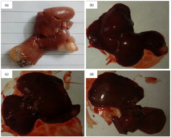 | |
| Fig. 1(a-d): | Effect of RT on the hepatic tissue of DEN control group rats, (a) Expansion of hepatic nodules (greyish in colour) and decolourization of the tissue, (b) DEN induced group rats treated with RT (25 mg kg–1) established the hepatic nodules (greyish in colour) and decolourization of the tissue, (c) DEN induced group rats treated with RT (50 mg kg–1) showed the hepatic nodules (greyish in colour) and decolourization of the tissue and (d) DEN induced group rats treated with RT (100 mg kg–1) showed the decolourization of the tissue |
Normal control group and normal control+RT (100 mg kg–1) groups rat did not show any sign and symptom of hepatic nodules and the macroscopical image of normal control and normal control+RT (100 mg kg–1) | |
| Table 1: | Effect of RT on the development of macroscopic hepatocytes nodules induced by DEN in rats |
 | |
Group I: Normal control and Group II: NC+RT (100 mg kg–1) did not show any visible hepatocytes nodule | |
| Table 2: | Effect of resveratrol on the size distribution and growth of hepatocyte nodules induced by DEN in rats |
 | |
Group I: Normal control and Group II: NC+RT (100 mg kg–1) did not show any visible hepatocytes nodule | |
The expansion of hepatic nodules (greyish in colour) and decolourization of the tissue was observed in Fig. 1a, DEN induced group rats treated with RT (25 mg kg–1) demonstrated the hepatic nodules (greyish in colour) and decolourization of the tissue (Fig. 1b). In Fig. 1c, DEN induced group rats treated with RT (50 mg kg–1) illustrated the hepatic nodules (greyish in colour) and decolourization of the tissue and Fig. 1d demonstrated that DEN induced group rats treated with RT (100 mg kg–1) exhibited the decolourization of the tissue. The data presented in Table 1 showed the total number of the nodule observed in the DEN control group rats, which suggested that the DEN group rats having the 100% tumour incidence DEN group rats treated with resveratrol (25, 50 and 100 mg kg–1) showed less nodules and suggested the hepatoprotective effect via inhibited the incidence of tumour formation and the data given in Table 2 exhibited that the average number of the nodules were greater in the DEN control group and resveratrol (25, 50 and 100 mg kg–1) treatment decreased the growth of hepatocyte nodules and also decreased by increasing the size while normal and normal received RT (100mg kg–1) did not show any visible hepatocytes nodule.
Effect of RT on the body weight: The data presented in Fig. 2a showed the effect of the RT of the body weight on the normal control and DEN-induced group rats. The initial body weight of the all group rats was almost similar. Normal control and normal control group rats received resveratrol (100 mg kg–1) showed the enhancement of the body weight at the end of the experimental study. DEN-induced rats illustrated the augmented body weight at end of the experimental study. On the other hand, resveratrol (25, 50 and 100 mg kg–1) received group rats exhibited the boosted body weight at dose dependent manner. Normal control and normal control received resveratrol (100 mg kg–1) demonstrated the almost similar liver and relative liver tissue weight. The DEN-induced HCC group rats exhibited the increased liver weight as a comparison to the normal and normal received resveratrol (100mg kg–1). Resveratrol-treated group rats showed the reduced liver weight at the dose level of 25, 50 and 100 mg kg–1 as compared to DEN control (Fig. 2b). A similar momentum was found in the relative body weight, DEN-induced group rats exhibited the increased relative hepatic tissue weight and resveratrol treatment demonstrated the reduced relative hepatic tissue weight in a dose-dependent manner.
Effect of resveratrol on hepatic parameters: The AFP is considered as the gold parameter and commonly used for the estimation of HCC. During the HCC, the level of hepatic parameters increased due to leakage into the serum. Normal control and normal control received resveratrol (100 mg kg–1) exhibited the almost similar hepatic parameters. The DEN-induced group rats showed the up-regulation of the AFP, AST, ALT and ALP, respectively (Fig. 3). The level of these parameters almost double as compared to the normal control and normal control received resveratrol (100 mg kg–1). Resveratrol (100 mg kg–1) treatment significantly (p<0.001) down-regulated the AFP, AST, ALT and ALP, respectively.
Effect of resveratrol on non-hepatic parameters: Similar hepatic parameters trends were observed in the non-hepatic parameters. The level of non-hepatic parameters such as; albumin, total protein, BUN, total bilirubin and direct bilirubin were found almost similar in the normal control and normal control treated with RT (100 mg kg–1) group rats (Fig. 4). On the other hand, DEN-induced HCC rats exhibited the alteration in the level of non-hepatic parameters including albumin, total protein, BUN, total bilirubin and direct bilirubin. DEN-induced rats treated with the resveratrol (100 mg kg–1) demonstrated the modulation in the level of non-hepatic parameters viz., albumin, total protein, BUN, total bilirubin and direct bilirubin, respectively (Fig. 4).
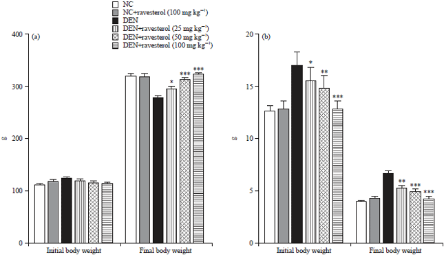 | |
| Fig. 2(a-b): | Body weight effect of RT on the normal control and DEN induced treated group rats, (a) Initial and final body weight of normal control and RT treated group rats and (b) Hepatic tissue and relative hepatic tissue weight |
RT: Ravesterol and DEN: Diethylnitrosamine. The comparisons were made by ANOVA followed by Dunnett’s test. *p<0.05 is considered as significant, **p<0.01 is considered as very significant, ***p<0.001 is considered as extremely significant | |
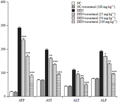 | |
| Fig. 3: | Effect of RT on the hepatic parameters on the normal control and DEN induced treated group rats as AFP, AST, ALT and ALP |
AFP: Alpha fetoprotein, AST: Aspartate transaminase, ALT: Alanine Aminotransferase, ALP: Alkaline phosphatase, RT: Ravesterol and DEN: Diethylnitrosamine. The comparisons were made by ANOVA followed by Dunnett’s test. *p<0.05 is considered as significant, **p<0.01 is considered as very significant, ***p<0.001 is considered as extremely significant | |
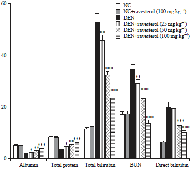 | |
| Fig. 4: | Effect of the RT on the non-hepatic parameters on the normal control and DEN induced treated group rats as albumin, total protein, total bilirubin, bun and direct bilirubin |
| BUN: Blood urea nitrogen, RT: Ravesterol and DEN: diethylnitrosamine. The comparisons were made by ANOVA followed by Dunnett’s test. *p<0.05 is considered as significant, **p<0.01 is considered as very significant, ***p<0.001 is considered as extremely significant | |
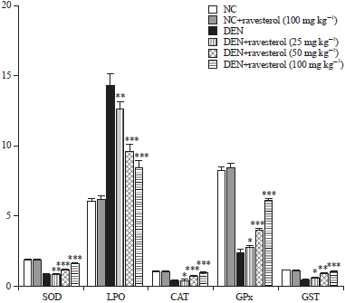 | |
| Fig. 5: | Effect of the RT on the antioxidant parameters on the normal control and DEN induced treated group rats as SOD, LPO, CAT, GPx and GST |
SOD: Superoxide dismutase, LPO: Lipid peroxidation, CAT: Catalase, Gpx: Glutathione peroxidise, GST: Glutathione-S-transferase, RT: Ravesterol and DEN: Diethylnitrosamine. The comparisons were made by ANOVA followed by Dunnett’s test. *p<0.05 is considered as significant, **p<0.01 is considered as very significant, ***p<0.001 is considered as extremely significant | |
Effect of resveratrol on antioxidant parameters: The data presented in Fig. 5 illustrated the antioxidant effect of resveratrol on the normal and DEN treated group rats. The resveratrol treatment restored the antioxidant defence via alteration in the level of the antioxidant parameter as a comparison to DEN group rats. Resveratrol successfully reduced the level of LPO and up-regulated the level of SOD, CAT, GPx and GST in comparison to DEN-induced toxic control group rats (Fig. 5).
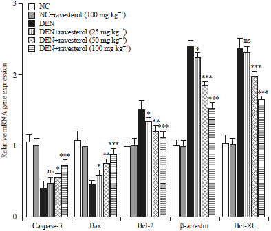 | |
| Fig. 6: | Effect of the RT on the mRNA expression of normal control and DEN induced treated group rats as Caspase-3, Bax, Bcl-2, β-arrestin and Bcl-xl |
RT: Ravesterol and DEN: Diethylnitrosamine. The comparisons were made by ANOVA followed by Dunnett’s test. *p<0.05 is considered as significant, **p<0.01 is considered as very significant, ***p<0.001 is considered as extremely significant | |
Resveratrol boosted apoptosis in hepatic tissue: The data presented in Fig. 6 represented the quantitative real-time PCR results showed the critical boosting level of mRNA expression of antiapoptotic proteins such as Bcl-xl, β-arrestin-2, Bcl-2, reduction in the level of caspase-3 and Bax level of DEN-induced control group rats. The DEN-induced HCC rats treated with resveratrol showed significantly (p<0.001) down-regulation of antiapoptotic proteins such as; Bcl-xl, β-arrestin-2, Bcl-2, up-regulation of caspase-3, Bax level as compared to DEN control group rats.
Effect of resveratrol on preneoplastic foci: The GST-P+foci were expanding in all groups rats treated with DEN. The DEN group rats showed the decreased number of GST-P+foci and also showed the reduced area of GST-P+foci as compared to the normal control and other group rats. Dose dependent treatment of resveratrol altered the preneoplastic foci at the end of the experimental study (Fig. 7a, b).
DISCUSSION
In the current experimental study, DEN was used for the induction of HCC. The DEN-induced group rats exhibited the formation of greyish white nodules and decolorization of hepatic tissue23,24. Resveratrol concentration dependent treatment demonstrated the less number of greyish white in colour hepatic nodules and the smooth tissue surface. It is already confirmed that greyish white colour hepatic nodules are the precursor of hepatic cancer23,24. Therefore, a large number of human hepatic cancer and experimental studies on the rodent clearly suggested that the relationship between the number and size of hepatocytes nodules and HCC. The reduction in the hepatic nodules formation via resveratrol suggested the protective effect against the HCC.
The AFP is the best parameter to estimate the expansion of HCC. During the HCC, the level of AFP significantly increased25,26. The shape and size of the AFP protein almost similar to the other serum protein and the concentration of its presence in minute quantity during the normal condition27,28. The similar minute concentration of AFP was found in the NC and NC received resveratrol (100 mg kg–1) and DEN-induced group rats exhibited the boosted level of AFP almost 100 times more as compared to NC and suggested the progression of HCC. Resveratrol significantly (p<0.001) down-regulated the level of AFP and suggested its chemoprotective effect. Transaminase enzymes viz., AST and ALT both are flooded in most of the human body tissue. These enzymes catalyse easily formed the amino groups from the amino acids and 1-oxoacids. Both the hepatic parameters such as ALT and AST generally used in the estimation of any changes in the hepatic tissue29.
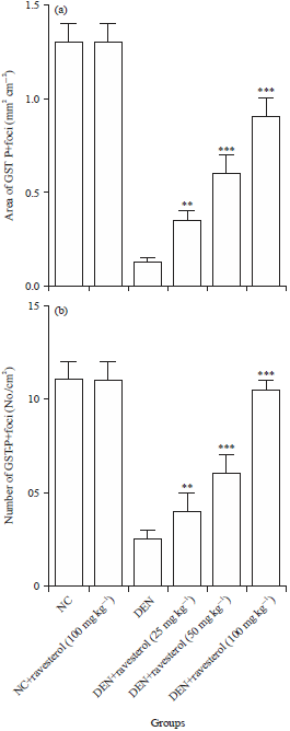 | |
| Fig. 7(a-b): | Effect of resveratrol on the number and area of placental glutathione S-transferase (GST-P) positive foci in DEN induced PH promoted hepatic carcinogenesis in rats. (a) Area of GST-P+foci and (b) Number of GST-P+foci |
Values are Mean±SE, Means with different letters are significantly different, p<0.05, whereas means with similar letters are not different from each other | |
Another hepatic enzyme ALP, also used as the indicator of hepatic damage. During the hepatic tissue injury, the level of ALP increased due to inhibition of conversion of amino acids into the keto acids and start the expansion of HCC30,31. The DEN-induced group rats showed the increased level of AST, ALT and ALP as compared to NC and NC received resveratrol and resveratrol reduced the level of hepatic enzymes and suggested the chemoprotective effect. Non-hepatic parameters were also used to identify the chemoprotective effect of the tested drug against the HCC. The reduced level of total protein and albumin is used as the indicator of reduction of protein biosynthesis due to dissociation and destruction of polyribosomes on the endoplasmic reticulum induced via DEN toxicant.
The liver is the significant site for the digestion of DEN and during digestion DEN-induced a lot of reactive oxygen species (ROS) and also plays a critical role to DEN prompted cancer-inducing effects. During DEN digestion, generation of free radical or ROS, they involved in the various stages of carcinogenesis via progression, expansion and initiation. Several researchers claimed that the DEN disturbs the cellular endogenous redox balance32-34.
It is believed that tumour initiation, expansion, metastasis and maintenance generally arbitrated via change in apoptosis-related proteins. Previous studies suggested that the dis-regulation of apoptosis is the significant basis for HCC progression35,36. Two major pathways played a crucial role in the process of apoptosis: first; mitochondrial arbitrated intrinsic pathway and second; death receptor arbitrated extrinsic apoptosis pathway. The Bcl-2 family protein, a known group of antiapoptotic proteins, which act via neutralizing the proapoptotic proteins viz., Bax protein and it also plays an important role in guiding the tumour cells to undergo apoptosis. Bax protein is commonly found in the cytosol and is shifted to the mitochondria to instigate apoptosis, yet the action of Bax also comes via antiapoptotic proteins such as; Bcl-xl and Bcl-2. Bcl-2 family proteins regulated the mitochondrial pathway, which is involved in the pro-anti-apoptotic member of proteins37-39. During the apoptosis process, pro-apoptosis protein (BAD and BAX) translocates to the mitochondrial outer membrane and boosts the secretion of cytochrome C40-42. On the other hand, the anti-apoptotic proteins such as Bcl-xl and Bcl-2 both reduce the secretion of cytochrome C43. Several researchers suggested that the number of cells removed from apoptosis via increased the expression of Bcl-xl and Bcl-240-42. The DEN-induced group rats showed the decreased expression of pro-apoptotic proteins (BAD and BAX) and up-regulation expression of anti-apoptotic (Bcl-2 and Bcl-xl) proteins. Resveratrol treatment exhibited the up-regulation of mRNA expression of pro-apoptotic expression along with down-regulation of expression of anti-apoptotic (Bcl-2 and Bcl-xl) protein expression in qRT-PCR. The implementation of apoptosis was performed via activation of caspase. The activation of caspase especially caspase-3 in two ways; intrinsic and extrinsic pathway. In the current experimental study, DEN group rats showed the decreased expression of caspase-3, while resveratrol treatment activated the caspase-3 expression. The HCC cells are directly linked to the increased expression of caspase-3 and Bax and reduced expression of Bcl-xl and Bcl-2. Resveratrol treatment initiated the apoptosis via reduced the Bcl-xl and Bcl-2 expression and increased of caspase-3 and Bax level might be considered as the crucial mechanism for preventing the HCC. Various researchers suggested that the β-arrestin-2 was first uncovered as an eliminator of G protein-coupled receptor signaling and it has been present to display the novel abilities as a signal transducer in diverse signaling pathways. The β-arrestin-2 found to obstruct the apoptosis signaling pathways via inactivation of the intrinsic apoptotic pathway44,45. The current experimental study showed the increased content of β-arrestin-2 in DEN-induced HCC group rats and dose-dependent treatment of resveratrol significantly (p<0.001) down-regulated the mRNA expression level and claim the apoptotic effect. The apoptotic effect of resveratrol also confirmed via an increased level of caspase-3 and Bax.
CONCLUSION
On the basis of the result, it can be concluded that the resveratrol exhibited the chemoprotective effect against the DEN-induced HCC. Resveratrol showed the chemoprotective effect via alteration of antioxidant effect and enlistment of apoptosis. Till date no successful treatment available for the HCC, current research recommends for the potential utilization of resveratrol in the chemoprevention of HCC.
SIGNIFICANCE STATEMENT
This study discovered the hepatoprotective effect of resveratrol that can be beneficial for hepatic cancer and this study will help the researchers to uncover the critical areas of hepatocellular carcinoma that many researchers were not able to explore. Thus a new theory on hepatocellular carcinoma may be arrived at in this experimental study.
REFERENCES
- El-Serag, H.B. and K.L. Rudolph, 2007. Hepatocellular carcinoma: Epidemiology and molecular carcinogenesis. Gastroenterology, 132: 2557-2576.
CrossRefDirect Link - Li, S. and M. Mao, 2013. Next generation sequencing reveals genetic landscape of hepatocellular carcinomas. Cancer Lett., 340: 247-253.
CrossRefDirect Link - Forner, A., M.E. Reig, C.R. de Lope and J. Bruix, 2010. Current strategy for staging and treatment: The BCLC update and future prospects. Semin. Liver Dis., 30: 61-74.
CrossRefDirect Link - Hwang, S., S.G. Lee and J. Belghiti, 2010. Liver transplantation for HCC: Its role: Eastern and Western perspectives. J. Hepato-Biliary-Pancreatic Sci., 17: 443-448.
CrossRefDirect Link - Scott, W.J., J. Howington, S. Feigenberg, B. Movsas and K. Pisters, 2007. Treatment of non-small cell lung cancer stage I and stage II: ACCP evidence-based clinical practice guidelines (2nd Edition). Chest, 132: 234S-242S.
CrossRefDirect Link - Dai, J. and R.J. Mumper, 2010. Plant phenolics: Extraction, analysis and their antioxidant and anticancer properties. Molecules, 15: 7313-7352.
CrossRefPubMedDirect Link - Montella, M., A. Crispo and A. Giudice, 2011. HCC, diet and metabolic factors: Diet and HCC. Hepatitis Monthly, 11: 159-162.
PubMedDirect Link - Gomaa, A.I., S.A. Khan, M.B. Toledano, I. Waked and S.D. Taylor-Robinson, 2008. Hepatocellular carcinoma: Epidemiology, risk factors and pathogenesis. World J. Gastroenterol., 14: 4300-4308.
CrossRefPubMedDirect Link - Liu, C.C., H. Yang, L.L. Zhang, Q. Zhang, B. Chen and Y. Wang, 2014. Biotoxins for cancer therapy. Asian Pac. J. Cancer Prevent., 15: 4753-4758.
Direct Link - Green, D.E. and H. Beinert, 1955. Biological oxidations. Annu. Rev. Biochem., 24: 1-44.
CrossRefDirect Link - Flores, E.R., 2016. Commentary on Apoptosis, p53 and tumor cell sensitivity to anticancer agents. Cancer Res., 76: 6763-6764.
Direct Link - Kumar, V., P.C. Bhatt, M. Rahman, F.A. Al-Abbasi, F. Anwar and A. Verma, 2017. Umbelliferon-α-d-glucopyranosyl-(2I→ 1II)-α-Dglucopyranoside ameliorates Diethylnitrosamine induced precancerous lesion development in liver via regulation of inflammation, hyperproliferation and antioxidant at pre-clinical stage. Biomed. Pharmacother., 94: 834-842.
CrossRefDirect Link - Khan, R., I. Kazmi, M. Afzal, F.A. Al Abbasi and G. Mushtaq et al., 2015. Fixed dose combination therapy loperamide and niacin ameliorates diethylnitrosamine-induced liver carcinogenesis in albino Wistar rats. RSC Adv., 5: 67996-68002.
CrossRefDirect Link - Wang, L. and C.L. Weller, 2006. Recent advances in extraction of nutraceuticals from plants. Trends Food Sci. Technol., 17: 300-312.
CrossRefDirect Link - Aggarwal, B.B., M.E. van Kuiken, L.H. Iyer, K.B. Harikumar and B. Sung, 2009. Molecular targets of nutraceuticals derived from dietary spices: Potential role in suppression of inflammation and tumorigenesis. Exp. Biol. Med., 234: 825-849.
CrossRefDirect Link - Youdim, K.A., A. Martin and J.A. Joseph, 2000. Essential fatty acids and the brain: Possible health implications. Int. J. Dev. Neurosci., 18: 383-399.
CrossRefDirect Link - Calder, P.C., 2009. Polyunsaturated fatty acids and inflammatory processes: New twists in an old tale. Biochimie, 91: 791-795.
CrossRefDirect Link - Singh, D., M. Singh, E. Yadav, N. Falls and U. Komal et al., 2018. Amelioration of diethylnitrosamine (DEN)-induced hepatocellular carcinogenesis in animal models via knockdown oxidative stress and proinflammatory markers by Madhuca longifolia embedded silver nanoparticles. RSC Adv., 8: 6940-6953.
CrossRefDirect Link - Afzal, M., I. Kazmi, R. Khan, P. Rana and V. Kumar et al., 2017. Thiamine potentiates chemoprotective effects of ibuprofen in DEN induced hepatic cancer via alteration of oxidative stress and inflammatory mechanism. Arch. Biochem. Biophys., 623: 58-63.
CrossRefDirect Link - Kumar, V., D. Ahmed, F. Anwar, M. Ali and M. Mujeeb, 2013. Enhanced glycemic control, pancreas protective, antioxidant and hepatoprotective effects by umbelliferon-α-D-glucopyranosyl-(2I → 1II)-α-D-glucopyranoside in streptozotocin induced diabetic rats. SpringerPlus, Vol. 2.
CrossRefDirect Link - Sadek, K.M., T.K. Abouzed, R. Abouelkhair and S. Nasr, 2017. The chemo-prophylactic efficacy of an ethanol Moringa oleifera leaf extract against hepatocellular carcinoma in rats. Pharm. Biol., 55: 1458-1466.
CrossRefDirect Link - Schiffer, E., C. Housset, W. Cacheux, D. Wendum and C. Desbois-Mouthon et al., 2005. Gefitinib, an EGFR inhibitor, prevents hepatocellular carcinoma development in the rat liver with cirrhosis. Hepatology, 41: 307-314.
CrossRefDirect Link - Heindryckx, F., I. Colle and H. van Vlierberghe, 2009. Experimental mouse models for hepatocellular carcinoma research. Int. J. Exp. Pathol., 90: 367-386.
CrossRefDirect Link - Boonacker, E. and C.J. van Noorden, 2001. Enzyme cytochemical techniques for metabolic mapping in living cells, with special reference to proteolysis. J. Histochem. Cytochem., 49: 1473-1486.
CrossRefDirect Link - El-Bashiti, T., H. Hamamci, H.A. Oktem and M. Yucel, 2005. Biochemical analysis of trehalose and its metabolizing enzymes in wheat under abiotic stress conditions. Plant Sci., 169: 47-54.
CrossRefDirect Link - Ajdukiewicz, A.B., P.C. Kelleher, E.L. Krawitt, C.J. Walters, P.B. Mason, R.S. Koff and L. Belanger, 1993. α-Fetoprotein glycosylation is abnormal in some hepatocellular carcinomas, including white patients with a normal α-fetoprotein concentration. Cancer Lett., 74: 43-50.
CrossRefDirect Link - Beneduce, L., F. Castaldi, M. Marino, N. Tono, A. Gatta, P. Pontisso and G. Fassina, 2004. Improvement of liver cancer detection with simultaneous assessment of circulating levels of free Alpha-Fetoprotein (AFP) and Afp-Igm complexes. Int. J. Biol. Markers, 19: 155-159.
CrossRefDirect Link - Prabu, S.M. and N.C. Sumedha, 2014. Ameliorative effect of diallyl trisulphide on arsenic-induced oxidative stress in rat erythrocytes and DNA damage in lymphocytes. J. Basic Clin. Physiol. Pharmacol., 25: 181-197.
CrossRefDirect Link - Ramaiah, S.K. and S. Rittling, 2007. Pathophysiological role of osteopontin in hepatic inflammation, toxicity and cancer. Toxicol. Sci., 103: 4-13.
CrossRefDirect Link - Dentin, R., F. Benhamed, I. Hainault, V. Fauveau and F. Foufelle et al., 2006. Liver-specific inhibition of ChREBP improves hepatic steatosis and insulin resistance in ob/ob mice. Diabetes, 55: 2159-2170.
CrossRefDirect Link - Gupta, S.C., D. Hevia, S. Patchva, B. Park, W. Koh and B.B. Aggarwal, 2012. Upsides and downsides of reactive oxygen species for cancer: The roles of reactive oxygen species in tumorigenesis, prevention and therapy. Antioxid. Redox Signal., 16: 1295-1322.
CrossRefDirect Link - Wang, J. and J. Yi, 2008. Cancer cell killing via ROS: To increase or decrease, that is the question. Cancer Biol. Ther., 7: 1875-1884.
CrossRefDirect Link - Circu, M.L. and T.Y. Aw, 2010. Reactive oxygen species, cellular redox systems, and apoptosis. Free Radical Biol. Med., 48: 749-762.
CrossRefDirect Link - Ashkenazi, A., P. Holland and S.G. Eckhardt, 2008. Ligand-based targeting of apoptosis in cancer: The potential of recombinant human apoptosis ligand 2/tumor necrosis factor-related apoptosis-inducing ligand (rhApo2L/TRAIL). J. Clin. Oncol., 26: 3621-3630.
CrossRefDirect Link - Tsujimoto, Y., 1998. Role of Bcl-2 family proteins in apoptosis: Apoptosomes or mitochondria? Genes Cells, 3: 697-707.
CrossRefDirect Link - Edlich, F., S. Banerjee, M. Suzuki, M.M. Cleland and D. Arnoult et al., 2011. Bcl-xL retrotranslocates Bax from the mitochondria into the cytosol. Cell, 145: 104-116.
CrossRefDirect Link - Hsu, Y.T., K.G. Wolter and R.J. Youle, 1997. Cytosol-to-membrane redistribution of Bax and Bcl-XL during apoptosis. Proc. Natl. Acad. Sci. USA., 94: 3668-3672.
CrossRefDirect Link - Chao, D.T. and S.J. Korsmeyer, 1998. BCL-2 family: Regulators of cell death. Annu. Rev. Immunol., 16: 395-419.
CrossRefDirect Link - Lindenboim, L., J. Yuan and R. Stein, 2000. Bcl-x S and Bax induce different apoptotic pathways in PC12 cells. Oncogene, 19: 1783-1793.
CrossRefDirect Link - Wu, C., H. Fujihara, J. Yao, S. Qi, H. Li, K. Shimoji and H. Baba, 2003. Different expression patterns of Bcl-2, Bcl-xl and Bax proteins after sublethal forebrain ischemia in C57Black/Crj6 mouse striatum. Stroke, 34: 1803-1808.
CrossRefDirect Link - Roy, S., M. Singh, A. Rawat, U. Devi and S. Gautam et al., 2018. GLA supplementation regulates PHD2 mediated hypoxia and mitochondrial apoptosis in DMBA induced mammary gland carcinoma. Int. J. Biochem. Cell Biol., 96: 51-62.
CrossRefDirect Link - Simon, H.U., A. Haj-Yehia and F. Levi-Schaffer, 2000. Role of Reactive Oxygen Species (ROS) in apoptosis induction. Apoptosis, 5: 415-418.
CrossRefDirect Link - Ott, M., V. Gogvadze, S. Orrenius and B. Zhivotovsky, 2007. Mitochondria, oxidative stress and cell death. Apoptosis, 12: 913-922.
CrossRefDirect Link