

Islam Nour
Department of Medical Biotechnology, Genetic Engineering and Biotech Institute, City for Scientific Research, New Borg Elarab, 21934, Alexandria, Egypt
Faiza Fattouh
Department of Microbiology, Faculty of Science, Alexandria University, Egypt
Hala El-Adawi
Department of Medical Biotechnology, Genetic Engineering and Biotech Institute, City for Scientific Research, New Borg Elarab, 21934, Alexandria, Egypt
International Journal of Pharmacology
Year: 2015 | Volume: 11 | Issue: 5 | Page No.: 440-447





ABSTRACT
Bacteriocin producing lactobacilli were obtained from different sources and were previously characterized outdoors. Lactobacillus bulgaricus 761N, Lactobacillus fermentum DSMZ 20049, Lactobacillus delbrueckii subsp. bulgaricus NCTC 12197 T and Lactobacillus delbrueckii subsp. bulgaricus DSMZ 20080T were the strains under study. Subcellular fractions were investigated for their proteolytic activity that results in production of bioactive peptides in the cell-free supernatant (bacteriocin). A single strain bacteriocin was selected depending upon cytotoxicity of the strain’s bacteriocin and antibacterial capacity. The cell-free supernatants of the tested strains were tested for their antibacterial capacity against a broad spectrum of pathogens involving Staphylococcus aureus, Pseudomonas aeruginosa, Burkholderia cepacia, Salmonella spp., Escherichia coli and Shigella spp. The selected candidate was Lactobacillus bulgaricus 761N. The bacteriocin was partially purified and assured via applying the semi-purified samples on SDS-PAGE. Advance to the purification process, the fractions selected were tested for their antibacterial capacity as well. Both fractions have been found to possess antibacterial activity but the lower molecular weight was over succeeded.
PDF Abstract XML References Citation
Article History
Received: February 26, 2015;
Accepted: May 25, 2015;
Published: June 19, 2015
How to cite this article
Islam Nour, Faiza Fattouh and Hala El-Adawi, 2015. Antibacterial Bioactivity of Selected Lactic Acid Bacterial Strains against some Human Pathogenic Bacteria. International Journal of Pharmacology, 11: 440-447.
DOI: 10.3923/ijp.2015.440.447
URL: https://scialert.net/abstract/?doi=ijp.2015.440.447
DOI: 10.3923/ijp.2015.440.447
URL: https://scialert.net/abstract/?doi=ijp.2015.440.447
INTRODUCTION
Bacteriocins were differentially defined by many scientists. Bacteriocins are proteinaceous compounds that are inhibitory towards sensitive strains and are produced by both gram-positive and gram-negative bacteria (Tagg et al., 1976). They were also termed as the ribosomally produced cationic proteins inhibiting other bacteria living in the same ecological niche (Riaz et al., 2010). Five required criteria were identified to call a chemical substance as a bacteriocin involving the presence of a biologically active part of protein nature, a spectrum of inhibitive activity narrow and centered on the homologues, a mode of bactericidal action, the adsorption to specific receivers and nature plasmodic genetic determiners coding for the production of the bacteriocin and for the immunity in this one (Tagg et al., 1976). During the last few years, a large number of new bacteriocins produced by Lactic Acid Bacteria (LAB) have been identified and characterized. LAB-bacteriocins comprise a heterogeneous group of physicochemically diverse ribosomally-synthesized peptides or proteins showing a narrow or broad antimicrobial activity spectrum against gram-positive bacteria (Cintas et al., 2001). Other studies showed that bacteriocins have also antimicrobial capability against gram negative bacteria, for instance, E. coli and Salmonella but usually only when the integrity of the outer membrane has been compromised, for example after osmotic shock or low pH treatment, in the presence of a detergent or chelating agent, or after pulsed electric field or high-pressure treatment (Stevens et al., 1991). The resistance of gram negative pathogenic bacteria to bacteriocins is attributed to the particular nature of their cellular envelope besides the fact that the mechanisms of action described for bacteriocins is owing to the phenomenon of adsorption. According to Bhunia et al. (1991), the pediocin (bacteriocins produced by Pediococcus acidilactici) interacts with lipoteichoic acids absent in gram negative bacteria. These variations of sensibility are due to the characteristics of indicators strains (presence or absence of receiving sites or immunoprotein) and thus in level of hurts caused by the inhibitive factor (Savadogo et al., 2004). Recently, It was proven that bacteriocins have the advantage over antibiotics even the third generation antibiotics owing to the fact that some microbial pathogens gain acquired immunity against these antibiotics due to their frequent uptake, for instance, bacteriocins produced by Lactobacilli strains isolated from yogurt was found to possess antimicrobial potential against cephalosporin resistant E. coli (Riaz et al., 2010). Also, bacteriocins obtained from L. fermentum were found to possess antimicrobial potential against methicillin resistant S. aureus (Nawaz et al., 2009). With respect to medical applications, antimicrobials produced by LAB might play a serious role during the in vivo interactions occurring in the human gastrointestinal tract, hence contributing to gut health (De Vuyst and Leroy, 2007). Thus, innovative approaches have been tried as alternative to antibiotics in treating gastrointestinal diseases and these include using live biotherapeutic agent (Daly and Davis, 1998; Soomro et al., 2002; Oyetayo et al., 2003).
MATERIALS AND METHODS
Isolation and identification of lactic acid bacteria: Lactobacillus delbrueckii subsp. bulgaricus DSMZ 20080T and NCTC 12197 T Lactobacillus fermentum DSMZ 20049, Lactobacillus acidophilus DSMZ 20079 T were transferred from reference strains purchased from culture collection. Another strain of Lactobacillus delbrueckii subsp. bulgaricus 761N was isolated from yogourt sample and identified up to PCR level at the laboratory of microbial biochemistry of dairy microorganisms (LMB), Alexandria University, Egypt.
Target pathogens preparation: Pathogens used involves Staphylococcus aureus, Pseudomonas aeruginosa, Burkholderia cepacia, Salmonella spp., Escherichia coli and Shigella spp. were obtained from microbiology Dept. faculty of medicine-Alexandria University. They were next sub-cultured on their specific media involving mannitol salt agar, Pseudomonas citrimide agar, brilliant green agar, Eosin methylene blue agar and triple sugar iron. All cultures were incubated for 24-48 h at 37°C then kept at 2-8°C for not more than 1 month. Prior use they were enriched on tryptic soya broth.
Subcellular fractionation of cell lysate: (1) Total cellular extracts: Total cellular extracts were obtained as described by Atlan et al. (1989). (2) Cell wall and cytoplasmic extracts: Proteins were extracted from the bacterial cell wall of Lactobacilli using a modification of the procedure previously developed to release APII from Lactobacillus delbrueckii subsp. bulgaricus (Atlan et al., 1989). After 4 h growth in 100 mL MRS or milk medium (A600 = 0.8-1.0), cells were harvested by centrifugation (10000 g, 10 min, 6°C), washed in 25 Mm-KH, PO, 10 mM-MgC12, (pH 5.8) and resuspended in 10 mL of the same buffer supplemented with 0.6 M-sucrose and treated with 0.1 or 1 mg mL-1 (for cells grown in MRS or milk medium, respectively) of lysozyme from chicken egg white (Sigma) for 30 min at 25 or 37°C (for cells grown in MRS or milk medium, respectively) with gentle shaking. Bacterial suspensions were centrifuged (10000 g, 10 min, 6°C) and the supernatants are referred to as lysozyme fluids. Lysozyme-treated bacteria were osmotically shocked by suspending them in 10 mL cold distilled water and maintained on ice for 10 min. Shocked cells were centrifuged (10000 g, 10 min, 6°C) and these supernatants are referred to as osmotic fluids. The bacterial pellets were resuspended in 10 mL cold distilled water and disrupted with a pulse sonicator 6°C for 30 min. The supernatants obtained after centrifugation of the broken cells (25000 g, 20 min, 6°C) are referred to as soluble cytoplasmic fluids.
Protease activity assessment: The protease assay was done according to the method of Kunitz (1947).
Antibacterial assay: Antibacterial assay was evaluated by microplate reader assay method according to Bechert et al. (2000) with some modifications. Aliquot of 100 μL of preinoculated pathogens (106 CFU μL-1) in LB broth was transferred to each well of 96 well plates, the same volume of extracellular lactic acid bacterial extracts added to each well in replica. The plates were incubated under microaerophilic conditions at 37°C for 24 h. After incubation, the absorbance of the plates was determined using automated ELIZA microplate reader adjusted at 620 nm. The inhibition percentage of lactic acid bacterial extracts was calculated according to the following equation:

where, A is absorbance of the treatment group, A1 is absorbance of the blank and A0 is absorbance of the control group.
Cytotoxicity test: PBMC cells were plated onto 96-well plates at a cell count of 5×10⁴ cell mL-1. After 24 h cells incubation at 37°C and 5% CO2 supply, the cells were sub-confluent. The medium was removed and a serial dilution of tested subcellular fraction in culture medium was added instead. The treated cells were then incubated for 3 days at 37°C and 5% CO2. Cells were fixed with 100 μL fixing solution (0.5% formaldehyde and 1% CaCl2) for 1 min then each well was supplemented with 100 μL solution of 50% ethanol with 1% acetic acid and shacked for 5 min.
Purification of the crude treatment: Skim milk medium containing the extracellular fraction (Crude enzyme that produces oligobioactive peptides) undergone ultrafiltration (150 kDa, VRR 5-20). The retentate is discarded, while the permeate undergone ultrafiltration (10 kDa, VRR 5) and the hydro lysate contains the bioactive peptides according to Konrad and Kleinschmidt (2008). The hydrolysate undergone several processing steps. First crude enzyme is inactivated by heating at 80°C for 20 min, Second separation of hydrolysate by centrifugation at 10000xg for 30 min, third Desalting (Demineralization) is achieved by using a negatively charged sephadex gel G50, Forth fractionation of peptide fraction is done by using size exclusion chromatography using sephadex G200. The purification process is followed by bioactivity testing and confirmed by performing SDS-PAGE (Konrad and Kleinschmidt, 2008).
Bioactivity testing: Bioactivity testing involves antibacterial assessment by Micro-broth dilution method.
RESULTS
Protease activity results of subcellular fractions: The extracellular fraction has the highest activity for all strains in comparison with inter-and intracellular fractions as shown in Table 1. The selected fraction for the four strains would be assessed for antibacterial activity and cytotoxicity.
Antibacterial activity assessment (expressed in inhibition percent using microplate reader assay method): The inhibition percent for all extracts of selected LAB strains against pathogenic bacteria increases with the increase of the concentration. The antibacterial activity of Lactobacillus bulgaricus 761N extracellular extract against different pathogenic bacteria under study was illustrated in Table 2. The maximum inhibition was recorded for Shigella spp, where it was elevated from 0 at 50% concentration to 89 at 100% concentration of the extracellular extract. However, the antibacterial effect of the extracellular extract of L. fermentum DSMZ 20049, illustrated in Table 3, the inhibition percent varied from 0 at 50% concentration of the extracellular extract against Shigella spp., to 96.89 at 100% concentration of the extracellular extract. Moreover, the antibacterial activity of L. delbrueckii subsp. bulgaricus NCTC 12197 T extracellular extract, shown in Table 4, illustrated the inhibition percent was found to be ranging from 0 at 50% concentration against P. aeruginosa to 85.6 at 100% concentration. The least antibacterial activity was that of the extracellular extract of L. delbrueckii subsp. bulgaricus DSMZ 20080T, clarified in Table 5. The inhibition percentage was the highest (70.17%) at 100% concentration of the extracellular extract against Salmonella spp.
| Table 1: | Protease activity of subcellular fractions of the four different Lactobacilli strains |
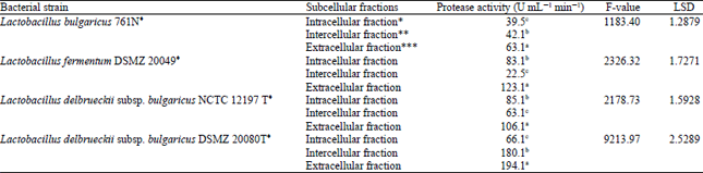 | |
| *Cytoplasmic fluids, **Osmotic fluids, ***Lysozyme fluids. Equation: y = 0.001x+0.0859, where, y = Test-control, x = Proteolytic activity (U mL-1 min-1). ♦α-significance = 0.05, p<0.0001 (i.e., highly significant difference). a, b, cSymbols to express significant difference in the statistical analysis (i.e., similar symbols means no significant difference while different symbols means that these data are significantly different), values are given in Mean±SD | |
| Table 2: | Antibacterial assay for Lactobacillus bulgaricus 761N extracellular extract tested against different pathogenic bacteria |
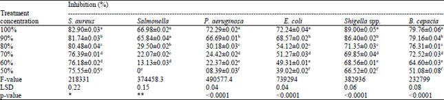 | |
| *No significant difference was observed for the extracellular extract of Lactobacillus bulgaricus 761N at concentrations 60 and 70% against Staphylococcus aureus (p>0.005), i.e., in the case of using the crude extract as a treatment against S. aureus at treatment concentration 60% instead of 70%, it would be better owing to gaining a less side effects, for example cytotoxicity and roughly the same influence as that of the latter treatment concentration. **No significant difference was observed for the extracellular extract of Lactobacillus bulgaricus 761N at concentrations 90 and 100% against Salmonella spp. (p>0.005), i.e., using the crude treatment against Salmonella at a concentration of 90% is better than using it at 100% concentration achieving less negative effects and roughly the same influence as that of the latter concentration | |
| Table 3: | Antibacterial assay for Lactobacillus fermentum DSMZ 20049 extracellular extract against some pathogenic bacteria |
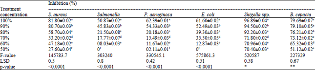 | |
| a,b,c,d,e and f: Significant difference in the statistical analysis, *: No significant difference was observed for the extracellular extract of Lactobacillus fermentum DSMZ 20049 at concentrations 50 and 60% against Shigella spp. (p>0.005), **: No significant difference was observed for the extracellular extract of Lactobacillus fermentum DSMZ 20049 at concentrations 90 and 100% against B. cepacia (p>0.005) | |
| Table 4: | Antibacterial assay for Lactobacillus delbrueckii subsp. bulgaricus NCTC 12197 T extracellular extract against some pathogenic bacteria |
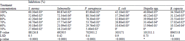 | |
| a,b,c,d,e and f: Significant difference in the statistical analysis, *: No significant difference was observed for the extracellular extract of Lactobacillus delbrueckii subsp. bulgaricus NCTC 12197 Tat concentration 80, 90 and 100% against Shigella spp. (p>0.005). Thus, using the crude extract as a treatment at 80% would be better than 100% treatment concentration for both safer utility and a nearly close anti-shigella activity | |
| Table 5: | Antibacterial assay for Lactobacillus delbrueckii subsp. bulgaricus DSMZ 20080T extracellular extract tested against some pathogenic bacteria |
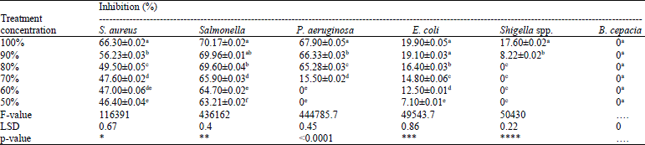 | |
| a,b,c,d,e and f: Significant difference in the statistical analysis, *: No significant difference was observed for the extracellular extract of Lactobacillus delbrueckii subsp. bulgaricus DSMZ 20080T between concentration 60 and 70% and between concentrations 50 and 60% against S. aureus (p>0.005), **: No significant difference was observed for the extracellular extract of Lactobacillus delbrueckii subsp. bulgaricus DSMZ 20080T between concentration 90 and 100% and between concentrations 80 and 90% against Salmonella spp. (p>0.005), ***: No significant difference was observed for the extracellular extract of Lactobacillus delbrueckii subsp. bulgaricus DSMZ 20080T at concentration 90 and 100% against E. coli (p>0.005), ****: No significant difference was observed for the extracellular extract of Lactobacillus delbrueckii subsp. bulgaricus DSMZ 20080T at concentration 50, 60, 70 and 80% against Shigella spp. (p>0.005) | |
| Table 6: | Cytotoxicity assay on human peripheral blood mononuclear cells |
 | |
| Values are given in Mean±SD | |
Cytotoxicity assay: The extracellular extract of Lactobacillus bulgaricus 761N was observed to possess the least cytotoxicity on PBMC (Table 6) and that 16% concentration would be the selected non-toxic dose.
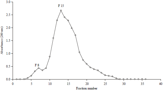 | |
| Fig. 1: | Bacteriocin fractionation by size exclusion chromatography (El-Adawi et al., 2015) |
| Table 7: | Antibacterial activity of fraction No. 8 against different pathogenic bacteria |
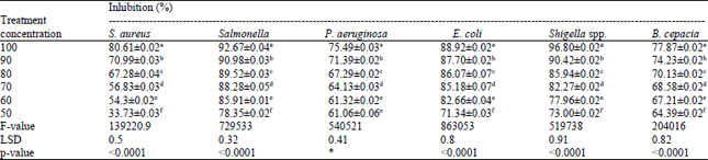 | |
| a,b,c,d,e and f: Significant difference in the statistical analysis, *No significant difference was observed for the fraction No. 8 (obtained from purification process of extracellular extract of Lactobacillus bulgaricus 761N) against P. aeruginosa at concentrations 50 and 60% (p>0.005) | |
Fractionation of extracellular extract by size exclusion column chromatography: Two fractions were obtained following the fractionation of the semi-purified extracellular extract of L. bulgaricus 761N using size exclusion chromatography which was fraction No. 8 (F8) and fraction No. 15 (F15) as reported previously by our group (El-Adawi et al., 2015) (Fig. 1). Both fractions were subjected to SDS-PAGE and their molecular weights are 14.4 and 45 kDa, respectively.
Bioactivity testing: After the fractionation process, the antibacterial activity of the bacteriocin extracts of L. bulgaricus 761N for both fractions (F8 and F15) were tested. In case of fraction 8, the inhibition against four (Salmonella, P. aeruginosa, E. coli and Shigella spp.) out of six pathogenic strains was elevated (Table 7). In case of fraction 15, the inhibition against only two (Salmonella and E. coli) out of six pathogenic strains was elevated (Table 8).
DISCUSSION
The proteolytic system of lactobacilli is complex and is composed of proteinases and exopeptidases with different subcellular locations (Atlan et al., 1989). It was found that the extracellular fraction for all strains under study possessed the highest proteolytic activity. This is in agreement with Kok and Venema (1988), who reported that the cell-wall-bound proteinases of LAB perform the first steps in the hydrolysis of casein. Seo et al. (2007), however, observed that the first step in casein degradation is mediated by proteases in the cell membrane. Further degradation into smaller peptides and amino acids that can pass through cell membrane, is achieved by peptidases of LAB (Wohlrab and Bockelmann, 1992; Shihata and Shah, 2000). The findings of Kabadjova-Hristova et al. (2006) approved that Lactobacillus (L. kefir DR22x) had cell-wall-bound proteinases with an extracellular location.
The antibacterial activity of extracellular extract referring to bacteriocins was mentioned in many literatures. Earlier reports (Tagg et al., 1976; Daeschel and Klaenhamner, 1985; Sanni et al., 1999) have shown that some bacteriocins produced by gram-positive bacteria have a broad spectrum of activity. However, it was generally observed that bacteriocin from the producer organism had no inhibitory effect on the organism producing it.
| Table 8: | Antibacterial activity of fraction No. 15 against different pathogenic bacteria |
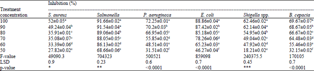 | |
| a,b,c,d,e and f: Significant difference in the statistical analysis, *: No significant difference was observed for the fraction No. 8 (obtained from purification process of extracellular extract of Lactobacillus bulgaricus 761N) against S. aureus at concentrations 70 and 80% (p>0.005), **: No significant difference was observed for the fraction No. 8 (obtained from purification process of extracellular extract of Lactobacillus bulgaricus 761N) against Salmonella at concentrations 90 and 100% (p>0.005), ***: No significant difference was observed for the fraction No. 8 (obtained from purification process of extracellular extract of Lactobacillus bulgaricus 761N) against Shigella spp. at concentrations 90 and 100% (p>0.005) | |
Tufail et al. (2011), have observed that culture supernatants that were obtained from the sixty out of hundred isolates of Lactobacillus spp. involving L. bulgaricus isolated from yogurt exhibited varying degrees of inhibitory activity against strains of Bacillus subtilis ATCC 6633, Escherichia coli ATCC 10536, Salmonella typhi ATCC 19430, Staphylococcus aureus ATCC 6538 and Vibrio cholerae ATCC 25870. This is in agreement with the current research findings that observed a significant antibacterial capability against a range of pathogenic bacteria involving E. coli, S. aureus, Pseudomonas aeruginosa, Salmonella sp., Burkholderia cepacia and Shigella sp. These results are also in agreement with previous work carried out by Erdogrul and Erbilir (2006) but on different strain types, in which supernatants obtained from Lactobacillus casei and L. bulgaricus exhibited varying degrees of inhibitory activity against strains of E. coli ATCC 8739, S. aureus ATCC 6538, Pseudomonas aeruginosa ATCC 9027, B. subtilis ATCC 6633, Klebsiella pneumoniae ATCC 18833, Salmonella typhimurium ATCC 13311 and Enterobacter cloacae ATCC 13047. The probiotic potential of these bacteria is also vastly investigated (Gilliland, 1990; Cleveland et al., 2001; Mojgani and Ashtiani, 2006; Diez-Gonzalez, 2007).
Akpinar et al. (2011) have shown that L. bulgaricus produces a bacteriocin called Bulgarican that is inhibitory towards both gram-positive and gram-negative bacteria. Some inhibitory compounds against Staphylococcus and Clostridium species have also been found (Erkus, 2007). Ocana et al. (1999) observed by using transmission electron microscopy that the bacteriocin obtained from L. salivarius subsp. salivarius CRL 1328 causes vesiculization of protoplasm, formation of pores and complete disintegration of cells of Enterococcus faecalis. According to Marteau et al. (2001), the electrostatic interactions between the LAB and the negative charged phosphate groups on target cell membranes are thought to contribute to the initial binding, forming pores and killing the bacterial cells after causing lethal damage and autolysin activation to digest the microbial cellular wall.
Regarding the cytotoxicity, bacteriocins obtained from LAB has been mentioned in many literatures to have a very low cytotoxicity that agree with the current research findings. EL-Adawi et al. (2012) observed that extracellular extract of several LAB were safe when conducted the cytotoxicity assay on PBMC and had no TC50 even in the maximum concentrations which agreed with the current study results. Low molecular weight bioactive proteins, termed as lactoferrin, that was tested for its cytotoxicity on both HepG2 and PBMC, was found to be safe (El-Fakharany et al., 2008) indicating a very low cytotoxic effect. These findings approved our study results that these low molecular weight bioactive peptides contained in the extracellular extract exert a low cytotoxicity on PBMC.
The investigations of the current study concerning the purification process of the crude extract revealed the presence of two semi purified fractions whose molecular weights 14.4 and 45 kDa. These low molecular weight peptides are produced owing to the proteolytic activity of L. bulgaricus 761N performed in a casein-derived media. This agreed with the findings of (Kim et al., 2004) who reported that L. bulgaricus produced a bacteriocin of molecular weight 14.4 kDa, determined by SDS-PAGE and overlay method and that the mode of action for the antibacterial activity was bacteriocidal.
These later results approved our findings in that the bacteriocin obtained from the fractionation process was 14.4 kDa which is nearly the same as that obtained by Kim et al. (2004) and in possessing High antibacterial activity as well (Table 7). However, the other semi-purified fraction of molecular weight 45 kDa was not mentioned before has lower antibacterial activity (Table 8). Such variation in the potentiality of antibacterial activity between the small peptide (14.4 kDa) and a long peptide (45 kDa ) might be explained if we take the three tables together, Table 2, 7 and 8. In case of gram-positive bacteria where the peptidoglycan makes up as much as 90% of the compact cell wall, the penetration of bacteriocin through the cell wall is much easier than penetration through the gram-negative bacteria where the cell wall is more chemically complex and less permeable. Accordingly, the antibacterial activity in case of small peptide fraction No.8 is higher against gram-ve bacteria (Salmonella, P. aeruginosa, E. coli and Shigella spp.) than both extracts, the crude extract (Table 2) and the fraction No. 15 (Table 8). On the other hand, the antibacterial activity against gram-+ve bacteria (S. aureus) is nearly the same for the crude extract (Table 2) and both fraction extracts (Table 7 and 8).
In brief, for broad antibacterial activity spectrum against both gram-ve and gram+ve bacteria, we suggest the small peptide fraction No.8 (14.4 kDa) as a promising candidate for such purpose.
REFERENCES
- Atlan, D., P. Laloi and R. Portalier, 1989. Isolation and characterization of aminopeptidase-deficient Lactobacillus bulgaricus mutants. Applied Environ. Microbial., 55: 1717-1723.
Direct Link - Bechert, T., P. Steinrucke and J.P. Guggenbichler, 2000. A new method for screening anti-infective biomaterials. Nat. Med., 6: 1053-1056.
CrossRefDirect Link - Bhunia, A.K., M.C. Johnson, B. Ray and N. Kalchayanand, 1991. Mode of action of pediocin AcH from Pediococcus acidilactici H on sensitive bacterial strains. J. Applied Bacteriol., 70: 25-33.
CrossRefDirect Link - Cintas, L.M., M.P. Casaus, C. Herranz, I.F. Nes and P.E. Hernandez, 2001. Review: Bacteriocins of lactic acid bacteria. Food Sci. Technol. Int., 7: 281-305.
CrossRefDirect Link - Cleveland, J., T.J. Montville, I.F. Nes and M.L. Chikindas, 2001. Bacteriocins: Safe, natural antimicrobials for food preservation. Int. J. Food Microbiol., 71: 1-20.
CrossRefPubMedDirect Link - Daeschel, M.A. and T.R. Klaenhamner, 1985. Association of a 13.6 megadalton plasmid in Pediococcus pentosaceus with Bacteriocin activity. Applied Environ. Microbiol., 50: 1538-1541.
PubMedDirect Link - Daly, C. and R. Davis, 1998. The biotechnology of lactic acid bacteria with emphasis on applications in food safety and human health. Agri. Food Sci. Finland, 7: 251-265.
Direct Link - EL-Adawi, H.I., M.A. Khalil, M.M. EL-Sheekh, N.M. El-Deeb and M.Z. Hussein, 2012. Cytotoxicity assay and antioxidant activities of the lactic acid bacterial strains. Afr. J. Microbiol. Res., 6: 1700-1712.
Direct Link - El-Fakharany, E.M., A. Tabll, A.A. El-Wahab, B.M. Haroun and E.M. Redwan, 2008. Potential activity of camel milk-amylase and lactoferrin against hepatitis C virus infectivity in HepG2 and lymphocytes. Hepatitis Monthly, 8: 101-109.
Direct Link - Erdogrul, O. and F. Erbilir, 2006. Isolation and characterization of Lactobacillus bulgaricus and Lactobacillus casei from various foods. Turk. J. Biol., 30: 39-44.
Direct Link - Gilliland, S.E., 1990. Health and nutritional benefits from lactic acid bacteria. FEMS Microbiol. Rev., 7: 175-188.
CrossRefPubMedDirect Link - Kim, H.J., J.H. Kim, H.S. Jeong, H.J. Seo, S.J. Park, N.S. Paek and S.K. Kim, 2004. Characterization of bacteriocin produced by Lactobacillus bulgaricus. J. Microbial. Biotechnol., 14: 503-508.
Direct Link - Kabadjova-Hristova, P.K., B. Bakalova, B. Gocheva and P. Moncheva, 2006. Evidence for proteolytic activity of lactobacilli isolated from kefir grains. Biotechnol. Biotechnol. Equip., 20: 89-94.
CrossRefDirect Link - Kok, J. and G. Venema, 1988. Genetics of proteinases of lactic acid bacteria. Biochimie, 70: 475-488.
CrossRefDirect Link - Konrad, G. and T. Kleinschmidt, 2008. A new method for isolation of native α-lactalbumin from sweet whey. Int. Dairy J., 18: 47-54.
CrossRefDirect Link - Marteau, P.R., M. De Vrese, C.J. Cellier and J. Schrezenmeir, 2001. Protection from gastrointestinal diseases with the use of probiotics. Am. J. Clin. Nutr., 73: 430S-436S.
PubMedDirect Link - Mojgani, N. and M.P. Ashtiani, 2006. In vitro inhibition of mastitis pathogens by bacteriocin RN 86 produced by an indigenous Lactobacillus casei isolate. J. Applied Sci., 6: 2629-2634.
CrossRefDirect Link - Nawaz, S.K., S. Riaz and S. Hasnain, 2009. Screening for anti-methicillin resistant Staphylococcus aureus (MRSA) bacteriocin producing bacteria. Afr. J. Biotechnol., 8: 365-368.
Direct Link - Riaz, S., S.K. Nawaz and S. Hasnain, 2010. Bacteriocins produced by L. fermentum and L. acidophilus can inhibit cephalosporin resistant E. coli. Braz. J. Microbiol., 41: 643-648.
CrossRefDirect Link - Ocana, V.S., A.A.P. de Ruiz Holgado and M.E. Nader-Macias, 1999. Characterization of a bacteriocin-like substance produced by a vaginal Lactobacillus salivarius strain Applied Environ. Microbiol., 65: 5631-5635.
PubMedDirect Link - Oyetayo, V.O., F.C. Adetuyi and F.A. Akinyosoye, 2003. Safety and protective effect of Lactobacillus acidophilus and Lactobacillus casei used as probiotic agent in vivo. Afr. J. Biotechnol., 2: 448-452.
CrossRefDirect Link - Sanni, A.I., A.A. Onilude, S.T. Ogunbanwo and S.I. Smith, 1999. Antagonistic activity of bacteriocin produced by Lactobacillus species from OGI an indigenous fermented food. J. Basic Microbiol., 3: 189-195.
CrossRef - Savadogo, A., C.A.T. Ouattara, I.H.N. Bassole and A.S. Traore, 2004. Antimicrobial activities of lactic acid bacteria strains isolated from Burkina Faso fermented milk. Pak. J. Nutr., 3: 174-179.
CrossRefDirect Link - Seo, J.M., G.E. Ji, S.H. Cho, M.S. Park and H.J. Lee, 2007. Characterization of a Bifidobacterium longum BORI dipeptidase belonging to the U34 family. Applied Environ. Microbial., 73: 5598-5606.
CrossRefDirect Link - Shihata, A. and N.P. Shah, 2000. Proteolytic profiles of yogurt and probiotic bacteria. Int. Dairy J., 10: 401-408.
CrossRef - Soomro, A.H., T. Masud and K. Anwaar, 2002. Role of Lactic Acid Bacteria (LAB) in food preservation and human health-A review. Pak. J. Nutr., 1: 20-24.
CrossRefDirect Link - Stevens, K.A., B.W. Sheldon, N.A. Klapes and T.R. Klaenhammer, 1991. Nisin treatment for inactivation of Salmonella species and other gram-negative bacteria. Applied Environ. Microbiol., 57: 3613-3615.
PubMedDirect Link - Tagg, J.R., A.S. Dajani and L.W. Wannamaker, 1976. Bacteriocins of gram-positive bacteria. Bacteriol. Rev., 40: 722-756.
CrossRefPubMedDirect Link - Tufail, M., S. Hussain, F. Malik, T. Mirza and G. Parveen et al., 2011. Isolation and evaluation of antibacterial activity of bacteriocin produced by Lactobacillus bulgaricus from yogurt. Afr. J. Microbiol. Res., 5: 3842-3847.
Direct Link - De Vuyst, L. and F. Leroy, 2007. Bacteriocins from lactic acid bacteria: Production, purification and food applications. J. Mol. Microbiol. Biotechnol., 13: 194-199.
CrossRefDirect Link - Wohlrab, Y. and W. Bockelmann, 1992. Purification and characterization of a dipeptidase from Lactobacillus delbrueckii sub sp. bulgaricus. Int. Dairy J., 2: 345-361.
CrossRefDirect Link