
Huynh Pham Phuong Nghi
Institute of Food and Biotechnology, Can Tho University, Can Tho, Vietnam
Pham Hieu Dan
Institute of Food and Biotechnology, Can Tho University, Can Tho, Vietnam
Bui Thai Hai Tran
Institute of Food and Biotechnology, Can Tho University, Can Tho, Vietnam
Tran Xuan Bao Ngoc
Institute of Food and Biotechnology, Can Tho University, Can Tho, Vietnam
Tran Thanh Men
College of Natural Sciences, Can Tho University, Can Tho, Vietnam
Do Tan Khang
Institute of Food and Biotechnology, Can Tho University, Can Tho, Vietnam
LiveDNA: 84.32587
ORCID: 0000-0001-5670-2536
Asian Journal of Plant Sciences
Year: 2023 | Volume: 22 | Issue: 2 | Page No.: 327-336


ABSTRACT
Background and Objective: In recent years, the trend of using natural compounds in cosmetics and drugs is increasing as it has proved to be effective with fewer side effects. Although many herbs have been studied, there has not really been a study on the Wedelia trilobata plant about the ability to treat melasma, more specifically, toxicity on the epithelial cancer cell line. This study aimed to evaluate the total content of flavonoids and phenolic in the Wedelia trilobata and specifically to evaluate the tyrosinase inhibitory activity, anti-UV activity and cytotoxicity of plant extract. Materials and Methods: In this study, Wedelia trilobata were planted and treated with salinity to obtain an ethanolic extract of the Wedelia trilobata plant in different experimental treatments. Quantitation of total flavonoid content (TFC) and total phenolic content (TPC) was done using the aluminum chloride colorimetric assay and Folin and Ciocalteu’s phenol reagent, respectively. The SPF value of the extracts was determined by ultraviolet spectrophotometry. Results: Three salinity treatments of Wedelia trilobata showed that the highest levels of flavonoids and phenolic were 3.48 mg QE g–1 and 2.81 mg GA g–1, respectively. At a sample concentration of 256 μg mL–1, three extract samples were found to have the strongest cytotoxicity on cancer cells with the inhibitory rate ranging from 17-25%. Extract samples grown in 0‰ salinity of this plant have shown a significant SPF value of 22.992. Furthermore, it also performs greatly in inhibiting tyrosinase with the lowest IC50 value (27.98 μg mL–1). The mentioned findings have proved that Wedelia trilobata can also be a promising source of organic yet inexpensive compounds for both cosmetology and pharmaceutical products. Conclusion: This paper provides the foundation for future research to find more potential botanical sources for the treatment of carcinoma.
PDF Abstract XML References Citation
Copyright: © 2023. This is an open access article distributed under the terms of the creative commons attribution License, which permits unrestricted use, distribution and reproduction in any medium, provided the original author and source are credited.
How to cite this article
Huynh Pham Phuong Nghi, Pham Hieu Dan, Bui Thai Hai Tran, Tran Xuan Bao Ngoc, Tran Thanh Men and Do Tan Khang, 2023. Evaluation of Tyrosinase and Carcinoma Inhibitory Activities from the Extract of Wedelia trilobata (L.) Hitchc. Asian Journal of Plant Sciences, 22: 327-336.
DOI: 10.3923/ajps.2023.327.336
URL: https://scialert.net/abstract/?doi=ajps.2023.327.336
DOI: 10.3923/ajps.2023.327.336
URL: https://scialert.net/abstract/?doi=ajps.2023.327.336
INTRODUCTION
Cancer and public health are issues of increasing concern in most countries around the world. Carcinoma is one of the most common cancers in many countries around the world including Vietnam. This type of cancer occurs when the skin is exposed to direct sunlight for a long time, especially under the effects of ultraviolet rays, the skin is easily damaged, which makes it susceptible to skin diseases such as melasma1. The number of people with carcinoma increases every year, so the need for skin care and protection is also increasing and has become one of the top healthcare priorities for everyone, not just women. According to the International Agency for Research on Cancer (IARC), there were a total of 325,000 new melanoma cases reported worldwide in 2020. With several 174,000 cases in males, 151,000 cases in females and an estimated 57,000 deaths (32,000 males, 25,000 females). A new study by their scientists also suggests that in 2040, the number of new cases of melanoma will increase by more than 50%, with the predicted number being 510 000 cases and 96 000 deaths2.
With the development of science and technology, people have found many methods and drugs to treat cancer but still, patients cannot avoid the side effects that drugs cause. Hence, it is a top concern to search for drugs capable of inhibiting many different types of cancer with minimal side effects. Apart from conventional radiation therapy and chemotherapy, natural compounds from plants have also been of interest in being employed as a treatment for cancer3.
After years of research, many vegetables have been reported to be toxic to cancer cells. One such vegetable is papaya4. The discovery further helps scientists believe that other vegetables can also bear this superior property. Among them, Wedelia trilobata (L.) Hitchc was also put to the test and gave promising results in some types of cancer. This plant is not too strange to Vietnamese people because it is grown as an ornamental plant at construction sites. It is also considered a wild plant because of its ability to adapt to many environments and its strong vitality. In traditional medicine, Wedelia trilobata is often used as a valuable medicine because it can cure many diseases in both adults and children. There are also quite a few studies that have determined that extracts from the stem, leaves and flowers of the plant can treat different types of cancer, such as prostate cancer, breast cancer and leukemia cancer5. In 2009, Tsai et al.6 reported Wedelia chinensis to have cytotoxic activity on prostate cancer cells (LNCaP/PC-3/22Rv1). Research by Mardina et al.7 showed that the hexane extract of this plant can inhibit the breast cancer line MCF-7. According to Mardina et al.7, they confirmed the potential of Wedelia biflora as an anticancer agent using the BSLT method (Brim Shrimp Lethal Test). However, in Vietnam, Wedelia trilobata (L.) Hitchc is still considered a common wild plant that has not been focused on for research. Although there have been many reports on the plant’s ability to treat cancer, there has not been any research on the ability to treat melasma, more specifically, its toxicity on carcinoma cell lines. For that reason, the topic "Evaluation of tyrosinase and carcinoma inhibitory activities from the extract of Wedelia trilobata (L.) Hitchc" has been suggested. This research aimed to evaluate the content of flavonoids and Phenolic in the Wedelia trilobata and specifically to evaluate the tyrosinase inhibitory activity, anti-UV activity and cytotoxicity of plant extract on carcinoma cell lines. Consequently, this research can lay the foundation for future research and the recognition of natural compounds that can treat other types of cancer.
MATERIALS AND METHODS
Study area: This study was conducted over six months from June/2022-November/2022.
Materials: Samples of the Wedelia trilobata were collected from various locations within the campus of Can Tho University. The sample was identified by MSc. Phung Thi Hang at the Plant Laboratory, Department of Biology Education, School of Education, Can Tho University.
Methods
Plant cultivation and treatment of sampled plant material: Healthy Wedelia trilobata with large leaves were selected for this experiment. The plant’s roots and stem were cut to a uniform length of 15–20 cm before being fixed on a hydroponic grow tray (84 holes) with cotton. All the root parts were dipped in the nutrient solution (Hydro Umat V). These samples were grown in an untreated hydroponic solution for 2 weeks before being transferred to a hydroponic solution with saline treatment. Experiment was conducted on three different salinity concentrations: 0‰ (control), 1‰ and 2‰, with four replicates for each treatment. The experiment was conducted in a net house with a temperature of 17-25°C and humidity of 70-80% under natural light conditions. The plants were harvested after 2 weeks of saline treatment. After removing any dry, yellow leaves from the plant, we cut off the roots. The rest was then washed, dried (<45°C) until the weight was constant and ground into powder.
Extract preparation: The plant powder was wrapped in a piece of cheesecloth and soaked in 96% ethanol in a jar. The powder-to-ethanol ratio was 1:10. Every 2 days, all the ethanol in the jar was removed to another container and the same amount of ethanol (96%) was added right after. Repeat this three times before discarding the powder. All the amount of ethanol was removed from the plant extract using a rotary vacuum evaporator. The leftovers were then dried at 45°C to evaporate any residual ethanol and put in an ultrasonic wave machine to separate the plant extract from the ethanol. All the plant extract samples were dried in a drying chamber for seven days to ensure no ethanol was left. After that, the sample was stored at 4-5°C for further analysis. Extraction efficiency was calculated based on the formula:
 |
| Where: | ||
| H | = | Extraction efficiency (%) |
| Mextract | = | Extracted mass (wet mass) obtained after evaporation of the solvent (g) |
| Mherbal samples | = | Weight of sample (after drying) extracted (g) |
Determination of total flavonoid contents: Total flavonoid contents were determined following the method of Marinova et al.8 (2005) with some slight modifications. About 1mg of Quercetin was mixed with 1mL of methanol to get a 1000 μg mL–1 standard solution and diluted to a range of concentrations. Weigh 2.5 g of AlCl3 and add it to 25 mL of distilled water to obtain a 10% AlCl3 solution. A mixture of 0.5 mL plant extract with a concentration of 1 mg mL–1 (mixed in methanol) and 0.5 mL of AlCl3 (2‰) was incubated for 15 minutes at room temperature (28-30oC). After incubation, the absorbance was measured at 430 nm. Quercetin was used as a control to construct the standard curve equation. The total flavonoid content of the plant extractions was determined based on the quercetin standard curve equation with mg quercetin equivalent (QE)/g extract.
Determination of total phenolic contents: The total phenolic content was determined using Folin-Ciocalteu according to Singleton et al.9 The Folin reagent was diluted with distilled water in a ratio of 1:9 (10%). The Na2CO3 10% solution was made by weighing 1 g of Na2CO3 and mixing it with 10 mL of distilled water. About 5 mg of gallic acid was weighed and dissolved in 5 mL of methanol to obtain a standard solution of 1000 μg mL–1. This standard solution was diluted to make a dilution series. The plant extract was diluted with methanol to reach a concentration of 1 g mL–1. Then, a 500 μL aliquot of this solution was mixed well with 500 μL of Folin-Ciocalteu reagent and incubated in the dark for five minutes at room temperature (28-30oC). After the first incubation, 500 μL of 10% Na2CO3 solution was added to the mixture and incubated for 30 minutes in a 40°C bath. The resulting product was measured at 765 nm using a 96-well spectrophotometer (Thermo Scientific, Finland), based on the absorbance of the reaction mixture (Abs). Gallic acid was used as a positive control and was diluted to make a total of 6 solutions in a dilution series (0, 5, 10, 15, 20 and 100 μg mL–1). Total phenolic contents of Wedelia Trilobata extracts were determined from a standard curve constructed with the equation y = ax+b, compared with gallic acid.
Tyrosinase inhibitory activity assay: The enzymatic inhibitory assay was performed according to Chintong et al.10 with slight modifications, using L-DOPA as the substrate. Enzyme tyrosinase (250 U) and plant extracts (200 μL, varying from 1 to 100 μg mL–1) were incubated for 10 min in phosphate buffer (1/15 M), pH 6.8 at 37°C. 100 μL L-DOPA (3 mg mL–1) was added up to a final reaction volume of 1 mL. The mixture was then incubated for 10 min at 37°C. The IC50 (concentration necessary for 50% inhibition of enzyme activity) was calculated by constructing a linear regression curve showing extracts concentrations (from 1 to X μg mL–1) on the x-axis and percentage inhibition on the y-axis.
A total of 100 μL L-DOPA (3 mg mL–1) was added to make a final reaction volume of 1 mL. The mixture was then incubated at 37°C for 10 min. The IC50 (concentration required for 50% inhibition of enzyme activity) was calculated based on a standard curve with the exact concentrations (from 1 to X g mL–1) on the x-axis and percentage inhibition on the y-axis. Kojic acid (solubilized in phosphate buffer) was used as a positive control, while phosphate buffer was used as the blank instead of extract. The percentage of enzyme inhibition was calculated using the following formula:
 |
| Where: | ||
| A | = | Absorbance of blank with the enzyme |
| B | = | Absorbance of the blank sample (substrate only, no extract and enzyme) |
| C | = | Absorbance of reaction between enzyme, substrate and extract |
| D | = | Absorbance of extracts and substrates (no enzymes) |
Cytotoxicity assay: The 3-(4,5-Dimethylthiazol-2-yl)-2,5-diphenyltetrazolium bromide (MTT) reduction assay was used to assess the cytotoxicity of the plant ethanolic extraction on cancer cells. The plant extraction samples were sent to be assayed at the laboratory of applied biochemistry, Vietnam Academy of Science and Technology. The MTT method was carried out as follows: Carcinoma cell lines were incubated in a plate for 3 days with chemical compounds at concentrations of 0.01, 0.1, 1, 10, 50 and 100 μM or with plant extraction (diluted with methanol) at different concentrations (0.01, 0.1, 1, 10, 50 and 100 μg mL–1).
After 3 days of incubation, 50 μL of MTT (2 mg mL–1) was added to each well and the cells were incubated at 37°C for an additional 4 hrs. At room temperature, the plates were centrifuged at 1000 rpm for five minutes and the supernatant was removed. Next, dimethylsulfoxide (150 μL) was added to each well to dissolve the formazan crystals. The OD result was measured at 540 nm on a microplate reader (Amersham Pharmacia Biotech, USA) and each concentration has three replicates. The results showed the percentage of cell inhibition when subjected to the reagents. There was a decrease in the adsorption intensity compared with the negative control. The standard curve equation was based on the concentration and the half-maximal inhibitory concentration (IC50) of each sample including positive control. The IC50 values < 100 μM for substances and IC50 values < 100 μg mL–1 for extracts were considered active.
Investigation of UV resistant ability: The UV resistance ability of the extract was evaluated using UV vis spectroscopy (with the UVB wavelength from 280-320 nm). The SPF (Sun protection factor) index is calculated based on the formula according to Ferrari et al.11:
 |
| Where: | ||
| EE (λ) | = | Erythemal effect spectrum at wavelength |
| I (λ) | = | Light transmittance measured at wavelength |
| CF (λ) | = | Correction factor (=10) |
| Abs | = | Absorbance of the sample |
The value of EE(λ)×I(λ) is a constant, calculated by Sayre et al.12 as mentioned in Table 1.
| Table 1: | Standard EE (λ)×I (λ) value in SPF index calculation |
| Wavelength (nm) | EE×I (standard) |
| 290 | 0.0150 |
| 295 | 0.0817 |
| 300 | 0.2874 |
| 305 | 0.3278 |
| 310 | 0.1864 |
| 315 | 0.0839 |
| 320 | 0.0180 |
Sample preparation and extraction: After four days of salinity treatment, the solution in the tray of the 0‰ treatment was relatively transparent, with no significant impurities and the plants were less susceptible to fungus. On the other hand, the solution in the 1‰ and 2‰ treatments was quite opaque, with impurities (scum, worms), the plant still looked healthy but a lot of fungi started to appear on the roots, especially in treatment 2‰. Briefly, 14 days after saline treatment, treatments 1‰ and 2‰ resulted in healthy plants, the leaves were relatively green and grew well. However, in the treatment of 0‰, the number of wilted plants in three trays was about 5%, the leaves withered and the roots were rotten. The remaining plants still grow well but the leaves were slightly yellow. During the developmental process, the plants were kept under net house conditions, so the possibility of being invaded by pests and diseases was significantly reduced. The pH of the hydroponic solution was determined using a pH meter, the results showed that the trays had a pH of about 6.1, within the standard pH range of 5.5-6.5 to ensure that the plants can easily absorb all the nutrients13. The experimental medium was strictly monitored and although some plants had fungi on the roots they continued to flourish after the fungi were removed. After that, the plants are harvested, washed with tap water, dried in natural conditions and used to extract.
Statistical analysis: Data were analyzed by the Minitab 16 software, the One-way ANOVA analysis of variance and the mean values were compared using Tukey’s Test with 95% individual confidence levels.
RESULTS AND DISCUSSION
Crude extraction yields from Wedelia trilobata in ethanol 96% were shown in Table 2. This table shows the extraction efficiency from the Wedelia trilobata in all treatments was ranging from 7-11%. The extraction efficiency of the 0‰ extraction treatment was 10.54%, the highest among the three salinity treatments. The lowest yield of 7.14% was extracted from the 1‰ salinity treatment sample and in the other treatment (2‰) the yield was 8.33%.
| Table 2: | Extraction efficiency | ||
| Experiment | Raw sample (g) | Crude (g) | Efficiency (%) |
| 0‰ | 247.3 | 26.05 | 10.54 |
| 1‰ | 482 | 34.40 | 7.14 |
| 2‰ | 512 | 41.63 | 8.33 |
| Extraction yield is based on the mass of 100 grams of dry sample | |||
The finding showed that salinity stress had a considerable impact on both fresh and dry plant weight. This situation was similar to Zhang et al.14 when growing hydroponic tomatoes under different salt treatments. The results suggest that salinity stress affected the plant’s ability to absorb water. This finding was in agreement with the results of Negrão et al.15, who reported that increasing salinity reduced water intake.
This situation might be the result of an osmotic and toxic action. Salt has disrupted the plant’s osmotic equilibrium, resulting in lower water intake and stomatal closure, which limits transpiration16. Salt induction results in the buildup of Na+ and Cl- ions in root tissues as well as an imbalance in the acquisition of other nutrients. It could be explained that under normal conditions, the plant absorbs enough water and nutrients for photosynthesis and synthesizes organic molecules for growth, therefore treatment 0‰ provides the highest extraction efficiency. While normal conditions permit plants to synthesize normally, salinity stress disrupts the plant’s physiological functions by causing the plants to absorb inadequate water. Insufficient water supply inhibited the synthesis of necessary substances, making the efficiency of treatment 1‰ and 2‰ lower than that of 0‰ samples. However, at 2‰, the extraction efficiency was greater than 1‰ because the higher salt concentration created an appropriate stress threshold, causing the plant to reduce the energy requirement for respiration and still conserve enough energy to produce necessary nutrients. On the other hand, at 1‰ salinity, it is possible that because the stress conditions have not reached the threshold for the plants to balance themselves, physiological and biochemical processes are interrupted leading to the lowest extraction efficiency.
Total phenolic and flavonoid contents
Total flavonoid content: The total flavonoid content was determined using the aluminum chloride colorimetric assay. The values in the table were determined based on the quercetin calibration curve (y = 0.0237x + 0.0858, R2 = 0.9865) with the calculation unit being “mg of quercetin/100g of dry medicinal herbs”. The results showed that the total flavonoid content (TFC) in the extract samples varied between the three treatments as shown in Fig. 1.
The content of flavonoid compounds in three methanol extracts ranged from 1.59 to 3.48 mg QE g–1. The 0‰ extract sample gave the greatest total flavonoid content of 3.48±0.01 mg QE g–1, which was more than two times higher than that of the 1‰ salinity treatment sample (1.59±0.02). The 2‰ extract sample gave an average flavonoid content of 2.86±0.04. The total flavonoid content of the 2‰ sample is larger than that of the 1‰ but smaller than that of the 0‰. Because in treatment 0‰, the plant absorbs enough water to synthesize substances necessary for the development such as flavonoids. With the presence of salt, the roots were unable to get water, so their ability to synthesize flavonoids may be reduced. However, the flavonoid concentration of sample 2‰ was higher than in the 1‰ sample, this increase may be due to the enhanced activities of phenylalanine ammonia-lyase, an enzyme responsible for the biosynthesis of phenolic17.
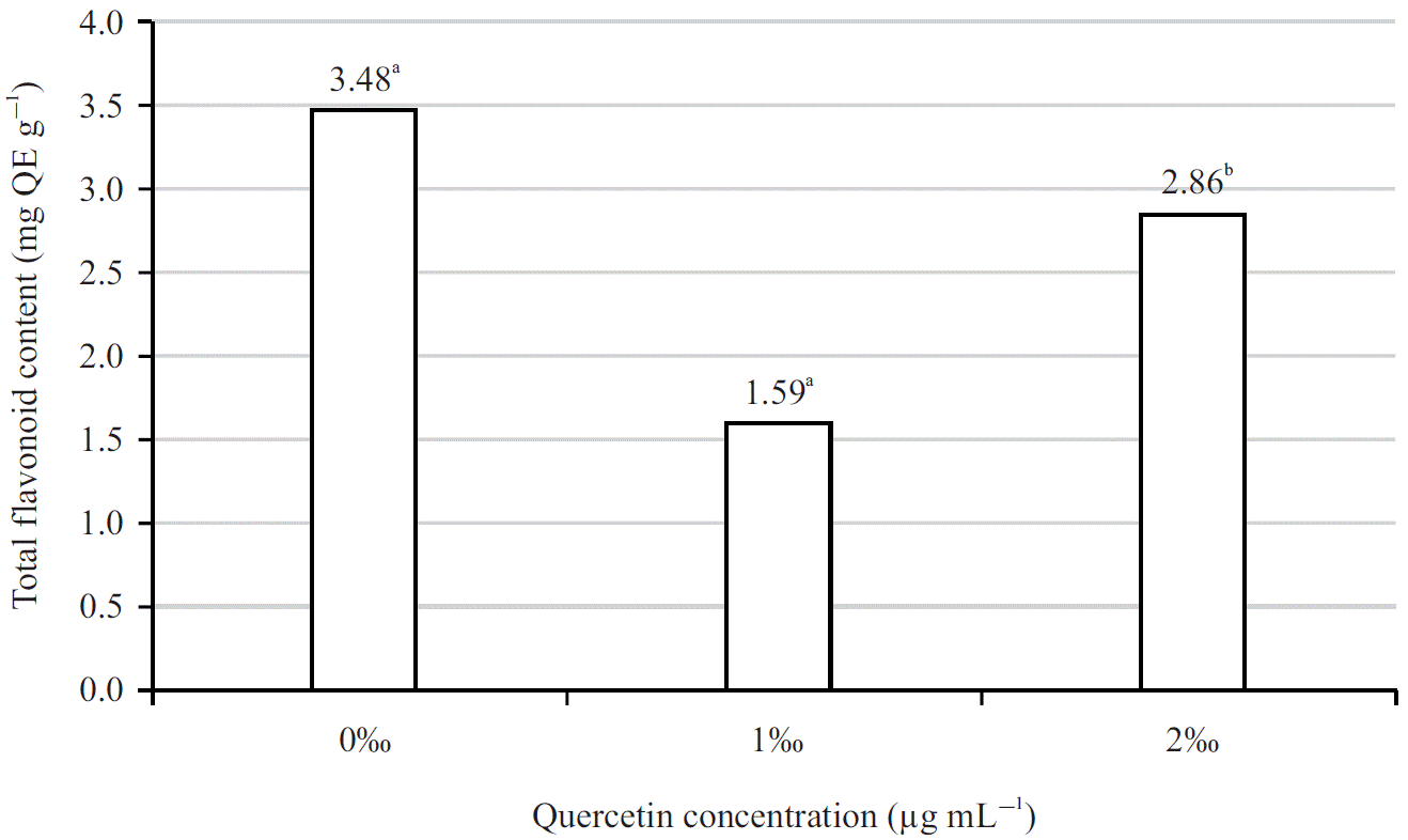 |
| Fig. 1: | Total flavonoid content of the ethanol extract The total flavonoid content is the mean of three replicates. This means that do not share a letter are significantly different |
Plants create reactive oxygen species (ROS) (H2O2 and O2) during salinity stress, which induces oxidative damage to cell organelles and membrane components and at high levels, could cause cell death. As a defense against ROS (caused by salinity), the plant produces non-enzymatic antioxidants (ascorbic acid and flavonoids)18.
In one research, Balekar et al.19 analyzed the ethanol extract from the leaves of Sphagneticola trilobata collected in Thailand and quantified the total flavonoid content of 16.67 ± 0.74 mg QE g–1. In addition, the study of Men et al.20 with the same solvent on Wedelia trilobata showed that the total flavonoid content obtained in the stem was 22.70 mg QE g–1. It can be seen that the results obtained in this paper are relatively low compared to those previously published on the same plant. The significant difference between the two research is due to the difference in climatic and soil conditions between Vietnam and Thailand as well as the fact that the plants have been treated with different concentrations of salt leading to differences in the content of bioactive compounds in plants described by Islam et al.21. Compared with the study of Men et al.20, Wedelia trilobata was harvested directly from the environment while, the Wedelia trilobata used in our research were treated with saline and grown hydroponically in a net house. Different conditions have affected the concentration of flavonoids obtained, the plants absorb a lot of water when growing hydroponically, causing the amount of plant matter to be reduced.
Quantitative concentrations of flavonoids and phenolic in Wedelia trilobata samples open up new paths in medicine. With outstanding biological activities, these compounds bring many benefits to human health such as antioxidant capacity, preventing the formation of single-molecule oxygen radicals and controlling cancer cell proliferation described by Abu Hazafa et al.22. The results suggested that all plant extracts contain flavonoids. Therefore, it is regarded as a suitable source of sunscreen.
Total phenolic content: The total phenolic content was determined using the Folin-Ciocalteu reagent and expressed in terms of mg gallic acid equivalent (GAE)/100 g extract. The results were derived from a calibration curve (y = 0.0398x + 0.1072, R2 = 0.9926). The results showed that the total phenolic content (TPC) in the extracts was different between the three treatments as shown in Fig. 2.
The results of Fig. 2 showed that the total phenolic content obtained from the three extracts ranges from 1.7 to 2.9 mg GA g–1. The 0‰ extract sample had the highest polyphenol content (2.81 mg GA g–1), while, the 1‰ extract sample (1.65 mg GA g–1) obtained the lowest content. The total phenolic content obtained from three samples gave higher results compared to the study of Govindappa et al.23, which investigated the total phenolic content of the Wedelia trilobata plant parts using distilled water as a solvent. In Govindappa’s research, the Wedelia trilobata plant was collected directly from the environment with total phenolic content measured in leaves and flowers after drying ranging from 1.8-2.6 mg GA g–1. However, due to the high extraction fraction and different solvents used, the data of Govindappa et al.23 was lower than current study. This showed that the extracted content depends on many factors such as medicinal nature, solvent and extraction conditions24.
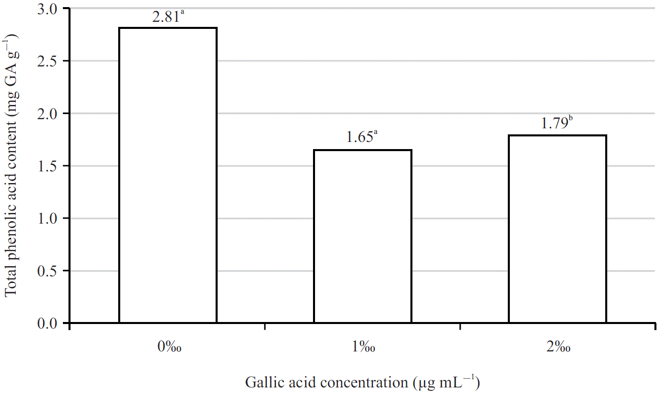 |
| Fig. 2: | Total phenolic content of the ethanol extract The total phenolic content is the mean of three replicates, means that do not share a letter are significantly different |
Fan et al.25 demonstrated that the higher the phenolic content of Lonicera, the higher its antioxidant capacity and tyrosinase inhibition would be.
Inhibitory activity on tyrosinase of Wedelia Trilobata extracts: In vitro tyrosinase inhibitory activity is indicated by the IC50 value shown in Fig. 3.
The positive control was kojic acid, a well-known tyrosinase inhibitor that is commonly utilized as a cosmetic skin-whitening treatment. Tyrosinase enzymatic activity was determined, providing the percentage of enzyme inhibition and IC50 values obtained by constructing standard curves and comparing them to kojic acid. Figure 3 shows IC50 value from 3 types of extract sample ranges from 27-31. In which, sample 0‰ had the strongest activity with the lowest IC50 value (IC50 = 27.884 μg mL–1) and sample 1‰ gave the lowest activity with the highest IC50 value (IC50 = 31.797 μg mL–1). Sample 2‰ showed moderate activity with IC50 =29.478 μg mL–1. At the same time, all three samples of Wedelia trilobata extract had lower tyrosinase inhibitory activity compared with the control Kojic acid (IC50 = 22.813 μg mL–1). The results of Zolghadri et al.26 implied that the methoxy and hydroxyl groups in the main flavonoid skeleton play an important role in tyrosinase inhibition. Therefore, in this study, flavonoids and phenolic were strongly correlated with tyrosinase inhibitory activity.
The inhibitory ability of the extract (indicated by IC50 value) was highest in treatment 0‰ and gradually decreased at 2‰ and 1‰ respectively, corresponding to the concentrations of flavonoids and phenolic obtained in the above 3 treatments (0‰>2‰>1‰). In addition, in the study of Jegal et al.27, flavonoids and Phenolic are a group of compounds containing many different groups, the difference in the structure of each substance can affect the tyrosinase enzyme inhibitory activity. Their research showed that hypolaetin 7-O-β-D-xylopyranoside (belongs to the flavonoid group) exhibited the highest tyrosinase inhibition with IC50 = 39.01 ± 3.36. This result was different from current findings, which can be explained that in the results of Jegal et al.27, they extracted and isolated each of the different flavonoid compounds and assigned each compound separately to inhibit tyrosinase. On the other hand, in this study, the obtained flavonoids consisted of many different subgroups and we did not address each flavonoid separately on which substances affect tyrosinase, leading to different data.
Effects of Wedelia trilobata extracts on skin carcinoma cell lines: Extracts of the Wedelia trilobata plant were examined for their cytotoxicity on carcinoma cells at four concentrations (0, 16, 64 and 256 μg mL–1). The results showed that each of the three salinity treatments exhibited different levels of cytotoxicity (Table 3).
| Table 3: | Percentage of KB carcinoma cell inhibition | |
| Cell inhibition (%) | ||
| Sample concentration | Sample concentration | |
| Experiment | (64 μg mL–1) | (256 μg mL–1) |
| 0‰ | 17±2.65a | 22±2.65a |
| 1‰ | 12±1.73a | 17±2.65a |
| 2‰ | 3±3.61b | 20±4.58a |
| Ellipticine (IC50 ) | 0.28±0.01 | |
| Percentage of KB inhibition is the mean of three replicates and Means that do not share a letter are significantly different | ||
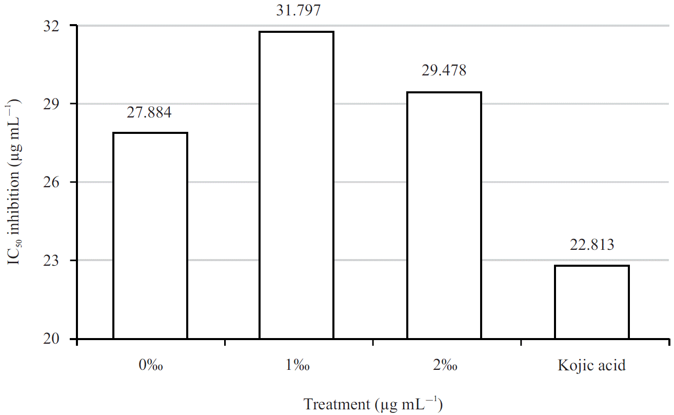 |
| Fig. 3: | IC50 inhibition of ethanol extract |
At a concentration of 256 μg mL–1, all three extract samples were found to have cytotoxicity on cancer cells, with values ranging from 17-25%. Of the three salinity treatments, the 0‰ salinity treatment showed the highest cytotoxic potential when inhibiting 25% of the cells. The lowest performance was exhibited in treatment 1‰ salinity when inhibiting only 17% of the cells at the same concentration of 256 μg mL–1. The 2‰ salinity sample exhibited moderate activity with 20% cell inhibition. While at concentrations of 4 and 16 μg mL–1, the extract did not show inhibitory activity on KB cancer cell lines.
The IC50 value of Ellipticine for KB is 1.67 ± 0.33 μM. The inhibitory effect on cancer cells of extract from treatment 0‰ increased from 0-25%, corresponding to an increase in extract concentration from 4 -256 μg mL–1. With the same increase in concentration, extract 1 ‰’s ability to inhibit KB cells increased from 0% to 17% and extract 2‰ increased from 0% to 20%. Based on the results, it is possible to say the sample concentration does affect the inhibitory effect of the KB carcinoma cell line. The higher the concentration of the extract, the higher the degree of toxicity to cancer cells.
In addition, the evaluation of anticancer results of three selected extracts can be based on IC50 results. In this experiment, the IC50 value was the concentration of the reagent that inhibited 50% of the growth of KB carcinoma cells. The IC50 values <20 μg mL–1 (with crude extract) and IC50 < 4 μg mL–1 (with purified substances) were evaluated as having a cytotoxic activity described by Vien et al.28. At a sample concentration of 256 μg mL–1, the inhibition of KB carcinoma cells was still less than 50%, so a higher concentration (>256 μg mL–1) was needed to inhibit 50% of the KB carcinoma cells. In a study by Khang et al.29, with the inhibitory object being the Hep2 hepatocyte cell line, Wedelia trilobata was able to inhibit 50% of cancer cells at a concentration as low as 4 μg mL–1 (38 ±1.5 μg mL–1) and at a concentration of 256 μg mL–1, it could inhibit 100% of cancer cells. The difference may occur depending on the growing conditions and the extraction method, which greatly influenced the investigation of the activities of the plants.
Sun protection factor evaluation: All plant extracts were able to protect the skin against ultraviolet (UV) light. Based on the Fig. 4, treatment 0‰ showed the highest SPF with of 22.992, while treatment 1‰ gave the lowest SPF of 19.710. A sample of treatment 2‰ represents an average SPF of 21.126.
 |
| Fig. 4: | Sun protection factor of ethanol extract The sun protection factor is the mean of three replicates. Means that do not share a letter are significantly different |
The SPF value classification according to the European Commission (EC) Recommendation in Osterwalder and Herzog (2009) is as follows:
| • | SPF 6-10 provides limited protection30 |
| • | SPF values of 15-25 provide moderate protection |
| • | SPF value of 30-50, provides high protection |
| • | 50+SPF value provides very high protection |
The higher the SPF value desired, the higher the required amount of sunscreen active ingredient31. In this study, all three treatments gave SPF in the range of 19-23, so based on the EC recommendation classification, it can be concluded that it provides moderate protection. The highest SPF results of the treatment 0‰ sample corresponded to the highest flavonoids and phenolic content. The number gradually decreased in treatment at 2‰ and was lowest in sample 1‰ salinity.
One study demonstrated that flavonoids and phenolic, which are antioxidants, have aromatic rings in their chemical structure that can absorb UVA and UVB radiation in the 200-400 nm wavelength range. As a result, they possess optical shielding characteristics and can be used as natural sunscreens32. Several other studies have shown a significant correlation between SPF and the phenolic and flavonoid content of nine medicinal plants33. Hence, it might be why differences in SPF of the three samples correlate to the differences in sample concentration of flavonoids and phenolic obtained in the three samples. High SPF values have been reported from leaf extract of Dracocephalum moldavica and flowering tops of Viola tricolor34, often considered to be due to high phenolic content. In addition, Cefali’s35 study on an extract from Ginkgo biloba with a concentration of 200 μg mL–1 gave an SPF of 7.06. Compared with their research, the highest SPF obtained in this study was 22.992 with a sample concentration of 1 mg mL–1. This difference may be due to the characteristics of each plant that have different concentrations of flavonoids and phenolic as well as geographical location and farming practices.
CONCLUSION
The results of the experiments showed that the inhibitory activity on carcinoma cells of three extracts was also investigated and showed that the 0‰ salinity extract had the strongest inhibitory activity. In this study, the highest concentration investigated was 256 μg mL–1, so studies with higher concentrations should be included for better cancer treatment outcomes. Assays on tyrosinase inhibition and skin protection also proved that W. trilobata extract at 0‰ salinity has relatively superior activity than other treatments. Moreover, this extract also exhibits a significant SPF of 22.991, while the SPF recommended by the Skin Cancer Foundation is 30. This shows the potential application of W. trilobata extracts as an ingredient for sunscreen while at the same time treating melasma. However, growing W. trilobata in saline environments has reduced its ability to synthesize secondary metabolites, which in turn reduces its ability to protect the skin. The potential of W. trilobata needs to be further studied for its ability to treat cancer as a safe, natural compound.
SIGNIFICANCE STATEMENT
Investigation of flavonoid, phenolic content, assessment of tyrosinase enzyme inhibitory activity, anti-UV activity and cytotoxicity of carcinoma extract of Wedelia trilobata (L.) Hitchc under saline conditions. There were differences in the synthesis of secondary compounds when growing Wedelia trilobata (L). Hitchc hydroponically with different salinity concentrations. The flavonoid and phenolic content of the 0‰ salinity treatment (control sample) were the highest and the lowest in the 1‰ salinity treatment. Specifically, the 0‰ salinity samples have the greatest ability to inhibit KB carcinoma cells as well as the tyrosinase enzyme inhibitory activity. The anti-UV activity of the 0‰ salinity treatment was highest and the lowest SPF index was in the 1‰ treatment.
ACKNOWLEDGMENT
The project was funded by Can Tho University, code: TSV2022-154.
REFERENCES
- Davis, L.E., S.C. Shalin and A.J. Tackett, 2019. Current state of melanoma diagnosis and treatment. Cancer Biol. Ther., 20: 1366-1379.
CrossRefDirect Link - Arnold, M., D. Singh, M. Laversanne, J. Vignat and S. Vaccarella et al., 2022. Global burden of cutaneous melanoma in 2020 and projections to 2040. JAMA Dermatol., 158: 495-503.
CrossRefDirect Link - Ma, L., M. Zhang, R. Zhao, D. Wang, Y. Ma and L. Ai, 2021. Plant natural products: Promising resources for cancer chemoprevention. Molecules, Vol. 26.
CrossRefDirect Link - Trinh, T.P., P.B. Linh and P.T. Tam, 2020. Potential anti-tumor activites of extract from fresh leaves of the male papaya (Carica papaya L.) collected in Ha Tinh Province. Vietnam J. Biotechnol., 18: 127-134.
CrossRefDirect Link - Chi, H.T., N.T.L. Thuong and B.T.K. Ly, 2021. Sphagneticola trilobata (L.) pruski (asteraceae) methanol extract induces apoptosis in leukemia cells through suppression of BCR/ABL. Plants, Vol. 10.
CrossRefDirect Link - Tsai, C.H., F.M. Lin, Y.C. Yang, M.T. Lee and T.L. Cha et al., 2009. Herbal extract of Wedelia chinensis attenuates androgen receptor activity and orthotopic growth of prostate cancer in nude mice. Clin. Cancer Res., 15: 5435-5444.
CrossRefDirect Link - Mardina, V., T. Harmawan, H. Halimatussakdiah, S. Ilyas and M. Tanjung, 2020. Anticancer activity of n-hexane extract from Sphagneticola trilobata (L.) J.F pruski against MCF-7 breast cancer cell. Elkawnie, Vol. 6.
CrossRefDirect Link - Marinova, D., F. Ribarova and M. Atanassova, 2005. Total phenolics and total flavonoids in Bulgarian fruits and vegetables. J. Univ. Chem. Technol. Metall., 40: 255-260.
Direct Link - Singleton, V.L., R. Orthofer and R.M. Lamuela-Raventos, 1999. Analysis of Total Phenols and Other Oxidation Substrates and Antioxidants by Means of Folin-Ciocalteu Reagent. In: Methods in Enzymology, Burslem, G.L. (Ed.), Academic Press, Cambridge, Massachusetts, ISBN: 9780121822002, pp: 152-178.
CrossRefDirect Link - Chintong, S., W. Phatvej, U. Rerk-Am, Y. Waiprib and W. Klaypradit, 2019. In vitro antioxidant, antityrosinase, and cytotoxic activities of astaxanthin from shrimp waste. Antioxidants, Vol. 8.
CrossRefDirect Link - Ferrari, M., M.S.C. Oliveira, A.K. Nakano and P.A. Rocha-Filho, 2007. In vitro and in vivo determinations of sun protection factor (SPF) of emulsions with andiroba oil (Carapa guianensis). Braz. J. Pharmacogn., 17: 626-630.
CrossRefDirect Link - Sayre, R.M., P.P. Agin, G.J. Levee and E. Marlowe, 1979. Comparison of in vivo and in vitro testing of sunscreening formulas. Photochem. Photobiol., 29: 559-566.
CrossRefDirect Link - Lammel, D.R., G. Barth, O. Ovaskainen, L.M. Cruz and J.A. Zanatta et al., 2018. Direct and indirect effects of a pH gradient bring insights into the mechanisms driving prokaryotic community structures. Microbiome, Vol. 6.
CrossRefDirect Link - Zhang, P., M. Senge and Y. Dai, 2016. Effects of salinity stress on growth, yield, fruit quality and water use efficiency of tomato under hydroponics system, Rev. Agric. Sci., 4: 46-55.
CrossRefDirect Link - Negrão, S., S.M. Schmockel and M. Tester, 2017. Evaluating physiological responses of plants to salinity stress. Ann. Bot., 119: 1-11.
CrossRefDirect Link - Orzechowska, A., M. Trtílek, K.M. Tokarz, R. Szymańska, E. Niewiadomska, P. Rozpądek and K. Wątor, 2021. Thermal analysis of stomatal response under salinity and high light. Int. J. Mol. Sci., Vol. 22.
CrossRefDirect Link - Yang, L., W.C. Li, F.l. Fu, J. Qu, F. Sun, H. Yu and J. Zhang, 2022. Characterization of phenylalanine ammonia-lyase genes facilitating flavonoid biosynthesis from two species of medicinal plant Anoectochilus. PeerJ, Vol. 10.
CrossRefDirect Link - Hasanuzzaman, M., M.R.H. Raihan, A.A.C. Masud, K. Rahman and F. Nowroz et al., 2021. Regulation of reactive oxygen species and antioxidant defense in plants under salinity. Int. J. Mol. Sci., Vol. 22.
CrossRefDirect Link - Balekar, N., N.G. Katkam, T. Nakpheng, K. Jehtae and T. Srichana, 2012. Evaluation of the wound healing potential of Wedelia trilobata (L.) leaves. J. Ethnopharmacol., 141: 817-824.
CrossRefDirect Link - Mến, T.T., N.Q. Cường, N.T.A. Thư, P.L.T. Quyên and P.C. Phương et al., 2019. Study on the ability to inhibit seed germination of extracts from three lobes (Wedelia trilobata (L.) Hitchc), (Vietnamese). Can Tho Univ. J. Sci., 55: 85-90.
CrossRefDirect Link - Islam, M.Z., B.J. Park and Y.T. Lee, 2019. Effect of salinity stress on bioactive compounds and antioxidant activity of wheat microgreen extract under organic cultivation conditions. Int. J. Biol. Macromol., 140: 631-636.
CrossRefDirect Link - Abu Hazafa, Khalil-Ur-Rehman, N. Jahan and Z. Jabeen, 2019. The role of polyphenol (Flavonoids) compounds in the treatment of cancer cells. Nutr. Cancer, 72: 386-397.
CrossRefDirect Link - Govindappa, M., N.S. Sravya, M.N. Poojashri, T.S. Sadananda and C.P. Chandrappa et al., 2011. Antimicrobial, antioxidant and in vitro anti-inflammatory activity and phytochemical screening of water extract of Wedelia trilobata (L.) Hitchc. J. Med. Plants Res., 5: 5718-5729.
CrossRefDirect Link - Silva, B., V. Cadavez, P. Ferreira-Santos, M. Alves and I. Ferreira et al., 2021. Chemical profile and bioactivities of extracts from edible plants readily available in Portugal. Foods, Vol. 10.
CrossRefDirect Link - Fan, Z., L. Li, X. Bai, H. Zhang, Q. Liu, H. Zhang, Y. Fu and R. Moyo, 2019. Extraction optimization, antioxidant activity, and tyrosinase inhibitory capacity of polyphenols from Lonicera japonica. Food Sci. Nutr., 7: 1786-1794.
CrossRefDirect Link - Zolghadri, S., A. Bahrami, M.T.H. Khan, J. Munoz-Munoz, F. Garcia-Molina, F. Garcia-Canovas and A.A. Saboury, 2019. A comprehensive review on tyrosinase inhibitors. J. Enzyme Inhib. Med. Chem., 34: 279-309.
CrossRefDirect Link - Jegal, J., P. Sang-a, K. Chung, H.Y. Chung and J. Lee et al., 2016. Tyrosinase inhibitory flavonoid from Juniperus communis fruits. Biosci. Biotechnol. Biochem., 80: 2311-2317.
CrossRefDirect Link - Vien, T.A., N.T.H. Van, D.T. Thao, T.T.N. Hang, N.A. Tuan and P.Q. Long, 2017. Antifungal, antibacterial and cytotoxic activities of some Ardisia species from Vietnam. Acta. J. Bio., 38: 75-80.
CrossRefDirect Link - Khang, D.T., H.D. Dieu, N.K. Dung, B.T. Gia and T.L.N. Thao et al., 2020. Anticancer and antioxidant of chloroform extracts from medical plants in the Mekong Delta, Vietnam. Asian J. Plant Sci., 19: 398-405.
CrossRefDirect Link - Osterwalder, U. and B. Herzog, 2009. Sun protection factors: World wide confusion. Br. J. Dermatol., 161: 13-24.
CrossRefDirect Link - Geoffrey, K., A.N. Mwangi and S.M. Maru, 2019. Sunscreen products: Rationale for use, formulation development and regulatory considerations. Saudi Pharm. J., 27: 1009-1018.
CrossRefDirect Link - Ng, S.Y., V.R.E. Suk and L.T. Gew, 2022. Plant polyphenols as green sunscreen ingredients: A systematic review. J. Cosmet. Dermatol., 21: 5409-5444.
CrossRefDirect Link - Khazaeli, P. and M. Mehrabani, 2008. Screening of sun protective activity of the ethyl acetate extracts of some medicinal plants. Iran. J. Pharm. Res., 7: 5-9.
CrossRefDirect Link - Hashemi, Z., M.A. Ebrahimzadeh and M. Khalili, 2021. Sun protection factor, total phenol, flavonoid contents and antioxidant activity of medicinal plants from Iran. Trop. J. Pharm. Res., 18: 1443-1448.
CrossRefDirect Link - Cefali, L.C., J.A. Ataide, A.R. Fernandes, E. Sanchez-Lopez and I.M. de Oliveira Sousa et al., 2019. Evaluation of in vitro solar protection factor (SPF), antioxidant activity, and cell viability of mixed vegetable extracts from Drmophandra mollis Benth, Ginkgo biloba L., Ruta graveolens L., and Vitis vinífera L. Plants, Vol. 8.
CrossRefDirect Link