
Faten A. Khorshid
Department of Biology, Faculty of Science, King Abdul Aziz University (KAU), P.O. Box 80216, Jeddah 21589, Kingdom of Saudi Arabia
LiveDNA: 966.3097
ORCID: 0000-0002-7298-6548
American Journal of Drug Discovery and Development
Year: 2011 | Volume: 1 | Issue: 3 | Page No.: 200-208


ABSTRACT
Breast cancer is the most common malignancy in the world among women. Many therapies have been designed to treat this disease. Mamectomy, chemotherapy and radiotherapy are still the main therapies of breast cancer. However, the results were unsatisfactory and still far from the ideal treatment. PM 701 is a natural product, has anticancer activity. The bioactive fraction PMF and subfraction PMFK had been isolated from PM 701. PM 701 and its fractions were proved to have a cytotoxic properties against different cancer cell lines. This study is directed for the further examination of lyophilized PM 701 and its active fractions on the growth of breast cancer cells (MCF-7). PM 701, PMF or PMFK were adding to the cultural medium, where MCF-7 is incubated. PM 701, PMF or PMFK were able to inhibit significantly the proliferation of MCF-7 cells. These new agents were proved to induce apoptosis of the breast cancer cells; through its direct effect on the nuclei.
PDF Abstract XML References Citation
Received: March 09, 2011;
Accepted: April 14, 2011;
Published: June 18, 2011
How to cite this article
Faten A. Khorshid, 2011. The Cytotoxic Effect of PM 701 and its Fractions on Cell Proliferation of Breast Cancer Cells, MCF7. American Journal of Drug Discovery and Development, 1: 200-208.
DOI: 10.3923/ajdd.2011.200.208
URL: https://scialert.net/abstract/?doi=ajdd.2011.200.208
DOI: 10.3923/ajdd.2011.200.208
URL: https://scialert.net/abstract/?doi=ajdd.2011.200.208
INTRODUCTION
The management of malignancies in humans still constitutes a major challenge for contemporary medicine (Coufal et al., 2007; He and Liu, 2007; Widodo et al., 2007; Feng et al., 2006). Although with progress in understanding cancer nature, many therapeutic anticancer have been developed which has relied on surgery, chemotherapy, radiotherapy, hormone therapy and more recently immunotherapy (Khorshid et al., 2010). However, all are still far from the ideal treatment, which selectively kill the malignant cells and sparing the normal healthy tissues and functions of vital organs (Grever and Chabner, 1997; Moshref, 2007).
Breast cancer is the most common malignancy in the world among women. Over a million women are diagnosed every year and 370,000 were died due to breast cancer (Schwartsmann et al., 2002). Many drugs and therapies have been designed to treat this disease. The identification and elucidation of the molecular components and signals that control different biological processes underlying the regulation of cell growth, differentiation and apoptosis of the mammary epithelium is important to lead to the development of new drugs and play an important role in designing an anti-cancer drug (Grever and Chabner, 1997; Moshref, 2007; Schwartsmann et al., 2002).
Hence, anti-cancer drug substitutes are actively sought after in the hopes of finding alternative ways to suppress the growth of breast cancer cells. The PM 701 an anticancer substrate (Khorshid et al., 2005; Khorshid, 2005, 2008; El-Shahawy et al., 2010; Moshref et al., 2006) was used in this study to test its effect on MCF-7 human breast cancer cell line. In addition to screening of its active fraction PMF (Khorshid et al., 2009, 2011) as a novel anti-cancer compounds for human breast cancer, this study also seeks to determine the effect of PMFK subfraction (Khorshid et al., 2009) on the MCF-7 proliferation. The study also determined the inhibitory concentration, IC50, upon investigation of these compounds on the MCF-7 human breast cancer cell line.
So we present herein the capacity of PM 701, PMF or PMFK in inhibit the proliferation of MCF-7 cell line in vitro.
MATERIALS AND METHODS
Media: Dulbecco's modified eagle medium (DMEM) and Dulbecco's Phosphate Buffered Saline (PBS) were purchased from MP Biomedicals Inc, USA. Fetal Calf Serum (FCS) was obtained from Gibco, Canada. Coomassie blue, pencillin-streptomycin, Trypan blue and Trypsin-EDTA were purchased from Sigma, USA.
Preparation of tested agents: The powder form of PM 701 and its fractions (PMF and PMFK) were prepared according to Khorshid et al. (2009) each fraction was dissolved in DMEM, 2% FBS at a concentration of 2 mg mL-1 before using. The solution was then filter sterilized using a 0.2 μ syringe filter to prepare the working solution.
Cell culture: Breast carcinoma cells (MCF-7) were purchased from National Cancer Institute, Cairo University, Egypt. These cells were preserved as cell line in tissue Culture Unit at King Fahad Medical Research Center (KFMRC) in King Abdulaziz University (KAU).
MCF-7 cells were cultured in DMEM supplemented with 10 % FCS, 100 U mL-1 penicillin and 100 μg mL-1 streptomycin. Cells were grown in 75 cm-2 tissue culture flasks in a 37°C incubator with a humidified mixture of 5% CO2 and 95% air (Ptak et al., 2009).
Cytotoxicity assay: Cells were detached with 0.025% trypsin-EDTA. The cells were resuspended in 10 mL of medium to make single cell suspension and viable cells were counted by trypan blue exclusion in haemocytometer and diluted with medium to give a final concentration of 1x10⁵ cells/well. The 1 mL well-1 of these cell suspensions were seeded in 24-well microtiter plates and incubated to allow for cell attachment.
Cell count using hemocytometer: A cell suspension fixed in a volume of cells (e.g.. 1 mL). 200 μL of this suspension was mix with an equal volume of trypan blue. Mixed solution was transferred using a pipette to a hemocytometer and live cells were counted. The numbers of cells were calculated per mL, using the following formula (Pollard and Walker, 1989; Khorshid et al., 2005).
Cell viability = Total viable cells (unstained) /Total cells x 104 x dilution factor (suspension cells: Trypan blue) |
Treatment with lyophilized PM 701 or it's fractions: Cytotoxicity assays were performed using short incubation for only 24 h in serials dilutions of examined drug, that were used to estimate the IC50 concentration. The long incubation for more than 72 h, with a concentration lower or higher than IC50 concentration, was also used with only crude substrate (PM 701) to confirm the cytotoxicity effect.
Short incubation for IC50 estimation: Lyophilized PM 701 or PMF, PMFK, were diluted in medium. Serial dilutions were prepared 0, 0.5 and 7.5 μg mL-1 of media each concentration was added to each well of the plate in 3 replicates of cancer cells (treated), after the selected time of treatment, the medium was aspirated. Then cells washed with PBS, trypsinized and counted by two methods counter coulter and Hemocytometer using trypan blue dye exclusion test (Khorshid et al., 2005; Moshref et al., 2006; Pollard and Walker, 1989; Khorshid and Moshref, 2006).
Fixing and staining cells: Each group of cells were plated onto Petri dishes in DMEM media for 24 h, then the media changed with examined media (with different concentrations) and control media and incubated at 37°C for 24 h.
Each group of cells were fixed in 4% formaldehyde for 5 min at room temperature after double washing with 1 X PBS each for 5 min. Then cells stained with Coomassie blue for 5-10 min followed by repeated washing with tap water (Khorshid et al., 2005).
Statistical analysis: The data were expressed as the Mean±SD of the optical density obtained from three independent experiments (each experiment was performed in three replicate wells). Statistical analysis was performed with SPSS and graph pad statistical programs.
RESULTS
The MCF-7 human breast cancer cell line was treated with PM 701 and its fractions at different concentrations. Cells were plated with 1x10⁵ cells/well in 24-well tissue culture plates and incubated in tested agent for 24 h at 37°C.
IC50 estimation (short incubation): This experiment was preformed to estimate the IC50 concentration of all tested agents.
The cytotoxic effect of lyophilized PM 701 was studied by incubated the human breast cancer cells, MCF-7 for 24 h in DMEM media with serial concentration of the drug. PM 701 and its fractions inhibited the proliferation of MCF-7 cells in a dose-dependent manner. The IC50 of PM 701 is 0.35 μg mL-1 of media, which was significantly low as compared to IC50 of PMF and PMFK (3 and 3.5 μg mL-1 of media, respectively).
The fixed and stained MCF-7 cells showed that they decreased in number when incubated in media containing the examined substrate for 24 h.
Effect of lyophilized PM 701 on MCF-7 cells: First results indicated that lyophilized PM 701 inhibited the proliferation of cancer cells and the IC50 was about 0.35 μg of PM 701 mL of media, (Table 1, Fig. 1-4).
Effect of PMF on MCF-7 cells: Treatment of MCF-7 with serial concentrations of PMF fraction in incubated media showed inhibition of the cell proliferation with IC50 near the concentration 3 μg of PMF mL-1 of media (Table 2, Fig. 2-4).
| Table 1: | The effect of lyophilized PM 701 on the growth of MCF-7 cancer cells after 24 h of incubation comparing with non treated cancer cells (control) |
 | |
| Table 2: | The effect of PMF on the growth of MCF-7 cancer cells after 24 h of incubation comparing with non treated cancer cells (control) |
 | |
 |
| Fig. 1: | The effect of lyophilized PM 701 on MCF-7 cell line after 24 h of incubation, PM 701 appeared cytotoxic to the cancer cells in a dose-dependent manner |
 |
| Fig. 2: | The effect of PMF on MCF-7 cell line after 24 h of incubation, PMF appeared cytotoxic to the cancer cells in a dose-dependent manner |
 |
| Fig. 3: | The effect of PMFK on MCF-7 cell line after 24 h of incubation, PMFK appeared cytotoxic to the cancer cells in a dose-dependent manner |
Effect of PMFK on MCF-7 cells: This part indicated that the serial concentrations of PMFK in incubated media inhibited the proliferation of MCF-7 cells and the IC50 was determined near the concentration 3.5 μg of PMFK mL-1 of media (Table 3, Fig. 3-4).
Long incubation: This experiment was carried on for only crude concentration of PM 701 to confirm the cytotoxic effect of this agent against MCF-7 cells after long incubation for more than 96 h, while the same result could apply to PMF and PMFK that separated from PM 701.
The incubation of MCF-7 cells in higher and lower concentrations than IC50 concentration of PM 701 confirmed the results of short incubation, long incubation of MCF-7 cells in 2 and 20 μg of PM 701 mL-1 of media inhibited the proliferation of cancer cells in both concentration compared with control non treated cancer cells, whereas Fig. 5 and 6 showed that MCF-7 cells decreased in numbers when PM 701 was added to the incubated media. It also showed that the cells incubated in 2 μg died mostly after incubation for 48 h.
| Table 3: | The effect of PMFK on the growth of MCF-7 cancer cells after 24 h of incubation comparing with non treated cancer cells (control) |
 | |
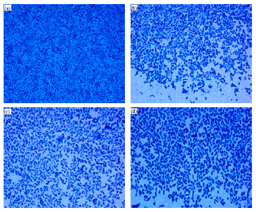 |
| Fig. 4: | MCF-7 cells imaged (x 10) after incubation with (b) PM 701, (c) PMF and (d) PMFK for 24 h, fixed and stained with coomassie blue. The numbers of cells were decreased in treated cultures compared with the number of (a) control non treated cells |
 |
| Fig. 5: | The confirmed effect of lyophilized PM 701 on MCF-7 cell line, where PM 701 appeared cytotoxic to the cancer cells in both concentration lower and higher than IC50 concentration of PM 701 even after long incubation for 24, 48, 72, 96 h |
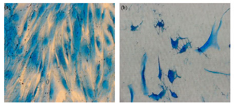 |
| Fig. 6: | Images illustrated MCF-7 incubated for 96 h in control media (a) and in treated media with PM 701 (b) (10X), note the scattered non healthy cells, with attack nuclei in treated culture compared with healthy and well defined nuclei in non treated culture |
DISCUSSION
The resistance of cancer cells to multiple chemotherapeutic agents poses a major problem in the successful treatment of breast cancer. Whether drug resistance is due to changes induced in the drug-exposed tumor cells (adaptation) or represents the selective growth of one or more drug-resistance clones present in initial tumor (selection) remains controversial. But some studies do provide evidence on the development of multidrug resistance (MDR) in a human breast cancer cell line, MCF-7, involving doxorubicin, vinblastine and colchicines (Devarajan et al., 2002). Our previous work in this area indicated that PM701 are increasingly being considered as a source of selective cytotoxic agent. The bioassay-guided fractionation was undertaken to isolate our new bioactive constituents PMF that contributed to this cytotoxic activity which exhibited a strong cytotoxic effect against human lung cancer cell line A549 by 80% after 72 h without any harmful effect on human foreskin cell line HFS, the same results was early proved using PM 701 on A549 and leukemic cancer cell line L1210 (Khorshid and Moshref, 2006; Moshref et al., 2006). PMF has shown to block cancer cells growth and induced them to undergo apoptosis in a dose-independent manner. PMF subfractionation led to isolate our new subfraction PMFK, which also demonstrated antiproliferative activity on cancer cells in a dose-independent manner. PMFK exhibits an antiproliferative effect by induction of apoptosis on human cancer cells (Khorshid et al., 2011). As apoptosis has become a new therapeutic target in cancer research, these results confirm the potential of PMF and PMFK as agents of cytotoxic therapy in human lung cancer cells. Khorshid et al. (2011) measured the metabolic activity colorimetrically by MTT assay (3- (4, 5-Dimethylthiazol-2-yl) -2, 5-Diphenyltetrazolium Bromide) in different cancer cell lines treated with PMF. Apoptotic cell death was also determined by the TUNEL method (Terminal deoxynucleotidyl Transferase (TdT)-biotin nick end-labeling). Their results showed that PMF induced apoptosis in association with increased number of TUNEL positive cells. MTT results showed that PMF decrease cell proliferation via inhibiting metabolic cell activities. Which conclude that PMF has anti-cancer effects by increasing apoptosis and altering cellular metabolic activities. All those results confirm the potential of PMF as an agent of cytotoxic therapy in human cancer cells (Khorshid et al., 2010).
Here this study confirm more the cytotoxic effect in more cancer cells (MCF-7), PM 701 and its fractions found to be cytotoxic to the MCF-7 cell line and were able to inhibit its proliferation. Moreover this work proved that PM 701 and its fractions inhibited the proliferation rate of human breast cancer cells in vitro in a dose dependent manner.
It is clear that PM 701 showed cytotoxic effect on MCF-7 human breast cancer cell line and this suggests the possibility that PMF plays a role in the oxygen transport of breast cancer cells, where earlier studies showed that the treatment of A549 cells with PM 701 increases the CH2 content (Ahmed et al., 2009). The assumption would then be that PMF may helps the cancer cells revert back to normal cells by enhancing oxygen transport (turning on aerobic metabolism) and once the cells become normal, they go through the normal process of apoptosis.
Other suggestion, that the chemoprevention and therapy by the use of PM 701 and its fractions may have offered new approaches to block tumor growth and progression via its polyphenolic component. Many studies reported that polyphenols are capable of inhibiting the growth of a variety of human cancer cells, via induction of apoptosis in vitro (Masuda et al., 2001, 2003; Huh et al., 2004; Chan et al., 2006).
On the other hand, apoptosis as an active process involves biochemical changes on three essential cellular components, DNA, protein and lipid. It should mentioned here that the amount of DNA decreases dramatically during the treatment of A549 lung cancer cell line with PM 701 (Ahmed et al., 2009) while chromatins condensation as a stage of apoptosis was also detected with PM 701 treatment in that study that where previously published (Ahmed et al., 2009). So the treatment of MCF-7 with PM 701 and its fractions resulted in the inhibition of both DNA synthesis and cell growth.
It also may that the anticancer effect of PM 701 and its fractions related to the potentially toxic or carcinogenic chemicals in it, which promotes the excretion of therapeutic agents reducing the ability of carcinogenic signals to react with cancer cells and damage their nucleic acids and proteins (Parkinson, 1996).
Ahmed et al. (2009) proved that PM 701 induced changes in plasma membrane permeability by altering the drug influx/efflux-due to change in membrane fluidity-system in cancerous cells, this result may implement on PM 701 fractions also.
PM 701 or one of its fractions may use as alternative therapy or in combination with doxorubicin or other regime of therapies. Whereas this preliminary in vitro study suggests that PMF may be developed to be a potent natural compound with anti-cancer properties.
CONCLUSION
In conclusion, the results showed that all tested fractions of PM 701 inhibited the growth of the MCF-7 cells; therefore, these results may lead to a finding for a successful alternative drug for breast cancer cells.
ACKNOWLEDGMENT
The authors gratefully acknowledged financial support of El-Zamel's scientific chair, Researches no "429/3/KBM", Research and Consultation Institute, King Abdulaziz University, Jeddah.
REFERENCES
- Chan, M.M., K.J. Soprano, K. Weinstein and D. Fong, 2006. Epigallocatechin-3-gallate delivers hydrogen peroxide to induce death of ovarian cancer cells and enhances their cisplatin susceptibility. J. Cell Physiol., 207: 389-396.
PubMed - Coufal, M., M.M. Maxwell, D.E. Russel, A.M. Amore and S.M. Altmann et al., 2007. Discovery of a novel small-molecule targeting selective clearance of mutant huntingtin fragments. J. Biomol. Screen, 12: 351-360.
PubMed - Devarajan, E., J. Chen, A.S. Multani, S. Pathak, A.A. Sahin and K. Mehta, 2002. Human breast cancer MCF-7 cell line contains inherently drug-resistance subclones with distinct genotypic and phenotypic features. Int. J. Oncol., 20: 913-920.
PubMed - El-Shahawy, A., N.M. Elsawi, W.S. Baker, F. Khorshid and N.S. Geweely, 2010. Spectral analysis, molecular orbital calculations and antimicrobial activity of PMF-G fraction extracted from PM-701. Int. J. Pharma Biosci., 1: 1-19.
Direct Link - Feng, L., L.F. Zhang, T.J. Yan, J. Jin and W.Y. Tao, 2006. Studies on active substance of anticancer effect in Polygonum cuspidatum. Zhong Yao Cai., 29: 689-691.
PubMed - He, X. and R.H. Liu, 2007. Triterpenoids isolated from apple peels have potent antiproliferative activity and may be partially responsible for apple's anticancer activity. J. Agric. Food Chem., 55: 4366-4370.
PubMed - Huh, S.W., S.M. Bae, Y.W. Kim, J.M. Lee and S. Eun et al., 2004. Anticancer effects of (-)-epigallocatechin-3- gallate on ovarian carcinoma cell lines. Gynecol. Oncol., 94: 760-768.
CrossRef - Khorshid, F.A., 2008. Preclinical evaluation of PM 701 in experimental animals. Int. J. Pharmacol., 4: 443-451.
CrossRef - Khorshid, F.A., 2009. Potential anticancer natural product against human lung cancer cells. Trends Med. Res., 4: 8-15.
CrossRefDirect Link - Khorshid, F.A., S.A. Rahimaldeen and J.S. Al-Amri, 2011. Apoptosis study on the effect of PMF on different cancer cells. Int. J. Biol. Chem., 5: 150-155.
CrossRefDirect Link - Khorshid, F., H. Alshazly, A. Al Jefery and A.M.M. Osman, 2010. Dose escalation phase I study in healthy volunteers to evaluate the safety of a natural product PM701. J. Pharmacol. Toxicol., 5: 91-97.
CrossRefDirect Link - Masuda, M., M. Suzui, J.T. Lim and I.B. Weinstein, 2003. Epigallocatechin-3-gallate inhibits activation of HER-2/neu and downstream signaling pathways in human head and neck and breast carcinoma cells. Clin. Cancer Res., 9: 3486-3491.
PubMed - Masuda, M., M. Suzui and I.B. Weinstein, 2001. Effects of epigallocatechin-3-gallate on growth, epidermal growth factor receptor signaling pathways, gene expression, an chemosensitivity in human head and neck squamous cell carcinoma cell lines. Clin. Cancer Res., 7: 4220-4229.
PubMed - Parkinson, A., 1996. Biotransformation of Xenobiotics. In: Casarett and Doull's Toxicology, The Basic Science of Poisons, Klaassen, C.D. (Ed.). McGraw-Hill, New York, pp: 113-186.
Direct Link - Ptak, A., G. Ludewig, A. Rak, W. Nadolna, M. Bochenek and E.L. Gregoraszczuk, 2010. Induction of cytochrome P450 1A1 in MCF-7 human breast cancer cells by 4-chlorobiphenyl (PCB3) and the effects of its hydroxylated metabolites on cellular apoptosis. Environ. Int., 36: 935-941.
CrossRefDirect Link - Ahmed, G.A.R., F.A.R. Khorshid and T.A. Kumosani, 2009. FT-IR spectroscopy as a tool for identification of apoptosis-induced structural changes in A549 cells dry samples treated with PM 701. Int. J. Nano Biomaterials, 2: 396-408.
CrossRefDirect Link - Schwartsmann, G., M.J. Ratain, G.M. Cragg, J.E. Wong and N. Saijo et al., 2002. Anticancer drug discovery and development throughout the world. J. Clin. Oncol., 20: 47-59.
Direct Link - Widodo, N., K. Kaur, B.G. Shrestha, Y. Takagi, T. Ishii, R. Wadhwa and S.C. Kaul, 2007. Selective killing of cancer cells by leaf extract of Ashwagandha: Identification of a tumor-inhibitory factor and the first molecular insights to its effect. Clin. Cancer Res., 13: 2298-2306.
CrossRefPubMedDirect Link