
J.D. Dabak
Department of Biochemistry, Faculty of Medical Sciences, University of Jos, P.M.B. 2084, Nigeria
S.Y. Gazuwa
Department of Biochemistry, Faculty of Medical Sciences, University of Jos, P.M.B. 2084, Nigeria
G.A. Ubom
Department of Biochemistry, Faculty of Medical Sciences, University of Jos, P.M.B. 2084, Nigeria
Asian Journal of Biotechnology
Year: 2009 | Volume: 1 | Issue: 1 | Page No.: 12-19


ABSTRACT
This study was designed to investigate the hepatoprotective potential of calcium and magnesium against lead and cadmium induced hepatotoxicity using a rat model. Varying concentrations of cadmium and lead salts in combination were used to induce liver damage. This was followed by adding varying concentrations of calcium and magnesium salts in combination to the same concentration used to induce liver damage. The degree of damage and protection were measured using biochemical parameters such as serum-glutamate-pyruvate transaminases (SGPT), serum-glutamate-oxaloacetate transaminases (SGOT), total protein, albumin and histopathological examination of the liver cells. Studies indicate that liver function is generally impeded particularly with respect to albumin synthesis and the cellular integrity of the organ is damaged as a function of elevations in cadmium and lead concentrations. The albumin concentration decreased as the concentrations of cadmium and lead were increased from control thus: 4.25±0.40 g/100 mL; 3.63±0.17 g/100 mL; 3.50±0.13 g/100 mL; 3.38±0.19 g/100 mL and 3.38±0.29 g/100 mL, respectively, while SGPT and SGOT were increasing. The addition of calcium and magnesium to the same combined concentrations of cadmium and lead which caused albumin synthesis impairment in the first phase of this research protected the liver as the albumin concentrations were not significantly different from control. The albumin concentrations were: 3.70±0.18 g/100 mL; 3.88±0.38 g/100 mL; 3.75±0.18 g/100 mL; 3.60±0.44 g/100 mL and 3.88±0.36 g/100 mL, respectively. Histopathological studies on the liver confirmed the protective potential of calcium and magnesium on the hepatotoxicity arising from cadmium and lead as the damage observed in phase 1 of this research using the same concentrations is obliterated.
PDF Abstract XML References Citation
How to cite this article
J.D. Dabak, S.Y. Gazuwa and G.A. Ubom, 2009. Hepatoprotective Potential of Calcium and Magnesium Against Cadmium and Lead Induced Hepatotoxicity in Wistar Rats. Asian Journal of Biotechnology, 1: 12-19.
DOI: 10.3923/ajbkr.2009.12.19
URL: https://scialert.net/abstract/?doi=ajbkr.2009.12.19
DOI: 10.3923/ajbkr.2009.12.19
URL: https://scialert.net/abstract/?doi=ajbkr.2009.12.19
INTRODUCTION
The fact that cadmium and lead are toxic to humans has been known for many years. These toxicities pose major environmental health problems in modern society, with potentially dangerous bioaccumulation through the food chain (Okoye, 1992). Cadmium and lead are among the non-essential trace elements that are wide spread in our environment because of contamination through anthropogenic activities (Gazuwa et al., 2006). Excessive levels of these non-essential toxic elements can have an unbalancing effect on the essential trace element balances in the body’s cells.
Recent research indicates that minerals may play a significant role against a variety of degenerative diseases and processes, prevent and reduce injury from environmental pollutants and enhance the ability to work and learn and can also protect the body from the effects of toxic elements (Perza et al., 1998). These discoveries should begin to make us view our daily intake of nutrients as performing dual roles: the role of preventing known mineral deficiencies and in optimizing the disease-preventing properties of these nutrients. The later role and the increasing evidence that supports it, makes it reasonable to believe that it will be possible to reduce the incidence of most life-limiting chronic diseases through the adoption of optimal daily nutrient intake levels if we have a thorough knowledge of how these trace elements interact in the cells (Hatton and McCarron, 1994).
Ubom (1991) showed that dietary iodine absorption and incorporation is reduced by calcium and magnesium in food and water. This informed our suspicion that calcium and magnesium, given their chemistry with cadmium and lead, may compete for receptor sites in cells. This research therefore was set out to test whether calcium and magnesium have protective effects on the hepatotoxicity of cadmium and lead. This is because the mining pond waters of Plateau State, Nigeria, have very high concentrations of cadmium and lead as well as very high concentrations of calcium and magnesium (Ubom, 1991).
MATERIALS AND METHODS
This study was conducted in 2005 to 2006 in the Biochemistry Laboratory of the University of Jos, Nigeria. The histopathological studies were done in the Anatomy Laboratory Department, University of Jos, Nigeria.
Experimental Animals
Fifty (50) adult male Wister rats weighing 336 g on the average were obtained from the animal house of the University of Jos, Nigeria. Commercial Feed Produced by Grand Cereal and Oil Mill Limited, Jos, Nigeria, was used.
Chemicals
Lead acetate and magnesium sulphate, both analar, were products of British Drug House (BDH), Poole, England. Cadmium chloride and calcium sulphate were products of May and Baker (M and B) Limited, Dagenham, England. Bovine Serum Albumin (BSA) was a product of Sigma Chemicals.
Experimental Design
The study was divided into two phases. Fifty rats were used in all. In the first phase, 25 rats were divided into 5 groups of 5 rats per group in cages. Varying concentration of the combination of cadmium (0.008, 0.013, 0.018 and 0.023 mg) and lead (0.020, 0.040, 0.060 and 0.080 mg), respectively in that order, were given to the rats, each group taking a particular concentration of the combination of the metals. Group 1 was the control and was placed on tap water only. Group 2 was placed on the combination of 0.008 mg of cadmium and 0.02 mg of lead; group 3 on 0.013 mg of cadmium and 0.040 mg of lead; group 4 on 0.018 mg of cadmium and 0.060 mg of lead; while group 5 was place on 0.023 mg of cadmium and 0.080 mg of lead.
In the second phase, 25 rats were divided into 5 groups of 5 rats per group as in the first phase. The same concentrations of cadmium and lead used in phase 1 were used with the addition of varying concentrations of calcium and magnesium of 0.054, 0.088, 0.122 and 0.154 mg to groups 2, 3, 4 and 5. The first group was the control and no metal was added.
The salts of these metals were made into solutions and given to the rats ad libitum. Their feed was also mashed with the same solutions meant for each group. Each group was placed on its solution for 14 days (Rodriguez de Fonsera et al., 2001).
Sample Collection and Preparation
Five to ten milliliters of blood was obtained from each rat by decapitation. To prevent mechanical lyses, the blood was allowed to flow at the walls of the tubes, which was brought close, to the bottom. The blood was allowed to clot at room temperature after which a gentle ringing was carried out to dislodge the clot from the walls of the tubes. The serum was then separated from whole blood by centrifugation using MSE Mistral 2L Centrifuge and kept frozen until required for the measurement of the following Biochemical parameters SGOT, SGPT, total proteins and albumin.
The liver was identified and fixed in 10% formal saline for histopathological studies.
Methods Used in the Determination of Liver-Specific Enzyme Activities, Total Proteins, Albumin and Immunoglobulin Concentrations
Liver-specific enzymes, SGPT and SGOT, were determined according to Reitham and Frankel method (1957), as in Biochemistry department laboratory practical manual of 2002, University of Jos, Nigeria. Serum protein concentration was determined by Biuret method; serum albumin by the dye-binding method i.e., the Bromocresol green (BCG) method. The histopathological examinations were done using the routine method for H and E staining protocol.
Statistical Analysis
The analysis of variance at 95% level of confidence was used to test for the significant differences in the activities of the liver-specific enzymes, total proteins, albumin and immunoglobulin concentrations.
RESULTS
Results obtained from the study show that enzyme activities for liver-specific enzymes increased as a function of elevations in the concentrations of cadmium and lead (Table 1). The results also show that the combined effect of cadmium and lead caused protein synthesis impairment as the concentrations of the metals are elevated leading to significant (p<0.05) decreases of albumin, a protein predominantly produced in the liver, at the higher concentrations. The level of globulins also increased as the concentrations of the metals are increased (Table 1). Histopathological examinations show that the damage is dose-dependent (Fig. 1, 2a, 3a, 4a and 5a).
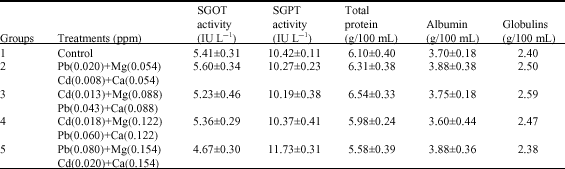
The results also indicate that the addition of calcium and magnesium to the different concentrations of cadmium and lead that caused liver damage in the first phase of this research was ameliorated (Table 2). This is reflected in the SGPT and SGOT activities, which were not significantly different (p>0.05) between the control and the various concentrations of cadmium and lead with the addition of calcium and magnesium. Total proteins for all the concentrations of the combination of lead and cadmium with the addition of calcium and magnesium were not significantly different (p>0.05) from control. There was no significant difference (p>0.05) between the albumin concentration of all the concentrations of the combination of cadmium and lead with the addition of calcium and magnesium.
Histopathological examinations of the hepatocytes of the rats in group 2-5 show that they were virtually the same and were not significantly different from the control group (Fig. 2b, 3b, 4b and 5b).
| Table 1: | Serum liver-specific enzyme activities, total proteins, albumin and globulin levels of rats treated with varying concentrations of the combination of cadmium and lead |
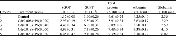 | |
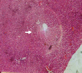 | |
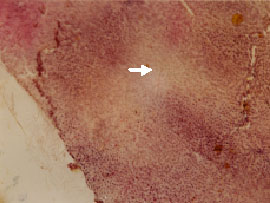
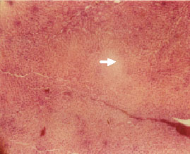
| Fig. 1: | L/S x 400. Representative renal biopsy of the control rats showing normal hepatocytes radiating from the central vein as spokes of a bicycle wheel (arrow) |
 | |
| Fig. 2a: | L/S x 400. Representative renal biopsy of the rats treated with 0.008 and 0.020 mg of cadmium and lead respectively in combination. The liver has patches of nodules. Within the nodules are cords of liver cells that look relatively normal (arrow) |
 | |
| Fig. 2b: | L/S x 400. Representative renal biopsy of the rats treated with 0.008 and 0.020 mg L-1 of cadmium and lead respectively in combination with the addition of calcium and magnesium. It shows relatively normal hepatocytes (arrow) |
 | |
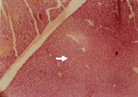
| Fig. 3a: | L/S x 400. Representative hepatic biopsy of the rats treated with 0.013 and 0.040 mg of cadmium and lead respectively in combination. The hepathocytes show mild damage with cords of regenerating cells staining deep purple (arrows) |
 | |
| Fig. 3b: | L/S x 400. Representative hepatic biopsy of the rats treated with 0.013 and 0.040 mg L-1 of cadmium and lead respectively in combination with the addition of calcium and magnesium. The hepatocytes are relatively normal (arrow) |
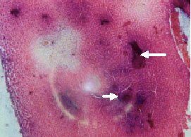 | |
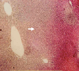
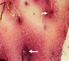
| Fig. 4a: | L/S x 400. Representative hepatic biopsy of the rats treated with 0.018 and 0.060 mg of cadmium and Lead respectively in combination. Throughout the liver there are very large number of small hepaplastic round nodules of regenerating cells. Between the nodules are cords of liver cells which look relatively normal apart from the sinusoids which are much wider |
 | |
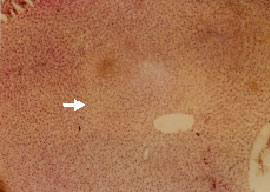
| Fig. 4b: | L/S x 400. Representative hepatic biopsy of the rats treated with 0.018 and 0.060 mg L-1 of cadmium and lead respectively in combination with the addition of calcium and magnesium. The hepatocytes are relatively normal (arrow) |
 | |
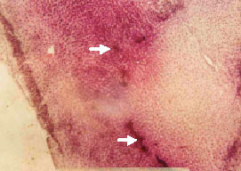
| Fig. 5a: | L/S x 400. Representative hepatic biopsy of the rats treated with 0.023 and 0.080 mg of cadmium and lead respectively in combination. The hepatocytes contain large number of small round bodies that stained deep purplish red which are present within the hepatocytes. This is mostly associated with drugs or ionization (arrows) |
 | |
| Fig. 5b: | L/S x 400. Representative hepatic biopsy of the rats treated with 0.023 and 0.080 mg L-1 of cadmium and lead respectively in combination with the addition of calcium and magnesium. The hepatocytes are relatively normal (arrow) |
| Table 2: | Serum liver-specific enzyme activities, total protein, albumin and globulin levels when magnesium and calcium where added to the same concentrations in Table 1 |
 | |
DISCUSSION
The results obtained from the study show that enzyme activities for liver-specific enzymes increased as a function of elevations in the concentrations of cadmium and lead. This indicates some damage to the liver which is dose dependent and this is supported by the histopathological studies which reveal that the damage is a function of concentration of the heavy metals used (Fig. 1, 2a, 3a, 4a and 5a). This confirms the hepatotoxicity of cadmium and lead.
Total protein is seen to be increasing as the concentrations of the metals are elevated. This observation may be as a result of the injury inflicted on the liver thereby making the proteins synthesized in the liver spill out into the blood. But when magnesium and calcium were added to all the different concentrations of the combination of cadmium and lead, total proteins were not significantly different from control. This also points to the fact that the damage caused by cadmium and lead had been ameliorated.
In contrast, albumin, a protein predominantly produced in the liver, decreased as the concentration of the metals were elevated. This suggests that the heavy metals, cadmium and lead, when present in toxic concentrations in the system impair protein synthesis in the liver. Results also show that the activities of the liver-specific enzymes were not significantly different (p>0.05) between the higher and lower concentrations of the combination of cadmium and lead with the addition of calcium and magnesium. The albumin levels were not significantly different (p>0.05) either, signifying that the damage observed when these same concentrations where administered without calcium and magnesium in phase 1 of this research, had been ameliorated (Table 2). The high concentration of calcium and magnesium might have been the factor that reduced the absorption of the two metals as Meredith et al. (1977) found out that calcium in domestic hard water significantly decreased absorption of lead in rats. These results give supporting evidence that in soft water regions, the uptake of cadmium and lead from drinking water could be increased because of the absence of calcium and magnesium in the drinking water, particularly when dietary calcium and magnesium are low.
The relationship between drinking water hardness and absorption of lead and cadmium could be important in view of general public health (Satarug et al., 2004). The insignificant difference (p>0.05) in the albumin concentration of the lower and higher concentration of lead and cadmium with the addition of calcium and magnesium as compared to control, suggest that protein synthetic capacity of the liver was not significantly affected for all the groups in the second phase of this research. This observation gives credence to the fact that there exist mutual exclusivity between cadmium/lead and calcium/magnesium at the level of intestinal absorption, distribution and excretion mechanisms (Neiboer and Fletcher, 1996). The knowledge obtained from the interactions between these metals is an important toxicological principle which if exploited would go a long way to improve public health (Neiboer and Fletcher, 1996).
Histopathological studies of the liver cells agree with the liver-specific enzyme activities, albumin concentrations, total proteins and the globulin concentrations. This is reflected in the fact that the histopathology of the liver of the rats in group 2, 3, 4 and 5, which were damaged as a function of elevation in the concentration of cadmium and lead were not significantly damaged with the addition of calcium and magnesium.
From the foregoing, it can be said that there is evidence that magnesium and calcium have hepatoprotective potential against cadmium and lead toxicities. This knowledge clearly points to the fact that nutrients function interactively both in the body and in the environment. The positive things that we can do to reduce lead and cadmium problems in our bodies include eating a wholesome diet with plenty of fresh fruits, vegetables and whole grains to obtain adequate minerals, avoiding refined foods and if recommended, take mineral supplement so as to competitively reduce cadmium and lead absorption. This can only be true in an uncontaminated environment as it is known that bioaccumulations of cadmium and lead in cereals and vegetables are high on contaminated soils (Silva et al., 2005).
CONCLUSION
This study has proved that cadmium and lead, when present in the environment can be hepatotoxic when ingested with food or water. The toxicity increases as the concentrations of the two metals are elevated. But the good news is that calcium and magnesium from this research can protect the liver from cadmium and lead toxicity well. Therefore, good levels of these minerals in the diets of subjects in polluted environments can reduce cadmium and lead toxicities as the results have shown.
REFERENCES
- Gazuwa, S.Y., J.D. Dabak and G.A. Ubom, 2006. Iron contamination of two local alcoholic drinks. J. Med. Trop., 8: 32-38.
Direct Link - Hatton D.C. and D.A. McCarron, 1994. Dietary calcium and blood pressure in expérimental models of hypertension. Nut. Rev., 52: 367-375.
PubMed - Meredith, P.A., M.R. Moore and A. Goldberg, 1977. The effect of calcium on lead absorption in rats. Biochem. J., 166: 531-537.
PubMedDirect Link - Peraza, M.A., F. Ayala-Fierro, D.S. Barber, E. Casarez and L.T. Rael, 1998. Effects of micronutrients on metal toxicity. Environ. Health Perspect., 10: 203-216.
Direct Link - Reitman, S. and S. Frankel, 1957. A colorimetric method for the determination of serum glutamic oxalacetic and glutamic pyruvic transaminases. Am. J. Clin. Pathol., 28: 56-63.
CrossRefPubMedDirect Link - De Fonseca, F.R., M. Navarro, R. Gomez, L. Escuredo and F. Nava et al., 2001. An anorexic lipid mediator regulated by feeding. Nature, 414: 209-212.
CrossRefDirect Link - Satarug, S., M. Nishijo, P. Ujjin, Y. Vanavanitkun, J.R. Baker and M.R. Moore, 2004. Evidence of concurrent effects of exposure to environmental Cadmium and Lead on Hepatic CYP2A6 Phenotype and Renal Function Biomakers in Nonsmokers. Environ. Health Perspect., 112: 1512-1518.
CrossRef - Silva, L.O., R.G.P. Barrocas and S.C. Jacob, 2005. Dietary intake and health effects of selected toxic elements. Braz. J. Plant Physiol., 17: 79-93.
CrossRef - Ubom, G.A., 1991. The goitre-soil-water-diet relationship: Case study in Plateau State, Nigeria. Sci. Total Environ., 107: 1-11.
PubMedDirect Link